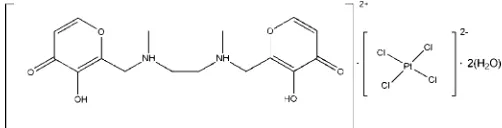
N
,
N
000-bis[(3-hydroxy-4(4
H
)-oxypyran-2-yl)methyl]-
N
,
N
000-dimethylethylene-1,2-diammonium tetrachloridoplatinate(II)
dihydrate
Vieri Fusi,aLuca Giorgi,aEleonora Macedi,bPaola Paolib* and Patrizia Rossib
aDepartment of Basic Sciences and Fundamentals, University of Urbino, I-61029 Urbino, Italy, andbDip. Energetica ‘Sergio Stecco’, University of Firenze, Via S. Marta 3, I-50139 Firenze, Italy
Correspondence e-mail: paolapaoli@unifi.it
Received 10 September 2012; accepted 28 September 2012
Key indicators: single-crystal X-ray study;T= 150 K; mean(C–C) = 0.005 A˚;
Rfactor = 0.023;wRfactor = 0.049; data-to-parameter ratio = 17.2.
The title compound (C16H22N2O6)[PtCl4]2H2O, shows anti-proliferative activity in eight tumor cell lines. The asymmetric unit consists of one solvent water molecule on a general position, and one half of each of the polyammonium cation and the tetrachloridoplatinate(II) anion, both of them located on centers of inversion. In the crystal, the cations are connected via hydrogen bonding between the carbonyl O atoms and the hydroxyl H atoms into zigzag chains that elongate in the c-axis direction. In addition, the carbonyl O atom is hydrogen-bonded to the water molecule which, in turn, interacts with the [PtCl4]2anion. Finally, the chains are
linked by N—H+ Cl interactions into a three-dimensional network.
Related literature
For the antitumor activity of maltol (systematic name: 3-hy-droxy-2-methyl-4-pyrone) and polyamines, see: Casero & Woster (2001); Lianget al.(2006); Murakamiet al.(2006). For background to the synthesis, solution behaviour, structural properties and biological activity of N,N0
-bis[(3-hydroxy-4-pyron-2-yl)methyl]-N,N0-dimethylethylendiamine (Malten),
see: Amatoriet al.(2010, 2012).
Experimental
Crystal data
(C16H22N2O6)[PtCl4]2H2O Mr= 711.28
Triclinic,P1
a= 6.4775 (4) A˚
b= 7.0037 (4) A˚
c= 13.1628 (8) A˚
= 88.810 (5)
= 87.033 (5)
= 71.927 (6)
V= 566.92 (6) A˚3 Z= 1
MoKradiation
= 6.71 mm1 T= 150 K
0.320.220.20 mm
Data collection
Oxford Diffraction Xcalibur3 diffractometer
Absorption correction: multi-scan (CrysAlis PRO; Oxford Diffraction 2009)
Tmin= 0.164,Tmax= 0.262
9431 measured reflections 2719 independent reflections 2694 reflections withI> 2(I)
Rint= 0.044
Refinement
R[F2> 2(F2)] = 0.023 wR(F2) = 0.049 S= 1.02 2719 reflections 158 parameters
H atoms treated by a mixture of independent and constrained refinement
max= 1.44 e A˚
3
min=1.14 e A˚
[image:1.610.44.295.646.710.2]3
Table 1
Hydrogen-bond geometry (A˚ ,).
D—H A D—H H A D A D—H A
O1—H1O O2i 0.73 (4) 1.98 (4) 2.655 (4) 154 (4) O1W—H1WB O2ii
0.79 (5) 2.07 (5) 2.853 (4) 171 (5) O1W—H1WA Cl2 0.84 (5) 2.45 (5) 3.282 (3) 174 (5) N1—H1N Cl1iii
0.74 (4) 2.79 (4) 3.380 (3) 139 (4) N1—H1N Cl1iv
0.74 (4) 2.79 (4) 3.362 (3) 136 (4)
Symmetry codes: (i) xþ2;yþ1;z; (ii) xþ2;y;z; (iii)
xþ2;yþ1;zþ1; (iv)x1;yþ1;z.
Data collection: CrysAlis PRO (Oxford Diffraction 2009); cell refinement: CrysAlis PRO; data reduction: CrysAlis PRO; program(s) used to solve structure:SIR97 (Altomare et al., 1999); program(s) used to refine structure:SHELXL97(Sheldrick, 2008); molecular graphics: ORTEP-3 (Farrugia, 1997); software used to prepare material for publication:PARST97(Nardelli, 1995).
The authors acknowledge CRIST (Centro di Cristallografia Strutturale, University of Firenze), where the data collection was performed, and the Italian Ministero dell’Istruzione dell’Universita` e della Ricerca (MIUR), PRIN2009, for financial support.
Supplementary data and figures for this paper are available from the IUCr electronic archives (Reference: NC2294).
References
Altomare, A., Burla, M. C., Camalli, M., Cascarano, G. L., Giacovazzo, C., Guagliardi, A., Moliterni, A. G. G., Polidori, G. & Spagna, R. (1999).J. Appl. Cryst.32, 115–119.
Amatori, S., Ambrosi, G., Fanelli, M., Formica, M., Fusi, V., Giorgi, L., Macedi, E., Micheloni, M., Paoli, P., Pontellini, R. & Rossi, P. (2012).J. Org. Chem. 77, 2207–2218.
Amatori, S., Bagaloni, I., Fanelli, M., Formica, M., Fusi, V., Giorgi, L. & Macedi, E. (2010).Br. J. Cancer,103, 239–248.
Casero, R. A. J. & Woster, P. M. J. (2001).Med. Chem.44, 1–26.
metal-organic compounds
Acta Cryst.(2012). E68, m1323–m1324 doi:10.1107/S1600536812040949 Fusiet al.
m1323
Acta Crystallographica Section EStructure Reports Online
Farrugia, L. J. (1997).J. Appl. Cryst.30, 565.
Liang, F., Wan, S., Li, Z., Xiong, X., Yang, L., Zhou, X. & Wu, C. (2006).Curr. Med. Chem.13, 711–727.
Murakami, K., Ishida, K., Watakabe, K., Tsubouchi, R., Naruse, M. & Yoshino, M. (2006).Toxicol. Lett.161, 102–107.
Nardelli, M. (1995).J. Appl. Cryst.28, 659.
Oxford Diffraction (2009).CrysAlis PRO. Oxford Diffraction Ltd, Abingdon, England.
supporting information
sup-1
Acta Cryst. (2012). E68, m1323–m1324
supporting information
Acta Cryst. (2012). E68, m1323–m1324 [doi:10.1107/S1600536812040949]
N
,
N
′
-bis[(3-hydroxy-4(4
H
)-oxypyran-2-yl)methyl]-
N
,
N
′
-dimethylethylene-1,2-diammonium tetrachloridoplatinate(II) dihydrate
Vieri Fusi, Luca Giorgi, Eleonora Macedi, Paola Paoli and Patrizia Rossi
S1. Comment
Maltol (3-hydroxy-2-methyl-4-pyrone) is a natural compound, which exhibits interesting antineoplastic activities
(Murakami et al., 2006). At the same time, linear polyamines are also known antitumor agents (Liang et al., 2006; Casero
& Woster, 2001). For these reasons we synthesized and studied compound N,N
′-bis((3-hydroxy-4-pyron-2-yl)methyl)-N,N′-dimethylethylendiamine (Malten) coupling two Maltol units to an aliphatic diamine. Malten has shown
antiproliferative activity in eight tumor cell lines (Amatori et al.,2010; Amatori et al., 2012). In the asymmetric unit of the
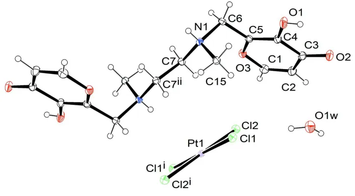
title compound half of the polyammonium cation [H2Malten]2+ and of the tetrachloroplatinate(II) counterion are present,
together with a crystallization water molecule. The two halves of each ion are related by a center of symmetry (Fig. 1).
The [H2Malten]2+ polyammonium chain, which joins the two aromatic rings, has an all-trans conformation and defines a
plane which forms an angle of 65.4 (2)° with each of them. In the crystal lattice the [H2Malten]2+ cations are each linked
by two pairs of complementary O—H···O hydrogen bonds into centrosymmetric dimers, which are further linked into
chains along the c axis (Fig. 2 and Table 1). Moreover, the carbonyl O atom (O2) is H-bonded to the lattice water
molecule, which is also linked to the (PtCl4)2- anion by O—H···Cl interactions. Finally, the cations and anions are linked
by N—H+···Cl interactions.
S2. Experimental
Malten.2HClO
4 was dissolved in H2O, K2PtCl4 was added and the pH adjusted to 3. Crystals suitable for X-ray analysis
formed in one day at room temperature.
S3. Refinement
The O—H and N—H H atoms were located in the Fourier difference map and refined with varying coordinates isotropic.
The C—H H atoms were introduced in calculated position and refined isotropic with Uiso(H) 1.2 times Ueq(C) (1.5 for
Figure 1
Crystal structure of the title compound with labelling and displacement ellipsoids drawn at the 30% probability level.
Symmetry codes: i) = -x + 2, -y, -z + 1; ii) = -x + 1, -y + 1, -z + 1.
Figure 2
Crystal structure of the title compound with view along the a axis. Intermolecular hydrogen bonding is shown as dashed
lines.
N,N′-bis[(3-hydroxy-4(4H)-oxypyran-2-yl)methyl]-N, N′-dimethylethylene-1,2-diammonium
tetrachloridoplatinate(II) dihydrate
Crystal data
(C16H22N2O6)[PtCl4]·2H2O
Mr = 711.28 Triclinic, P1 Hall symbol: -P 1 a = 6.4775 (4) Å b = 7.0037 (4) Å c = 13.1628 (8) Å α = 88.810 (5)° β = 87.033 (5)°
γ = 71.927 (6)° V = 566.92 (6) Å3
Z = 1 F(000) = 346 Dx = 2.083 Mg m−3
Mo Kα radiation, λ = 0.71073 Å Cell parameters from 7279 reflections θ = 4.1–29.2°
[image:4.610.123.489.316.490.2]supporting information
sup-3
Acta Cryst. (2012). E68, m1323–m1324
T = 150 K
Prismatic, light yellow
0.32 × 0.22 × 0.20 mm
Data collection
Oxford Diffraction Xcalibur3 diffractometer
Radiation source: Enhance (Mo) X-ray Source Graphite monochromator
Detector resolution: 16.4547 pixels mm-1
ω scans
Absorption correction: multi-scan
(CrysAlis PRO; Oxford Diffraction 2009) Tmin = 0.164, Tmax = 0.262
9431 measured reflections 2719 independent reflections 2694 reflections with I > 2σ(I) Rint = 0.044
θmax = 29.3°, θmin = 4.1°
h = −8→8 k = −9→9 l = −17→17
Refinement
Refinement on F2 Least-squares matrix: full R[F2 > 2σ(F2)] = 0.023
wR(F2) = 0.049
S = 1.02 2719 reflections 158 parameters 0 restraints
Primary atom site location: structure-invariant direct methods
Secondary atom site location: difference Fourier map
Hydrogen site location: inferred from neighbouring sites
H atoms treated by a mixture of independent and constrained refinement
w = 1/[σ2(F
o2) + (0.0253P)2] where P = (Fo2 + 2Fc2)/3 (Δ/σ)max < 0.001
Δρmax = 1.44 e Å−3 Δρmin = −1.14 e Å−3
Special details
Geometry. All s.u.'s (except the s.u. in the dihedral angle between two l.s. planes) are estimated using the full covariance matrix. The cell s.u.'s are taken into account individually in the estimation of s.u.'s in distances, angles and torsion angles; correlations between s.u.'s in cell parameters are only used when they are defined by crystal symmetry. An approximate (isotropic) treatment of cell s.u.'s is used for estimating s.u.'s involving l.s. planes.
Refinement. Refinement of F2 against ALL reflections. The weighted R-factor wR and goodness of fit S are based on F2, conventional R-factors R are based on F, with F set to zero for negative F2. The threshold expression of F2 > 2σ(F2) is used only for calculating R-factors(gt) etc. and is not relevant to the choice of reflections for refinement. R-factors based on F2 are statistically about twice as large as those based on F, and R- factors based on ALL data will be even larger.
Fractional atomic coordinates and isotropic or equivalent isotropic displacement parameters (Å2)
x y z Uiso*/Ueq
Pt1 1.0000 0.0000 0.5000 0.01531 (6)
Cl1 1.25762 (12) 0.12554 (11) 0.42237 (6) 0.01926 (15)
Cl2 0.82520 (13) 0.02992 (11) 0.34889 (6) 0.02098 (16)
O1 0.8029 (5) 0.6477 (4) 0.1284 (2) 0.0298 (6)
O2 0.8598 (4) 0.3386 (4) −0.00821 (17) 0.0278 (5)
O3 0.3953 (4) 0.4312 (3) 0.22157 (16) 0.0218 (5)
N1 0.5757 (4) 0.6487 (4) 0.3876 (2) 0.0169 (5)
C1 0.4294 (6) 0.2757 (5) 0.1565 (3) 0.0261 (7)
H1 0.3424 0.1924 0.1649 0.031*
C2 0.5823 (6) 0.2365 (5) 0.0809 (3) 0.0263 (7)
H2 0.5998 0.1263 0.0394 0.032*
C3 0.7197 (5) 0.3601 (5) 0.0625 (2) 0.0224 (7)
C5 0.5256 (5) 0.5510 (5) 0.2108 (2) 0.0187 (6)
C6 0.4731 (5) 0.7161 (5) 0.2869 (2) 0.0190 (6)
H6A 0.3166 0.7682 0.2984 0.023*
H6B 0.5233 0.8244 0.2595 0.023*
C7 0.4568 (5) 0.5266 (5) 0.4470 (2) 0.0181 (6)
H7A 0.4719 0.4039 0.4103 0.022*
H7B 0.3033 0.6018 0.4532 0.022*
C15 0.8152 (5) 0.5442 (5) 0.3735 (2) 0.0181 (6)
H15A 0.8821 0.6290 0.3351 0.027*
H15B 0.8389 0.4207 0.3376 0.027*
H15C 0.8778 0.5159 0.4388 0.027*
O1W 1.1440 (5) −0.0251 (4) 0.1423 (2) 0.0343 (6)
H1N 0.559 (6) 0.742 (6) 0.417 (3) 0.028 (11)*
H1O 0.888 (7) 0.623 (6) 0.088 (3) 0.024 (11)*
H1WA 1.062 (9) −0.020 (8) 0.194 (4) 0.059 (16)*
H1WB 1.128 (7) −0.105 (7) 0.104 (3) 0.040 (13)*
Atomic displacement parameters (Å2)
U11 U22 U33 U12 U13 U23
Pt1 0.01339 (9) 0.01279 (9) 0.02021 (9) −0.00476 (6) 0.00072 (6) −0.00306 (6)
Cl1 0.0169 (4) 0.0185 (4) 0.0241 (4) −0.0084 (3) 0.0032 (3) −0.0031 (3)
Cl2 0.0223 (4) 0.0208 (4) 0.0219 (4) −0.0092 (3) −0.0040 (3) −0.0009 (3)
O1 0.0347 (15) 0.0327 (14) 0.0275 (13) −0.0200 (12) 0.0137 (12) −0.0108 (11)
O2 0.0310 (14) 0.0309 (13) 0.0237 (12) −0.0136 (11) 0.0074 (10) −0.0087 (10)
O3 0.0236 (12) 0.0246 (12) 0.0197 (11) −0.0112 (10) −0.0005 (9) −0.0021 (9)
N1 0.0197 (14) 0.0154 (13) 0.0168 (12) −0.0074 (11) 0.0002 (10) −0.0020 (10)
C1 0.0284 (19) 0.0261 (18) 0.0283 (17) −0.0143 (15) −0.0056 (15) 0.0001 (14)
C2 0.0310 (19) 0.0242 (17) 0.0271 (17) −0.0134 (15) 0.0003 (15) −0.0062 (13)
C3 0.0251 (18) 0.0222 (16) 0.0196 (15) −0.0064 (13) −0.0035 (13) −0.0020 (12)
C4 0.0236 (17) 0.0208 (16) 0.0191 (15) −0.0096 (13) −0.0003 (13) −0.0030 (12)
C5 0.0218 (16) 0.0177 (15) 0.0172 (14) −0.0065 (13) −0.0041 (13) 0.0014 (11)
C6 0.0220 (16) 0.0170 (15) 0.0170 (14) −0.0047 (12) −0.0027 (12) 0.0018 (11)
C7 0.0171 (15) 0.0176 (15) 0.0200 (15) −0.0060 (12) 0.0006 (12) 0.0009 (11)
C15 0.0158 (15) 0.0189 (15) 0.0194 (14) −0.0051 (12) 0.0007 (12) −0.0008 (11)
O1W 0.0380 (17) 0.0383 (16) 0.0315 (15) −0.0187 (13) −0.0001 (13) −0.0064 (12)
Geometric parameters (Å, º)
Pt1—Cl1i 2.3018 (8) C2—C3 1.431 (5)
Pt1—Cl1 2.3018 (8) C2—H2 0.9300
Pt1—Cl2i 2.3132 (7) C3—C4 1.462 (4)
Pt1—Cl2 2.3132 (7) C4—C5 1.348 (5)
O1—C4 1.345 (4) C5—C6 1.491 (4)
O1—H1O 0.73 (4) C6—H6A 0.9700
O2—C3 1.243 (4) C6—H6B 0.9700
O3—C1 1.356 (4) C7—C7ii 1.526 (6)
supporting information
sup-5
Acta Cryst. (2012). E68, m1323–m1324
N1—C15 1.499 (4) C7—H7B 0.9700
N1—C7 1.501 (4) C15—H15A 0.9600
N1—C6 1.514 (4) C15—H15B 0.9600
N1—H1N 0.74 (4) C15—H15C 0.9600
C1—C2 1.337 (5) O1W—H1WA 0.84 (5)
C1—H1 0.9300 O1W—H1WB 0.79 (5)
Cl1i—Pt1—Cl1 180.00 (4) O1—C4—C3 119.7 (3)
Cl1i—Pt1—Cl2i 90.28 (3) C5—C4—C3 121.2 (3)
Cl1—Pt1—Cl2i 89.72 (3) C4—C5—O3 121.9 (3)
Cl1i—Pt1—Cl2 89.72 (3) C4—C5—C6 124.4 (3)
Cl1—Pt1—Cl2 90.28 (3) O3—C5—C6 113.7 (3)
Cl2i—Pt1—Cl2 180.0 C5—C6—N1 112.9 (2)
C4—O1—H1O 115 (3) C5—C6—H6A 109.0
C1—O3—C5 118.6 (3) N1—C6—H6A 109.0
C15—N1—C7 113.3 (2) C5—C6—H6B 109.0
C15—N1—C6 111.5 (2) N1—C6—H6B 109.0
C7—N1—C6 110.8 (2) H6A—C6—H6B 107.8
C15—N1—H1N 109 (3) N1—C7—C7ii 111.9 (3)
C7—N1—H1N 107 (3) N1—C7—H7A 109.2
C6—N1—H1N 106 (3) C7ii—C7—H7A 109.2
C2—C1—O3 123.1 (3) N1—C7—H7B 109.2
C2—C1—H1 118.5 C7ii—C7—H7B 109.2
O3—C1—H1 118.5 H7A—C7—H7B 107.9
C1—C2—C3 121.5 (3) N1—C15—H15A 109.5
C1—C2—H2 119.2 N1—C15—H15B 109.5
C3—C2—H2 119.2 H15A—C15—H15B 109.5
O2—C3—C2 125.6 (3) N1—C15—H15C 109.5
O2—C3—C4 120.7 (3) H15A—C15—H15C 109.5
C2—C3—C4 113.7 (3) H15B—C15—H15C 109.5
O1—C4—C5 119.1 (3) H1WA—O1W—H1WB 109 (5)
Symmetry codes: (i) −x+2, −y, −z+1; (ii) −x+1, −y+1, −z+1.
Hydrogen-bond geometry (Å, º)
D—H···A D—H H···A D···A D—H···A
O1—H1O···O2iii 0.73 (4) 1.98 (4) 2.655 (4) 154 (4)
O1W—H1WB···O2iv 0.79 (5) 2.07 (5) 2.853 (4) 171 (4)
O1W—H1WA···Cl2 0.84 (5) 2.45 (5) 3.282 (3) 174 (5)
N1—H1N···Cl1v 0.74 (4) 2.79 (4) 3.380 (3) 139 (4)
N1—H1N···Cl1vi 0.74 (4) 2.79 (4) 3.362 (3) 136 (4)