The plant-parasitic nematodes are the most important pests around the world. Many species of the plant-parasitic nematodes cause high losses of crop yield and many of them have a quarantine status. The stem and bulb nematode Ditylenchus dipsaci (Kühn 1857) Filipjev 1836 is a migratory endoparasite nematode of over five hundred vas-cular plant species. The stem nematode D. dipsaci is prevalent in a wide range of climatic condi-tions, where moisture regimes enable nematode infection, multiplication and dispersal. The main method of D. dipsaci control is crop rotation, but the presence of morphologically indistinguishable host races with different host preferences makes rotation difficult (Wendt et al. 1993). The
biologi-cal races exhibit different degrees of reproductive isolation, such as partial or complete reproduc-tive incompatibility (Erikson 1974). Each biologi-cal race is able to complete its life cycle only on a specific plant host. Among the most important plant hosts of D. dipsaci in Central Europe are bulb vegetables (onion, garlic and leek), carrot, alfalfa, clover, sugar beet, chicory, potatoes, strawberry, ornamental bulb plants (Narcissus spp.), and also many common weeds. Symptoms on host plants are not always specific as for appearance. Early infested plants and low infested seeds show no symptoms. Nematodes cause swellings and distor-tion of aerial plant parts and necrosis or rotting of stem bases, bulbs, tubers and rhizomes. The
Conversion of sequence-characterized amplified region
(SCAR) bands into high-throughput DNA markers based
on RAPD technique for detection of the stem nematode
Ditylenchus dipsaci
in crucial plant hosts
M. Zouhar
1, M. Marek
1, O. Douda
2, J. Mazáková
1, P. Ryšánek
11
Faculty of Agrobiology, Food and Natural Resources, Czech University of Life Sciences
Prague, Czech Republic
2
Nematological Laboratory, Plant Medicine Division, Research Institut of Crop Production,
Czech Republic
ABSTRACT
Ditylenchus dipsaci, the stem nematode, is a migratory endoparasite of over 500 species of angiosperms. The main method of D. dipsaci control is crop rotation, but the presence of morphologically indistinguishable host races with different host preferences makes rotation generally ineffective. Therefore, a sensitive, rapid, reliable, as well as cost effective technique is needed for identification ofD. dipsaci in biological samples. This study describes the develop-ment of species-specific pairs of PCR oligonucleotides for detection and identification of theD. dipsaci stem ne-matode in various plant hosts. Designed DIT-2 primer pair specifically amplified a fragment of 325 bp, while DIT-5 primer pair always produced a fragment of 245 bp in all D. dipsaci isolates.Two developed SCAR primer pairs were further tested using template DNA extracted from a collection of twelve healthy plant hosts; no amplification was however observed. The developed PCR protocol has proved to be quite sensitive and able to specifically detect
D. dipsaci in artificially infested plant tissues.
Keywords:Ditylenchus dipsaci; stem nematodes; quarantine organism; SCAR; diagnostics; detection; specific PCR;
RAPD
stem nematode is resistant to low temperatures. In Central Europe this nematode overwinters without any damages. The stem nematode D. dipsaci is one of the most destructive plant-parasitic nematodes in the temperate climate zone. The host plants could be completely destroyed without a crop control (Brzecki 1998). The pivot element in the control of the stem nematodes should be fast, accurate and reliable diagnostics, including moni-toring of its geographic occurrence. Diagnostic methods of plant-parasitic nematodes are usually based on anatomy and morphology characteris-tics comparable with the original descriptions. Diagnostics using light microscopy technique is time-consuming and it depends too strongly on personal experiences (Ebrahimi et al. 2004).
During the last several years DNA-diagnos-tics or population study methods including RFLP (Curran et al. 1985), DNA hybridization (Burrows and Perry 1988), RAPD (Folkertsma et al. 1994, Williamson et al. 1997, Esquibet et al. 1998 and Zhang et al. 1998), allele specific-PCR (Zouhar et al. 2000), SCAR (Zijlstra 2000) and RAPD (Samal et al. 2003, Sedlak et al. 2004), have been developed for characterization and identification of plant-parasitic nematodes. Specific PCR assays based on ribosomal RNA gene cluster for detection of D. dipsaci were developed (Marek et al. 2005, Subbotin et al. 2005). Nevertheless, Subbotin et al. (2004) characterized nucleotide sequences of evolutionary divergent ITS1-5.8S-ITS2 cistron for a number of gall-forming and stem nematodes. These results showed a high ITS rDNA sequence homology among the stem nematode D. dipsaci and several members of Heteroanquina, Mesoanquina
and Subanquina genera. Therefore, an alternative molecular detection technique is needed to be developed for a reliable molecular detection and determination of the stem nematode D. dipsaci in biological materials, such as host plant tis-sues, soil samples etc. Alternative DNA markers for a differentiation of giant and normal types of D. dipsaci were previously characterized by SCAR and AFLP experimental approaches (Esquibet et al. 1998, Esquibet et al. 2003). However, these authors did not verify the developed SCAR and AFLP markers for routine molecular diagnostics purposes.
Hence, the main aim of the present study was the production of the appropriate collection of species-specific RAPD markers, their conversion to specific PCR-based assay for the rapid and sensitive identification of D. dipsaci normal type of biological races in plant organs and tissues of majority plant hosts in Central Europe.
MATERIAL AND METHODS
[image:2.595.62.535.572.764.2]Nematodes isolates and plant hosts. Populations of nematodes from free field cultures were deter-mined by morphological characters and cultivated on corresponding host plants. In this report geo-graphically different origin of D. dipsaci popula-tions were used (Table 1). Plants were placed in a cultivation chamber with controlled temperature and day length. Nematode inoculation was done by suspension of nematodes mix with dicarbo-xymethylcellulose (saturated solution) of final concentration 2%. Temperature was decreased on
Table 1. Origin of biological races of Ditylenchus dipsaci used in this study
Species Host plant Study code Geographic origin Origin
Ditylenchus dipsaci
Allium sativum As-B Blatnice Czech Republic
Allium sativum As-M Marefy Czech Republic
Allium sativum As-Bo Boškůvky Czech Republic
Allium sativum As-L Litomyšl Czech Republic
Allium sativum As-O Olomouc Czech Republic
Medicago sativa Ms-P Piešťany Slovakia
Cichorium inthybus Ci-L Ljubljana Slovenia
Allium cepa Ac-B Bari Italy
Daucus carota Dc-B Bari Italy
14–16°C for a better penetration and multiplica-tion of nematodes for two weeks. Nematodes were extracted from sliced plants with the Baerman funnel and several times rinsed in sterile water; subsequently they were used for DNA isolation.
DNA isolation and purification. DNA was iso-lated from nematodes, nematode-infested plant tissue and nematode free plant tissue (Table 4). In this part of work biological races of D. dip-saci normal type originated from garlic, carrot, alfalfa and chicory were used. For the isolation approximately 10 individuals or 0.5–1.0 g plant tissue of artificially inoculated plant with 10 in-dividuals were used. These kinds of biological material were crushed in microtube containing glass beads (250 µm diameter) using mini grinder (Pellet Pestle ®Motor, Sigma) and homogenized in 300 µl lysis buffer (100mM Tris-HCl (pH 8.0), 5mM EDTA, 200mM NaCl, 0.2% SDS and 0.4 mg/ml proteinase K). The mixture was incubated for 1 h at 55°C with shaking and finally denatured for 5 min at 85°C. The homogenate was mixed 1:1 with phenol (pH 8.0)-chloroform-isoamylalcohol (25:24:1), vortexed for 15 min and centrifuged at 7 000 × g. Each lysate (water phase) was transferred to a new tube to which equal volume chloroform was added and extraction was repeated. DNA was precipitated with equal volume of isopropanol in –20°C overnight or in liquid nitrogen for 20 min and centrifuged at 10 000 × g for 10 min. The supernatant was discarded and the remaining pellets were vacuum-dried. The pellets for each sample were resuspended in 50 µl TE (10mM Tris, 1mM EDTA (pH 8.0) or ddH2O. DNA was stored at –20°C for a long-term storage. The working stock of DNA for RAPD amplification was di-luted to approximately 50 ng/µl after quantifica-tion using the Helios Gamma spectrophotometer (ThermoSpectronic, USA).
RAPD fingerprinting protocol. Isolated nema-todes’ DNA was used for the amplification by three random primer sets (Operon Technology sets A, B and C). All RAPD reactions were performed in 25 µl volumes including: 100 ng of DNA, 200µM dNTPs, 20 pmol primer, 1.5mM MgCl2 and 1.5 U Taq DNA polymerase (Fermentas, Lithuania). Amplification conditions were as follows: an initial denaturation at 94°C for 2 min, after which 2 cycles of denatura-tion (1 min at 94°C), primer annealing (1 min at 35°C) and primer extension (2 min at 72°C) after 34 cycles of denaturation (30 sec at 94°C), primer annealing (30 sec at 35°C) and primer extension (1.5 min at 72°C), final extension (25 min at 72°C). Amplification was performed in the thermal
cy-cler (PTC 200 MJ Research Inc. USA). Aliquots (5.0 µl) of RAPD products were separated by the horizontal electrophoresis in 1.5% (w:v) agarose gel, with 1 × TBE buffer, stained with ethidium bromide (0.5 µg/ml) and analysed under ultraviolet (UV) light. The length of the DNA fragments was estimated by comparison with MassRuler 100 bp DNA ladder (Fermentas, Lithuania).
RAPD fragments selection and cloning. From obtained RAPD fingerprints different fragments were selected. These bands were cut and elu-ated from agarose gel by using the QIAEX II Gel Extraction Kit 150 (Qiagen). DNA fragments were directly cloned to pTZ57R/T vector using 3’-A overhangs generated by the Taq polymer-ase (InsT/Aclone™PCR Product Cloning Kit, Fermentas, Lithuania), following the protocol provided by the supplier and transformed into E. coli DH5. Blue-white selection was used for de-tection of positive bacterial colonies. Plasmid isola-tion from overnight-cultivated bacterial cultures was made with the Perfectprep Plasmid Mini Kit (Eppendorf, Germany). Plasmids were cleaved by restriction endonucleases (KpnI and PstI or BamHI and EcoRI) for verification of predicted cloned DNA fragment. Fragments were sequenced using the automatic sequencing system (ABI Prism 377, Perkin Elmer, USA). The sequence data were com-pared with other nucleotide sequences available throughout the National Center for Biotechnology Information (NCBI, USA) databases.
DNA amplification of plant hosts genomic DNA, Rbcl primer pair was used, which specifically ampli-fied partial sequence of plant chloroplast gene for ribulose-1,5-bisphosphate carboxylase. Aliquots (5.0 µl) of the PCR products were resolved by electrophoresis in 1.2% agarose gels and visualized as already stated above.
RESULTS AND DISCUSSION
DNA profiling by RAPD approach
To find species-specific fragments, RAPD-PCRs were performed with genomic DNA of some plant-parasitic nematodes, e.g. Meloidogyne fal-lax and M. hapla, M. chitwoodi (Zijlstra 2000). In our experiments, three sets of random prim-ers (Operon technology sets A, B and C) were analyzed in RAPD reactions using D. dipsaci ge-nomic DNA to find identical DNA bands among all tested biological races. Generated RAPD patterns were largely characteristic for each of biological races. Moreover, we also uncovered inter- and intra-population genetic diversity by experimental RAPD approach (data not shown). Nevertheless, we identified six requisite uniform DNA markers for all tested biological races of D. dipsaci gener-ated by OPA-18, OPA-19, OPA-20 and OPB-16 random decameric primers. Using the primers OPA-18, OPA-19, OPA-20, OPB-16, following fragments – 955 bp, 522 bp and 1103 bp, 703 bp
and 1007 bp, and 951 bp – were amplified strongly from all proved biological races of D. dipsaci, respectively (Figures 1–4). These 955, 522, 1103, 703, 1007 and 951 bp fragments were chosen for their uniformity, strong intensity and size, which would facilitate their cloning and sequencing. None of the RAPD fragments selected as specifically or more highly PCR amplified in DNA samples was polymorphic between the number of different D. dipsaci isolates.
Characterization of selected RAPD fragments
[image:4.595.101.255.504.685.2]The selected species-specific RAPD fragments were isolated, cloned and sequenced. Each ob-tained sequence was compared by the BLASTN algorithm with sequences within the GenBank, DDJB and EMBL databases. The closest database similarities for each RAPD sequence are shown in Table 3. Three RAPD markers were similar to bacterial organisms, one marker matched to Bubalus bubalis microsatellite BBMS27and last two show no significant similarity to any sequences from the GenBank database. From the obtained sequences, six specific primer pairs were designed by using Primer3 software. Nucleotide sequences of specific primers con-versed from RAPD markers and its PCR product size and established PCR conditions are shown in Table 2.
Figure 1. DNA fingerprint of four Ditylenchus dipsaci
biological races after PCR with primer OPA-18. Bands that were sequenced are marked. The MassRuler 100 bp DNA ladder (Fermentas, Lithuania) was used as mo-lecular marker
Figure 2. DNA fingerprint of four Ditylenchus dipsaci
biological races after PCR with primer OPA-20. Bands that were sequenced are marked. The MassRuler 100 bp DNA ladder (Fermentas, Lithuania) was used as mo-lecular marker
A
s-O
M
s-P
C
i-L
D
c-B
Kbp
0.9
0.7
0.5
A
s-O
M
s-P
C
i-L
D
c-B
Kbp
0.9
0.7
[image:4.595.328.501.505.685.2]Development of specific PCR-based assay
The specificity of the designed primer pairs was tested by attempting amplification using ge-nomic DNA of all isolates of proved D. dipsaci biological races as template. Only primer pairs DIT-2 and DIT-5 generated species-specific PCR
products for all isolates of our D. dipsaci collec-tion (Table 1). The DIT-2 primer pair specifically amplified a fragment of 325 bp, while DIT-5 primer pair always produced a fragment of 245 bp in all D. dipsaci isolates.
Unlike, other four primer pairs (DIT-1, DIT-3, DIT-4 and DIT-6) produced unspecific and
non-Figure 3. DNA fingerprint of four Ditylenchus dipsaci
[image:5.595.344.498.71.297.2]biological races after PCR with primer OPA-19. Bands that were sequenced are marked. The MassRuler 100 bp DNA ladder (Fermentas, Lithuania) was used as mo-lecular marker
Figure 4. DNA fingerprint of four Ditylenchus dipsaci
[image:5.595.104.263.73.297.2]biological races after PCR with primer OPB-16. Bands that were sequenced are marked. The MassRuler 100 bp DNA ladder (Fermentas, Lithuania) was used as mo-lecular marker
Table 2. Designed primers and PCR conditions used in this study
RAPD marker
RAPD product
size (bp)
Specific PCR oligonucleotides Specific PCR product size
(bp)
PCR condition
sense sequence 5´→3´ cycles No. temperature (°C)annealing
DIT-1OPA-18 955 forward reverse ACGTGGTGGATACGGCTATTT CATTTCGAGACGCACATTCTC 460 30 58
DIT-2 OPA-19 522 forward reverse CTGTCTGTGATTTCACGGTAGACGCAATGCACAGGTGGATAAAG 325 30 60
DIT-3 OPB-16 951 forward reverse GAATAATCAGCAGAGCGGTGA ATTCGATCACCTGTCCCACTT 449 30 57
DIT-4 OPA-20 703 forward reverse TTATCATGTTGGGGCTCTGTC CGGTCCAAAGGTGAACAAA 568 30 62
DIT-5 OPA-19 1103 forward reverse GAAAACCAAAGAGGCCGTAAC ACCTGATTCTGTACGGTGCAA 245 30 60
DIT-6 OPA-20 1007 forward reverse GAACAACCAGAATGGCGGTAT TGTACCTGGGTATTGCCTTTG 892 30 59
A
s-O
M
s-P
C
i-L
D
c-B
Kbp
0.9
0.7
0.5
A
s-O
M
s-P
C
i-L
D
c-B
Kbp
0.9 0.7
[image:5.595.63.533.544.762.2]reproducible PCR products with the use of genomic DNA of D. dipsaci. Therefore, these primer pairs were not suitable for a reliable molecular detec-tion of D. dipsaci and were rejected. Moreover, the
[image:6.595.65.533.77.204.2]specificity of primer pairs DIT-2 and DIT-5 was also examined using template genomic DNA of twelve plant hosts important for central Europe cli-mate conditions (Table 4). The primer pairs DIT-2
Table 3. Results of bioinformatic analyses of sequences acquired from RAPD
RAPD marker Bioinformatic predicted sequence homologyBLASTN E-value
DIT-1OPA-18 AY059069.1, Serratia marcescens aconitase gene 1e-24 DIT-2OPA-19 CP000152.1, Burkholderia sp. 383 chromosome 2 0.001
DIT-3OPB-16 No significant homology
DIT-4OPA-20 No significant homology
DIT-5OPA-19 AE008693.1, Salmonella typhimurium LT2 7e-11 DIT-6OPA-20 AY912156.1, Bubalus bubalis microsatellite BBMS27 2e-04
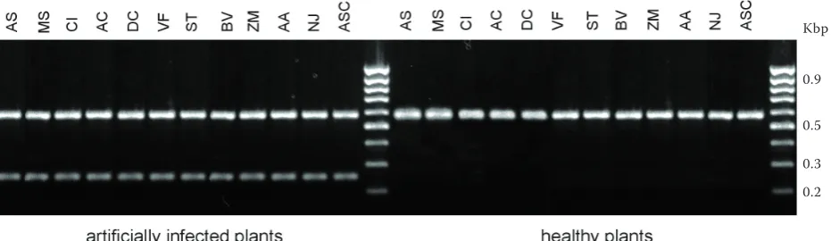
[image:6.595.68.527.316.436.2]Figure 6. PCR products from twelve different healthy and artificially infected plant species described in Table 4 amplified by primer pairs DIT-5 and Rbcl. Species-specific primer pair DIT-5 generated the fragment of 245 bp length and Rbcl which specifically amplified partial sequence of plant chloroplast gene for ribulose-1,5-bispho-sphate carboxylase approximately 590 bp. Biological race (study code As-B) was used in this experiment. Gen-erally, all isolates described in Table 1 and used for the same assay provided identical results (data not shown). The MassRuler 100 bp DNA ladder (Fermentas, Lithuania) was used as molecular marker
Figure 5. PCR products from twelve different healthy and artificially infected plant species described in Table 4 amplified by primer pairs DIT-2 and Rbcl. Species-specific primer pair DIT-2 generated the fragment of 325 bp length and Rbcl which specifically amplified partial sequence of plant chloroplast gene for ribulose-1,5-bispho-sphate carboxylase approximately 590 bp. Biological race (study code As-B) was used in this experiment. Gener-ally, all the isolates described in Table 1 and used for the same assay provided identical results (data not shown). The MassRuler 100 bp DNA ladder (Fermentas, Lithuania) was used as molecular marker
Kbp
0.9
0.5
0.3 0.2
Kbp
0.9
0.5
[image:6.595.68.534.541.676.2]and DIT-5 did not amplify any fragment where the genomic DNA of any plant hosts was used as template (Figures 5 and 6). Thirty cycles of PCR amplification using specific primers produced a sufficient amount of the predicted-size frag-ments (325 bp for DIT-2 and 245 bp for DIT-5) to visualize them on the ethidium bromide-stained gels, with one-fifth of the PCR reaction volume (5.0 µl) loaded on the gel. Previously, we described a PCR-based technique for sensitive identification of the stem nematode D. dipsaci in plant material (Marek et al. 2005). This technique was based on evolutionary divergent ITS1 and ITS2 sequences of the ribosomal RNA gene cluster (rDNA). The rDNA is multicopy, tandemly repeated array ac-cording in the nucleolar organizer region at one or several chromosomal sites (Szalanski et al. 1997), and is therefore very suitable for DNA amplification methods. However, Subbotin et al. (2004) pointed on the closest ITS rDNA sequence similarities between the stem nematode D. dipsaci and some species of Heteroanquina, Mesoanquina and Subanquina genera, which complicates de-velopment of molecular detection based on the rDNA cistron. Hence, this report shows the use-fulness of converting alternative RAPD markers into reliable SCAR markers. This study is the only second report of SCAR analysis on D. dipsaci. Esquibet et al. (1998) did previous SCAR analyses on the stem nematode D. dipsaci, but the authors did not solve DNA-diagnostics problems. In this study, we developed the first species-specific
PCR oligonucleotides useful for a sensitive and reliable detection and identification of the stem nematode D. dipsaci in plant host tissues. Some authors developed specific DNA probes for detec-tion of D. dipsaci by the Southern-blot technique (Palmer et al. 1991, Wendt et al. 1993). In each case, these probes were labeled radioactively; although radioisotope labeling could be avoided using non-radioactive detection methods (Allefs et al. 1990), relatively large amounts of DNA are required for hybridization.
It thus seems that this PCR-based method can be employed when the phytosanitary laboratories are asked to verify the health of the batch of plant materials. Moreover, in the case of quarantined fields or greenhouses this PCR assay would serve to ensure whether the quarantine procedures had been effective. As our data showed, the PCR-based detection of D. dipsaci in plant tissue would also markedly speed up this process and indicate in a short time whether specific precautions are required to prevent the spread of this stem nema-tode. In this study SCAR primers were designed to identify D. dipsaci, but the method offers great possibilities to create markers at species level, serving a much wider range of identification levels and facilitating genetic, molecular biology and phytopathological research.
Acknowledgement
The authors thank to Genomac Inc. for DNA sequencing services and to all the workers of the phytopathological team at the Department of Plant Protection in the Czech University of Agriculture in Prague, with particular thanks to Dr. J. Eberová for excellent discussion and advice during experimental work at molecular biology techniques.
REFERENCES
Allefs J.J.H.M., Salentijn E.M.J., Krens F.A., Rouwendal G.J.A. (1990): Optimization of non-radioactive South-ern blot hybridization: single copy detection and reuse of blots. Nucl. Acids Res., 18: 3099–3100.
Brzeski M.W. (1998): Nematodes of Tylenchina in Po-land and Temperate Europe. Muzeum i Instytut Zoo-logii Polska Akademia Nauk, Warszawa.
Burrows P.R., Perry R.N. (1988): Two cloned DNA frag-ments which differentiate Globodera pallida from
[image:7.595.63.291.90.315.2]G. rostochiensis. Rev. Nematol., 11: 441–445. Table 4. Plant species used in this study for testing
specificity of DIT-2 and DIT-5 primer pairs
Plant species Study code
Allium sativum AS
Medicago sativa MS
Cichorium inthybus CI
Allium cepa AC
Daucus carota DC
Vicia faba VF
Solanum tuberosum ST
Beta vulgaris BV
Zea mays ZM
Allium ampeloprasum porrum AA
Narcissius juncifolius NJ
Curran J., Baillie D.L., Webster J.M. (1985): Use of ge-nomic DNA restriction length differences to identify nematode species. Parasitology,90: 137–144. Ebrahimi N., Kheiri A., Pakniat M. (2004): Occurrence
of plant parasitic nematodes (Tylenchina) in sugar beet fields in Fars Province, Iran. Commun. Agr. Appl. Biol. Sci., 69: 397–401.
Erikson K.B. (1974): Intraspecific variation in Dity-lenchus dipsaci I. Compatibility tests with races. Nematologica, 20: 147–162.
Esquibet M., Bekal S., Castagnone-Sereno P., Gauthier J.P., Rivoal R., Caubel G. (1998): Differentiation of normal and giant Vicia faba populations of the stem nematode
Ditylenchus dipsaci: agreement between RAPD and phenotypic characteristics. Heredity, 81: 291–298. Esquibet M., Grenier E., Plantard O., Andaloussi F.A.,
Caubel G. (2003): DNA polymorphism in the stem nematode Ditylenchus dipsaci: Development of diag-nostic markers for normal and giant races. Genome,
46: 1077–1083.
Folkertsma R.T., Jeroen N.A., Rouppe Van Dert V., Marga P.E. (1994): Inter- and intraspecific variation between populations of Globodera rostochiensis and
G. pallida revealed by random amplified polymorphic DNA. Phytopathology, 84: 807–811.
Marek M., Zouhar M., Rysanek P., Havranek P. (2005): Analysis of ITS sequences of nuclear rDNA and de-velopment of a PCR-based assay for the rapid iden-tification of the stem nematode Ditylenchus dipsaci
(Nematoda: Anguinidae) in plant tissues. Helmin-thologia, 42: 49–56.
Palmer H.M., Atkinson H.J., Perry R.N. (1991): The use of DNA probes to identify Ditylenchus dipsaci. Rev. Nematol., 14: 625–628.
Samal S., Rout G.R., Lenka P.C. (2003): Analysis of genetic relationships between populations of cashew (Anacardium occidentale L.) by using morphologi-cal characterisation and RAPD markers. Plant Soil Environ., 49: 176–182.
Sedlák P., Melounová M., Skupinová S., Vejl P., Domkářová J. (2004): Study of European and Czech populations of potato cyst nematodes (Globodera
rostochiensis and G. pallida) by RAPD method. Plant Soil Environ., 50: 70–74.
Subbotin S.A., Krall E.L., Riley I.T., Chizhov V.N., Stae-lens A., De-Loose M., Moens M. (2004): Evolution of the gall-forming plant parasitic nematodes (Tylench-ida: Anguinidae) and their relationships with hosts as inferred from internal transcribed spacer sequences of nuclear ribosomal DNA. Mol. Phylogenet. Evol.,
30: 226–235.
Subbotin S.A., Madani M., Krall E., Sturhan D., Moens M. (2005): Molecular diagnostics, taxonomy, and phylogeny of the stem nematode Ditylenchus dipsaci species complex based on the sequences of the internal transcribed spacer-rDNA. Phytopathol-ogy, 95: 1308–1315.
Szalanski A.L., Sui D.D., Harris T.S., Powers T.O. (1997): Identification of cyst nematodes and agronomic regu-latory concern with PCR-RFLP of ITS. J. Nematol.,
29: 255–267.
Wendt K.R., Vrain T.C., Webster J.M. (1993): Separa-tion of three species of Ditylenchus and some host races of D. dipsaci by restriction fragment length polymorphism. J. Nematol., 25: 555–563.
Williamson V.M., Cashwell-Chen E.P., Westerdahl B.B., Wu F.F., Caryl G. (1997): A PCR assay to identify and distinguish single juveniles of Meloidogyne hapla and
M. chitwoodi. J. Nematol., 29: 9–15.
Zhang L., Dean R.A., Knap H.T., Lewis S.A. (1998): Diversity among a Heterodera glycines field isolate and derived inbreds based on RAPD analysis and reproduction on soybean genotypes. J. Nematol.,
30: 477–484.
Zijlstra C. (2000): Identification of Meloidogyne chit-woodi, M. fallax and M. hapla based on SCAR-PCR: a powerful way of enabling reliable identification of populations or individuals that share common traits. Eur. J. Plant Pathol., 106: 283–290.
Zouhar M., Rysanek P., Kocova M. (2000): Detection and differentiation of the potato cyst nematodes by PCR. Plant Protec. Sci., 36: 81–84.
Received on May 23, 2006
Corresponding author:
Ing. Miloslav Zouhar, Ph.D., Česká zemědělská univerzita v Praze, Fakulta agrobiologie, potravinových a přírodních zdrojů, 165 21 Praha 6-Suchdol, Česká republika