research communications
Acta Cryst.(2017). E73, 1875–1877 https://doi.org/10.1107/S2056989017016231
1875
Received 3 November 2017Accepted 10 November 2017
Edited by H. Ishida, Okayama University, Japan
Keywords:crystal structure; pyridine-2,6-di-carboxylate; pyrazole; square-pyramidal CuII
complex.
CCDC reference:1584872
Supporting information:this article has supporting information at journals.iucr.org/e
Crystal structure of aqua(1
H
-pyrazole-
j
N
2)-(pyridine-2,6-dicarboxylato-
j
3O
2,
N
,
O
6)copper(II)
dihydrate
Daeyoung Kim and Sung Kwon Kang*
Department of Chemistry, Chungnam National University, Daejeon 34134, Republic of Korea. *Correspondence e-mail: skkang@cnu.ac.kr
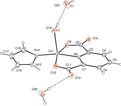
In the title compound, [Cu(C7H3NO4)(C3H4N2)(H2O)]2H2O, the Cu II
atom is coordinated by three O atoms and two N atoms, provided by a tridentate pyridine-2,6-dicarboxylate (pdc), one pyrazole and one water ligand, forming a slightly distorted square-pyramidal geometry [range of O—Cu—O and O— Cu—N bond angles = 79.55 (8)–166.22 (10)]. The water molecule is positioned at the apical position. In the crystal, the complex molecule and the two crystallographically independent non-coordinating water molecules are linked into a supramolecular layer structure parallel to theabplaneviaO—H O and N—H O hydrogen bonds.
1. Chemical context
Metal complexes with the tridentate ligand 2,6-bis[(1H -pyrazol-1-yl)methyl]pyridine are known to be catalysts of polyethylene polymerization (Singhet al., 2003; Watsonet al., 1987; Son et al., 2014; Kim & Kang, 2015). 2,6-Bis[(1H -pyrazol-1-yl)methyl]pyridine was oxidized to pyridine-2,6-di-carboxylate (pdc) by metal nitrate (Unuigboje & Anyile, 2007). The pdc molecule has been recognized as a component of bacterial spores, and is also useful in a variety of processes as an enzyme inhibitor, plant preservative and food sanitizer (Cui et al., 2011). The pdc molecule has been selected as a primary dibasic tridentate ligand and a metal complex with pdc was reported to be a new chemical sensor (Mistri et al., 2013). Attention has been paid to the design of various N -donor ligands with special structural properties in order to investigate the specific stereochemical requirements of a particular metal-binding site (Mukherjee, 2000). Various substituted N-donor heterocyclic ligands such as imidazole and pyrazole have been selected as a second ligand, so that the structural and electronic effects on the biologically important Cu—N bond could be probed (Ang et al., 1991; Chen et al., 2011; Lin et al., 2009; Liu et al., 2005). As part of these continuing studies, the title complex has been synthesized and characterized by single crystal X-ray diffraction.
2. Structural commentary
cated by thevalue of 0.113 (Addisonet al., 1984). The CuII atom lies in the center of the basal plane defined by two nitrogen atoms (N2 from pdc and N14 from pyrazole) and two oxygen atoms (O9 and O12 from pdc). The plane including the CuIIatom is almost planar, with an r.m.s. deviation of 0.0847 A˚ from the corresponding least-squares plane defined by the five constituent atoms. The pyrazole ring is twisted by 66.61 (10) from the basal plane. The apical Cu1—O19 bond length of 2.217 (2) A˚ is much longer than those of the basal Cu—O lengths [Cu1—O9 = 2.026 (2) A˚ and Cu1—O12 = 2.058 (2) A˚].
3. Supramolecular features
In the crystal, O—H O hydrogen bonds (O19— H19B O21, O20—H20B O13 and O20—H20A O10iii; symmetry code as in Table 1) link the complex molecule to the
non-coordinating water molecules (Fig. 1). Two crystal-lographically independent non-coordinating water molecules are also linked to each other by O—H O hydrogen bonds (O21—H21A O20iv and O21—H21B O20v; Table 1). Adjacent complex molecules are connected by other O— H O and N—H O hydrogen bonds (N15—H15 O12i and O19—H19A O9ii; Table 1). The above-mentioned intermolecular interactions stabilize and link the components into a two-dimensional network parallel to the ab plane (Fig. 2).
4. Database survey
A search of the Cambridge Structural Database (Version 5.37 with two updates, Groomet al., 2016) returned 1448 entries for crystal structures related to the name pyridine-2,6-di-carboxylato. Most of them are crystal structures of metal complexes. However, there are only four entries with a secondary ligand of a pyrazolyl derivative bonded to a tran-sition metal,viz. a Cu complex (Linet al., 2009; Wang et al., 2014) and Zn and Co complexes (Zhanget al., 2011).
1876
Kim and Kang [Cu(C7H3NO4)(C3H4N2)(H2O)]2H2O Acta Cryst.(2017). E73, 1875–1877
[image:2.610.313.566.85.178.2] [image:2.610.47.297.197.385.2]research communications
Table 1
Hydrogen-bond geometry (A˚ ,).
D—H A D—H H A D A D—H A
O19—H19B O21 0.75 (4) 2.09 (5) 2.831 (5) 169 (4)
O20—H20B O13 0.70 (5) 2.12 (5) 2.807 (4) 172 (6)
N15—H15 O12i 0.93 (4) 1.93 (4) 2.832 (3) 164 (4)
O19—H19A O9ii 0.70 (5) 2.12 (5) 2.805 (3) 165 (5)
O20—H20A O10iii 0.78 (5) 2.01 (5) 2.784 (4) 171 (5)
O21—H21A O20iv 0.91 (6) 2.05 (6) 2.933 (5) 163 (5)
O21—H21B O20v 0.81 (6) 2.17 (6) 2.938 (5) 161 (6)
Symmetry codes: (i)xþ1;y;z; (ii)x1;y;z; (iii)x1;yþ1;z; (iv)x;y1;z; (v)
[image:2.610.134.547.482.697.2]xþ1;y1;z.
Figure 2
Part of the packing diagram of the title compound, showing molecules linked by intermolecular O—H O and N—H O hydrogen bonds (dashed lines).
Figure 1
[image:2.610.46.294.485.703.2]5. Synthesis and crystallization
A solution of copper nitrate trihydrate (0.072 g, 0.3 mmol) in acetonitrile (5 ml) was added to a solution of 2,6-bis[(1H -pyrazol-1-yl)methyl]pyridine (0.072 g, 0.3 mmol) in aceto-nitrile (5 ml) in a pressure vessel. After sealing the high-pressure vessel, the resulting solution was stirred for three days at 403 K. The precipitate formed was removed by filtra-tion, and the filtrate was washed with acetonitrile and di-chloromethane to get a dark-green powder. Single crystals of the title compound were obtained from its aqueous solution by slow evaporation of the solvent at 333 K within five days.
6. Refinement
Crystal data, data collection and structure refinement details are summarized in Table 2. H atoms of the water molecules and the NH group were located in a difference-Fourier map and refined freely [refined distances; O—H = 0.70 (5)– 0.91 (6) A˚ and N—H = 0.93 (4) A˚]. Other H atoms were positioned geometrically and refined using a riding model, with C—H = 0.93 A˚ , and withUiso(H) = 1.2Ueq(C).
Funding information
This work was supported by research fund of Chungnam National University.
References
Addison, A. W., Rao, T. N., Reedijk, J., van Rijn, J. & Verschoor, G. C. (1984).J. Chem. Soc. Dalton Trans.pp. 1349–1356.
Ang, H. G., Kwik, W. L., Hanson, G. R., Crowther, J. A., McPartlin, M. & Choi, N. (1991). J. Chem. Soc. Dalton Trans. pp. 3193– 3201.
Bruker (2012).SADABS, SAINT andSMART. Bruker AXS Inc., Madison, Wisconsin, USA.
Chen, T. T., Shang, Y. F., Xi, X., Zhang, Y. H. & Wang, N. P. (2011).
Acta Cryst.E67, m809.
Cui, G.-H., Liu, T.-F. & Peng, X. (2011).J. Chem. Crystallogr.41, 322– 327.
Farrugia, L. J. (2012).J. Appl. Cryst.45, 849–854.
Groom, C. R., Bruno, I. J., Lightfoot, M. P. & Ward, S. C. (2016).Acta Cryst.B72, 171–179.
Kim, D. & Kang, S. K. (2015).Acta Cryst.E71, m79–m80.
Lin, Y.-Y., Yu, Y.-P., Liu, B.-X. & Zhang, L.-J. (2009).Acta Cryst.E65, m279.
Liu, S.-H., Li, Y.-Z. & Meng, Q.-J. (2005).Acta Cryst.E61, m1183– m1184.
Mistri, S., Zangrando, E. & Manna, S. C. (2013).Inorg. Chim. Acta, 405, 331–338.
Mukherjee, R. (2000).Coord. Chem. Rev.203, 151–218. Sheldrick, G. M. (2008).Acta Cryst.A64, 112–122. Sheldrick, G. M. (2015).Acta Cryst.C71, 3–8.
Singh, S., Mishra, V., Mukherjee, J., Seethalekshmi, N. & Mukherjee, R. (2003).Dalton Trans.pp. 3392–3397.
Son, K., Woo, J. O., Kim, D. & Kang, S. K. (2014).Acta Cryst.E70, o973.
Unuigboje, A. & Anyile, N. (2007).J. Chem. Soc. Nigeria,32, 203– 210.
Wang, Y.-F., Li, Z., Sun, Y.-C. & Zhao, J.-S. (2014). Synth. React. Inorg. Met.-Org. Nano-Met. Chem.44, 277–281.
Watson, A. A., House, D. A. & Steel, P. J. (1987).Inorg. Chim. Acta, 130, 167–176.
Zhang, C. S., Li, J., Hou, K. L., Xing, Y. H. & Shi, Z. (2011).
Spectrochim. Acta Part A,78, 777–782.
research communications
Acta Cryst.(2017). E73, 1875–1877 Kim and Kang [Cu(C
[image:3.610.314.560.90.394.2]7H3NO4)(C3H4N2)(H2O)]2H2O
1877
Table 2Experimental details.
Crystal data
Chemical formula [Cu(C7H3NO4)(C3H4N2
)(-H2O)]2H2O
Mr 350.77
Crystal system, space group Triclinic,P1
Temperature (K) 296
a,b,c(A˚ ) 5.2171 (9), 8.9249 (16), 15.309 (3)
,,(
) 105.289 (8), 94.523 (8), 93.295 (9)
V(A˚3) 683.2 (2)
Z 2
Radiation type MoK
(mm1) 1.64
Crystal size (mm) 0.250.230.12
Data collection
Diffractometer Bruker SMART CCD
area-detector
Absorption correction Multi-scan (SADABS; Bruker,
2012)
Tmin,Tmax 0.546, 0.726
No. of measured, independent and observed [I> 2(I)] reflections
15587, 3312, 3110
Rint 0.024
(sin/ )max(A˚
1) 0.667
Refinement
R[F2> 2(F2)],wR(F2),S 0.035, 0.088, 1.15
No. of reflections 3312
No. of parameters 214
H-atom treatment H atoms treated by a mixture of
independent and constrained refinement
max,min(e A˚
3) 0.68,0.53
Computer programs:SMARTandSAINT(Bruker, 2012),SHELXS2013(Sheldrick, 2008), SHELXL2013 (Sheldrick, 2015) and ORTEP-3 for Windows and WinGX
supporting information
sup-1
Acta Cryst. (2017). E73, 1875-1877
supporting information
Acta Cryst. (2017). E73, 1875-1877 [https://doi.org/10.1107/S2056989017016231]
Crystal structure of aqua(1
H
-pyrazole-
κ
N
2)(pyridine-2,6-dicarboxylato-κ
3O
2,
N
,
O
6)copper(II) dihydrate
Daeyoung Kim and Sung Kwon Kang
Computing details
Data collection: SMART (Bruker, 2012); cell refinement: SAINT (Bruker, 2012); data reduction: SAINT (Bruker, 2012);
program(s) used to solve structure: SHELXS2013 (Sheldrick, 2008); program(s) used to refine structure: SHELXL2013
(Sheldrick, 2015); molecular graphics: ORTEP-3 for Windows (Farrugia, 2012); software used to prepare material for
publication: WinGX (Farrugia, 2012).
Aqua(1H-pyrazole-κN2)(pyridine-2,6-dicarboxylato-κ3O2,N,O6)copper(II) dihydrate
Crystal data
[Cu(C7H3NO4)(C3H4N2)(H2O)]·2H2O Mr = 350.77
Triclinic, P1 a = 5.2171 (9) Å b = 8.9249 (16) Å c = 15.309 (3) Å α = 105.289 (8)° β = 94.523 (8)° γ = 93.295 (9)° V = 683.2 (2) Å3
Z = 2 F(000) = 358 Dx = 1.705 Mg m−3
Mo Kα radiation, λ = 0.71073 Å Cell parameters from 9694 reflections θ = 2.4–28.2°
µ = 1.64 mm−1 T = 296 K Block, green
0.25 × 0.23 × 0.12 mm
Data collection
Bruker SMART CCD area-detector diffractometer
Radiation source: fine-focus sealed tube φ and ω scans
Absorption correction: multi-scan (SADABS; Bruker, 2012) Tmin = 0.546, Tmax = 0.726 15587 measured reflections
3312 independent reflections 3110 reflections with I > 2σ(I) Rint = 0.024
θmax = 28.3°, θmin = 2.4° h = −6→6
k = −11→11 l = −20→20
Refinement
Refinement on F2 Least-squares matrix: full R[F2 > 2σ(F2)] = 0.035 wR(F2) = 0.088 S = 1.15 3312 reflections 214 parameters 0 restraints
Hydrogen site location: mixed
H atoms treated by a mixture of independent and constrained refinement
w = 1/[σ2(F
o2) + (0.0181P)2 + 1.271P] where P = (Fo2 + 2Fc2)/3
supporting information
sup-2
Acta Cryst. (2017). E73, 1875-1877 Special details
Geometry. All esds (except the esd in the dihedral angle between two l.s. planes) are estimated using the full covariance
matrix. The cell esds are taken into account individually in the estimation of esds in distances, angles and torsion angles; correlations between esds in cell parameters are only used when they are defined by crystal symmetry. An approximate (isotropic) treatment of cell esds is used for estimating esds involving l.s. planes.
Fractional atomic coordinates and isotropic or equivalent isotropic displacement parameters (Å2)
x y z Uiso*/Ueq
Cu1 0.72303 (6) 0.46457 (4) 0.26827 (2) 0.02692 (10)
N2 0.6036 (4) 0.3633 (2) 0.14415 (14) 0.0245 (4)
C3 0.7285 (5) 0.2455 (3) 0.09966 (17) 0.0262 (5)
C4 0.6564 (6) 0.1715 (3) 0.00900 (19) 0.0332 (6)
H4 0.7444 0.0895 −0.0224 0.040*
C5 0.4473 (6) 0.2238 (3) −0.03373 (19) 0.0363 (6)
H5 0.3942 0.1762 −0.0947 0.044*
C6 0.3182 (6) 0.3459 (3) 0.01379 (19) 0.0328 (6)
H6 0.1778 0.3807 −0.0143 0.039*
C7 0.4036 (5) 0.4151 (3) 0.10435 (17) 0.0253 (5)
C8 0.9466 (5) 0.2086 (3) 0.16040 (18) 0.0288 (5)
O9 0.9876 (4) 0.3056 (2) 0.23975 (13) 0.0327 (4)
O10 1.0648 (5) 0.0935 (3) 0.13212 (15) 0.0443 (5)
C11 0.2971 (5) 0.5503 (3) 0.16952 (18) 0.0282 (5)
O12 0.4307 (4) 0.6002 (2) 0.24738 (13) 0.0319 (4)
O13 0.0961 (4) 0.6013 (3) 0.14620 (15) 0.0417 (5)
N14 0.8880 (4) 0.6023 (3) 0.38160 (16) 0.0330 (5)
N15 1.1120 (5) 0.6903 (3) 0.39067 (18) 0.0409 (6)
H15 1.218 (8) 0.680 (5) 0.344 (3) 0.066 (12)*
C16 1.1553 (7) 0.7888 (4) 0.4734 (2) 0.0530 (9)
H16 1.2963 0.8616 0.4944 0.064*
C17 0.9590 (8) 0.7642 (5) 0.5214 (2) 0.0583 (10)
H17 0.9378 0.8150 0.5814 0.070*
C18 0.7951 (7) 0.6469 (4) 0.4620 (2) 0.0492 (8)
H18 0.6417 0.6053 0.4765 0.059*
O19 0.4765 (5) 0.3250 (3) 0.33442 (17) 0.0397 (5)
H19A 0.356 (9) 0.304 (5) 0.309 (3) 0.061 (15)*
H19B 0.521 (8) 0.246 (5) 0.333 (3) 0.057 (14)*
O20 0.0431 (7) 0.9056 (3) 0.2509 (2) 0.0657 (8)
H20A 0.061 (10) 0.952 (6) 0.215 (4) 0.079*
H20B 0.042 (10) 0.828 (6) 0.226 (4) 0.079*
O21 0.5720 (7) 0.0147 (4) 0.3314 (3) 0.0839 (11)
H21A 0.420 (11) −0.039 (7) 0.303 (4) 0.101*
H21B 0.676 (12) −0.028 (7) 0.299 (4) 0.101*
Atomic displacement parameters (Å2)
U11 U22 U33 U12 U13 U23
supporting information
sup-3
Acta Cryst. (2017). E73, 1875-1877
N2 0.0246 (10) 0.0259 (10) 0.0219 (10) 0.0018 (8) 0.0023 (8) 0.0047 (8)
C3 0.0272 (12) 0.0239 (11) 0.0268 (12) 0.0010 (9) 0.0047 (10) 0.0053 (9)
C4 0.0398 (15) 0.0276 (13) 0.0290 (13) 0.0014 (11) 0.0078 (11) 0.0011 (10)
C5 0.0445 (16) 0.0381 (15) 0.0217 (12) −0.0033 (12) −0.0001 (11) 0.0023 (11)
C6 0.0340 (14) 0.0364 (14) 0.0279 (13) −0.0002 (11) −0.0028 (11) 0.0107 (11)
C7 0.0239 (12) 0.0269 (12) 0.0253 (12) 0.0010 (9) 0.0025 (9) 0.0075 (9)
C8 0.0277 (13) 0.0292 (13) 0.0306 (13) 0.0043 (10) 0.0054 (10) 0.0089 (10)
O9 0.0276 (9) 0.0372 (10) 0.0306 (10) 0.0083 (8) −0.0014 (7) 0.0042 (8)
O10 0.0511 (13) 0.0405 (12) 0.0425 (12) 0.0220 (10) 0.0085 (10) 0.0083 (9)
C11 0.0279 (13) 0.0290 (12) 0.0294 (13) 0.0057 (10) 0.0036 (10) 0.0097 (10)
O12 0.0311 (10) 0.0336 (10) 0.0277 (9) 0.0107 (8) 0.0012 (7) 0.0012 (8)
O13 0.0366 (11) 0.0424 (12) 0.0448 (12) 0.0166 (9) −0.0044 (9) 0.0089 (9)
N14 0.0282 (11) 0.0387 (13) 0.0280 (11) −0.0003 (9) 0.0003 (9) 0.0029 (10)
N15 0.0372 (14) 0.0487 (15) 0.0299 (13) −0.0047 (11) 0.0072 (10) −0.0011 (11)
C16 0.053 (2) 0.054 (2) 0.0371 (17) −0.0137 (16) 0.0027 (15) −0.0095 (15)
C17 0.059 (2) 0.072 (2) 0.0294 (16) −0.0077 (19) 0.0098 (15) −0.0112 (16)
C18 0.0427 (18) 0.068 (2) 0.0308 (15) −0.0053 (16) 0.0080 (13) 0.0026 (15)
O19 0.0297 (12) 0.0496 (14) 0.0428 (13) 0.0017 (10) 0.0000 (10) 0.0188 (11)
O20 0.096 (2) 0.0400 (14) 0.0618 (19) 0.0135 (16) 0.0016 (16) 0.0158 (13)
O21 0.070 (2) 0.0589 (19) 0.106 (3) 0.0035 (16) −0.021 (2) 0.0010 (18)
Geometric parameters (Å, º)
Cu1—N2 1.913 (2) C11—O13 1.231 (3)
Cu1—N14 1.944 (2) C11—O12 1.288 (3)
Cu1—O9 2.0255 (19) N14—C18 1.329 (4)
Cu1—O12 2.0577 (19) N14—N15 1.347 (3)
Cu1—O19 2.217 (2) N15—C16 1.331 (4)
N2—C3 1.328 (3) N15—H15 0.93 (4)
N2—C7 1.333 (3) C16—C17 1.346 (5)
C3—C4 1.382 (4) C16—H16 0.9300
C3—C8 1.519 (4) C17—C18 1.388 (5)
C4—C5 1.394 (4) C17—H17 0.9300
C4—H4 0.9300 C18—H18 0.9300
C5—C6 1.384 (4) O19—H19A 0.70 (5)
C5—H5 0.9300 O19—H19B 0.75 (4)
C6—C7 1.386 (4) O20—H20A 0.78 (5)
C6—H6 0.9300 O20—H20B 0.70 (5)
C7—C11 1.513 (4) O21—H21A 0.91 (6)
C8—O10 1.226 (3) O21—H21B 0.81 (6)
C8—O9 1.286 (3)
N2—Cu1—N14 166.22 (10) O10—C8—O9 125.9 (3)
N2—Cu1—O9 80.44 (8) O10—C8—C3 119.8 (2)
N14—Cu1—O9 100.39 (9) O9—C8—C3 114.3 (2)
N2—Cu1—O12 79.55 (8) C8—O9—Cu1 114.62 (16)
N14—Cu1—O12 97.91 (9) O13—C11—O12 125.8 (2)
supporting information
sup-4
Acta Cryst. (2017). E73, 1875-1877
N2—Cu1—O19 98.60 (9) O12—C11—C7 114.5 (2)
N14—Cu1—O19 95.05 (10) C11—O12—Cu1 114.43 (16)
O9—Cu1—O19 94.79 (9) C18—N14—N15 105.1 (2)
O12—Cu1—O19 92.87 (9) C18—N14—Cu1 129.0 (2)
C3—N2—C7 122.1 (2) N15—N14—Cu1 125.41 (19)
C3—N2—Cu1 118.43 (17) C16—N15—N14 111.1 (3)
C7—N2—Cu1 119.42 (17) C16—N15—H15 126 (3)
N2—C3—C4 121.0 (2) N14—N15—H15 122 (3)
N2—C3—C8 111.8 (2) N15—C16—C17 108.0 (3)
C4—C3—C8 127.3 (2) N15—C16—H16 126.0
C3—C4—C5 117.7 (3) C17—C16—H16 126.0
C3—C4—H4 121.1 C16—C17—C18 105.3 (3)
C5—C4—H4 121.1 C16—C17—H17 127.4
C6—C5—C4 120.6 (3) C18—C17—H17 127.4
C6—C5—H5 119.7 N14—C18—C17 110.5 (3)
C4—C5—H5 119.7 N14—C18—H18 124.7
C5—C6—C7 118.3 (3) C17—C18—H18 124.7
C5—C6—H6 120.9 Cu1—O19—H19A 110 (4)
C7—C6—H6 120.9 Cu1—O19—H19B 115 (3)
N2—C7—C6 120.3 (2) H19A—O19—H19B 101 (5)
N2—C7—C11 111.7 (2) H20A—O20—H20B 103 (6)
C6—C7—C11 128.0 (2) H21A—O21—H21B 102 (5)
C7—N2—C3—C4 0.4 (4) C4—C3—C8—O9 173.6 (3)
Cu1—N2—C3—C4 −177.8 (2) O10—C8—O9—Cu1 −172.0 (2)
C7—N2—C3—C8 −179.6 (2) C3—C8—O9—Cu1 7.5 (3)
Cu1—N2—C3—C8 2.3 (3) N2—C7—C11—O13 −172.6 (2)
N2—C3—C4—C5 −0.5 (4) C6—C7—C11—O13 7.5 (4)
C8—C3—C4—C5 179.4 (3) N2—C7—C11—O12 6.4 (3)
C3—C4—C5—C6 0.1 (4) C6—C7—C11—O12 −173.5 (3)
C4—C5—C6—C7 0.5 (4) O13—C11—O12—Cu1 171.0 (2)
C3—N2—C7—C6 0.2 (4) C7—C11—O12—Cu1 −7.9 (3)
Cu1—N2—C7—C6 178.3 (2) C18—N14—N15—C16 −1.2 (4)
C3—N2—C7—C11 −179.7 (2) Cu1—N14—N15—C16 170.8 (3)
Cu1—N2—C7—C11 −1.6 (3) N14—N15—C16—C17 1.2 (5)
C5—C6—C7—N2 −0.6 (4) N15—C16—C17—C18 −0.7 (5)
C5—C6—C7—C11 179.2 (3) N15—N14—C18—C17 0.7 (4)
N2—C3—C8—O10 173.0 (3) Cu1—N14—C18—C17 −170.9 (3)
C4—C3—C8—O10 −6.9 (4) C16—C17—C18—N14 0.0 (5)
N2—C3—C8—O9 −6.5 (3)
Hydrogen-bond geometry (Å, º)
D—H···A D—H H···A D···A D—H···A
O19—H19B···O21 0.75 (4) 2.09 (5) 2.831 (5) 169 (4)
O20—H20B···O13 0.70 (5) 2.12 (5) 2.807 (4) 172 (6)
N15—H15···O12i 0.93 (4) 1.93 (4) 2.832 (3) 164 (4)
supporting information
sup-5
Acta Cryst. (2017). E73, 1875-1877
O20—H20A···O10iii 0.78 (5) 2.01 (5) 2.784 (4) 171 (5)
O21—H21A···O20iv 0.91 (6) 2.05 (6) 2.933 (5) 163 (5)
O21—H21B···O20v 0.81 (6) 2.17 (6) 2.938 (5) 161 (6)