OCULAR DEFECT REHABILITATION USING DIGITAL PHOTOGRAPHY
1,*
Jaiswal, N.,
1, 2
Department of Prosthodontics
3
Department of Orthodontics, Bharati Vidyapeet Dental College and Hospital, Khargar,
ARTICLE INFO ABSTRACT
The disfigurement associated with the loss of an eye or any other facial structures affect the physical, emotional and psychological wellbeing of a person. Rehabilitation with max
relieves this psychological trauma by restoring lost facial structure and esthetic of the patient. Replacing a natural eye with an artificial prosthesis is the most critical as it is the first thing to be noticed by an observer.
color of the eyes. This articles aims at describing a simplified technique to fabricate custom made ocular prosthesis with the help of digital photography to achieve life like appearance
Copyright © 2018, Jaiswal et al. This is an open access distribution, and reproduction in any medium, provided
INTRODUCTION
Eye is most sensitive among all vital organs. Loss of an eye can occur due to trauma, congenital malformation and surgical removal of an uncontrolled growth of ocular tissue or for the need of histological confirmation of suspected diagnosis
(Perman and Baylis, 1988). Loss of an eye has crippling effect
on the psychology of the patient leading to significant emotional and physical problems (Newton
Prosthetic rehabilitation of patients following enucleation with an ocular prosthesis at the earliest is necessary to promote physical and psychological healing for the patient and to
improve social acceptance (Artopulou et al.,
prosthesis should mimic the natural eye as closely as possible, mainly regarding the iris, which determines color of the eyes. The reproduction of the prosthetic iris is a critical step during the construction of ocular prosthesis. It has been accomplished with technical and artistic resources available
2008). Several studies in the literature proposed prosthetic reproduction of iris, using several materials like
pigments and papers, such as white cardboard paint or black cardboard with acrylic or oil paint
al., 1945; Brown, 1970).
*Corresponding author:Jaiswal, N.
Department of Prosthodontics and Crown and Bridge, Govt. Dental College and Hospital, Mumbai, Maharashtra, India
ISSN: 0975-833X
Article History:
Received 19th December, 2017
Received in revised form
22nd January, 2018
Accepted 01st February, 2018
Published online 28th March, 2018
Citation: Jaiswal, Gangurde and Gangurde, 2018.
Research, 10, (03), 66614-66618.
Key words:
Artificial eye, Ocular prosthesis, Digital photography, Enucleation, iris.
p
RESEARCH ARTICLE
OCULAR DEFECT REHABILITATION USING DIGITAL PHOTOGRAPHY
Jaiswal, N.,
2Gangurde, A. and
3Gangurde, P.
Department of Prosthodontics and Crown and Bridge, Govt. Dental College and Hospital,
Mumbai, Maharashtra, India
Department of Orthodontics, Bharati Vidyapeet Dental College and Hospital, Khargar,
Navi Mumbai, Maharashtra, India
ABSTRACT
The disfigurement associated with the loss of an eye or any other facial structures affect the physical, emotional and psychological wellbeing of a person. Rehabilitation with max
relieves this psychological trauma by restoring lost facial structure and esthetic of the patient. Replacing a natural eye with an artificial prosthesis is the most critical as it is the first thing to be noticed by an observer. The ocular prosthesis must resemble the natural iris, which determines the color of the eyes. This articles aims at describing a simplified technique to fabricate custom made ocular prosthesis with the help of digital photography to achieve life like appearance
access article distributed under the Creative Commons Attribution License, the original work is properly cited.
Eye is most sensitive among all vital organs. Loss of an eye can occur due to trauma, congenital malformation and surgical removal of an uncontrolled growth of ocular tissue or for the need of histological confirmation of suspected diagnosis Loss of an eye has crippling effect on the psychology of the patient leading to significant
(Newton et al., 1999).
Prosthetic rehabilitation of patients following enucleation with earliest is necessary to promote physical and psychological healing for the patient and to ., 2006). The ocular prosthesis should mimic the natural eye as closely as possible, mines color of the eyes. The reproduction of the prosthetic iris is a critical step during the construction of ocular prosthesis. It has been accomplished
with technical and artistic resources available (Reis et al.,
literature proposed prosthetic using several materials like paints, white cardboard with water color
black cardboard with acrylic or oil paint (Murphey et
Department of Prosthodontics and Crown and Bridge, Govt. Dental College and Hospital, Mumbai, Maharashtra, India.
But most of these techniques depend on the artistic skill and color sciences of the operator and is time consuming. purpose of this article is to describe a simple technique for the fabrication of custom made ocular prosthesis using digital photography in an attempt to avoid excessive time consumption.
CASE REPORT
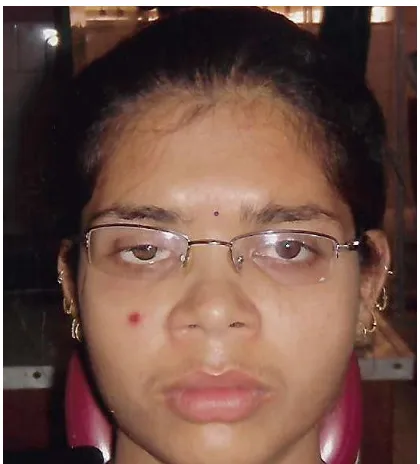
An 18 year old female patient reported to the department of Prosthodontics, Govt. Dental College and Hospital for the restoration of missing right eye. Her past medical history revealed that she had undergone enucleation of the right eye to treat septicemia resulting from an injury one year back (fig.1). Examination of the socket revealed complete healing of the eye socket and adequate depth between upper and lower fornices for the retention of the prosthesis. All possible treatment options were considered and explained to the patient. An informed consent was obtained from the patient prior to the procedure. Our treatment plan was to restore the defect with customized acrylic ocular prosthesis that would appear as similar as to the patient’s natural eye and
comfortable fit. An impression of the right enucleated socket was made using light body addition silicone impression material which was injected into the socket through the disposable syringe. Remaining material was loaded onto the ocular shaped cold cure acrylic resin tray
International Journal of Current Research
Vol. 10, Issue, 03, pp.66614-66618, March, 2018
“Ocular defect rehabilitation using digital photography- A case report
Available online at http://www.journalcra.com
z
OCULAR DEFECT REHABILITATION USING DIGITAL PHOTOGRAPHY- A CASE REPORT
and Crown and Bridge, Govt. Dental College and Hospital,
Department of Orthodontics, Bharati Vidyapeet Dental College and Hospital, Khargar,
The disfigurement associated with the loss of an eye or any other facial structures affect the physical, emotional and psychological wellbeing of a person. Rehabilitation with maxillofacial prosthesis relieves this psychological trauma by restoring lost facial structure and esthetic of the patient. Replacing a natural eye with an artificial prosthesis is the most critical as it is the first thing to be cular prosthesis must resemble the natural iris, which determines the color of the eyes. This articles aims at describing a simplified technique to fabricate custom made ocular prosthesis with the help of digital photography to achieve life like appearance.
License, which permits unrestricted use,
But most of these techniques depend on the artistic skill and sciences of the operator and is time consuming. The purpose of this article is to describe a simple technique for the fabrication of custom made ocular prosthesis using digital photography in an attempt to avoid excessive time
An 18 year old female patient reported to the department of Govt. Dental College and Hospital for the restoration of missing right eye. Her past medical history revealed that she had undergone enucleation of the right eye to a resulting from an injury one year back (fig.1). Examination of the socket revealed complete healing of the eye socket and adequate depth between upper and lower fornices for the retention of the prosthesis. All possible treatment and explained to the patient. An informed consent was obtained from the patient prior to the procedure. Our treatment plan was to restore the defect with customized acrylic ocular prosthesis that would appear as patient’s natural eye and at same time provide An impression of the right enucleated socket was made using light body addition silicone impression which was injected into the socket through the disposable syringe. Remaining material was loaded onto the ular shaped cold cure acrylic resin tray with multiple
INTERNATIONAL JOURNAL OF CURRENT RESEARCH
perforations in it and placed over previously injected impression material (Fig 2).
[image:2.595.325.545.97.422.2]Figure 1. Pre-operative photograph
Figure 2. Impression of enucleated socket with light body silicone impression material with the help of disposable syringe and
custom made acrylic tray
The patient was instructed to sit upright and look straight with eyes open in order to allow the tissues involved in the defect to be recorded in natural drape. Set impression was removed from the socket by applying slight oblique outward pr Sectional pouring of impression was done. Initially the impression was poured in the base of the flask
contour with type IV dental stone to obtain the internal shape of the eye socket. Three orientation holes
surface of the set stone.
perforations in it and placed over previously injected
operative photograph
Impression of enucleated socket with light body silicone impression material with the help of disposable syringe and
The patient was instructed to sit upright and look straight with eyes open in order to allow the tissues involved in the defect to Set impression was removed from outward pressure. Sectional pouring of impression was done. Initially the impression was poured in the base of the flask till the height of contour with type IV dental stone to obtain the internal shape were made on the
Then a second layer was poured in two
external surface stone mould for fabrication of wax pattern 3).
Figure 3. Split cast obtained from the impression
Wax conformer was made in the split cast with ivory wax. The wax sclera blank was smoothened and tried in the ocular socket and the fullness of the eyelid was adjusted (
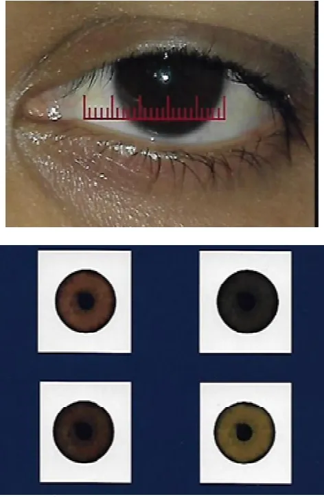
taken that the height of convexity of the wax pattern should be centered over the pupil and the palpebral opening should be same as the natural eye. A high quality digital photograph of patient’s contra lateral iris was obtained using digital camera (Nikon COOLPIX P90, New York, U.S.A., 12.1 Megapixel, 24 optical zoom). The photograph wa
natural iris. Adjustment of the color, brightness, contrast, hue of the image was done by using graphics software (Coral draw and Adob photoshop cs4). The final image was printed on good quality photopaper in different size and sha
the patient’s natural iris (Fig 5).
Then a second layer was poured in two halves to obtain the external surface stone mould for fabrication of wax pattern (Fig
Figure 3. Split cast obtained from the impression
was made in the split cast with ivory wax. The wax sclera blank was smoothened and tried in the ocular socket and the fullness of the eyelid was adjusted (Fig 4). Care was taken that the height of convexity of the wax pattern should be upil and the palpebral opening should be same as the natural eye. A high quality digital photograph of lateral iris was obtained using digital camera (Nikon COOLPIX P90, New York, U.S.A., 12.1 Megapixel, 24 optical zoom). The photograph was compared with patient natural iris. Adjustment of the color, brightness, contrast, hue of the image was done by using graphics software (Coral draw The final image was printed on good quality photopaper in different size and shade to match
[image:2.595.349.523.630.803.2]Figure 4. Wax conformer tried in patient’s enucleated socket for the fit, comfort and contour
Figure 5. Digital mapping of the patient’s natural contralateral iris and printed images of it
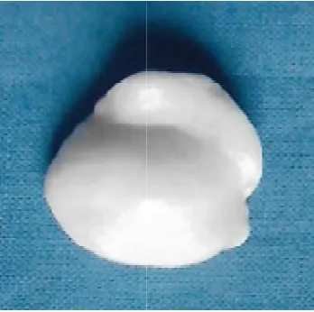
[image:3.595.328.547.61.435.2]Meanwhile, wax conformer was flasked and processed using mixture of clear heat polymerized acrylic resin and zinc oxide powder (Fig 6). The acrylic sclera blank was tried in, with the patient sitting erect and looking straight forward and by fixing her gaze on a predetermined object. The supra orbital fold, margins of the lower eyelid and iris plane were evaluated to resemble the natural eye. During scleral blank trial, distance from the inner canthus of the eye to medial periphery of the natural iris was measured and same was marked on the scleral blank (Fig 7).
Figure 6. Wax pattern invested and processed with the mixture of clear heat cure acrylic resin and zinc oxide powder
Figure 7. Centre of iris transferred onto the acrylic scleral blank by fixation of gauge
Diameter of natural iris was measured to customize the photographic iris which was obtained from the digital photograph of the patient. The artificial iris was tried on the scleral blank by keeping the distance between the inner canthus and the medial margin of the iris identical on either side. An acrylic resin of 1 mm was removed from the anterior scleral curvature for better adaptation of photographic iris on the scleral blank and to create space for the clear acrylic (Fig 8).
[image:3.595.47.283.286.648.2] [image:3.595.317.552.474.663.2]Figure 8 Reduction of scleral blank to create space for clear acrylic
[image:4.595.44.284.317.489.2]Characterization of scleral blank was done by using red silk fibers as veins and permanent colors to impart natural look. Photographic iris was finally attached to the scleral blank using cyanoacrylate adhesive (Fig 9).
Figure 9. Characterization of scleral blank and fixation of photographic iris
Figure 10. Customized ocular prosthesis in situ at the time of delivery
The position of iris was again rechecked by asking the patient to fix his gaze by looking straight forward. A protective coating was applied over the iris (G-Coat plus, GC America Inc.) to protect the color of printed iris. Later, the lost thickness of sclera was replaced with clear heatcure acrylic resin using the preserved mold. Finishing and polishing of prosthesis was carried out. The customized ocular prosthesis was then inserted and instruction for insertion and removal of the prosthesis was given to the patient (Fig 10).
DISCUSSION
Various techniques have been described in the literature for the fabrication of ocular prosthesis. Stock eye prosthesis was advocated by Laney and Gardner (Laney and Gardner 1979). In comparision to custom occular prosthesis, stock prosthesis has several disadvantages, for example poor fit, constant tissue irritation due to bacterial growth in the accumulated fluid in tissue prosthesis interface and compromise esthetic outcome (Sykes, 1956; Weldon and Niiranen 1996; Smith 1995; Bartlett and Moore, 1973). Authors have also suggested modifying existing stock ocular prosthesis by use of relining material to
achieve acceptable fit (Ow and Amrith 1997; Mathewet al,
2000). The custom ocular prosthesis provides good fit, enhanced esthetic, proper eyelid fullness, accurate sclera contour and iris colour match and positioning. Benson has suggested a classic technique which is the starting point of several techniques where in wax sclera blank is created and after the addition of iris button to it, the pattern is invested and processed (Benson, 1977). The general consensus among authors is that close matching with the natural eye is the key to mask the loss and achieve an aesthetic outcome for patients
(Reis et al., 2008). Many clinicians have concluded that an iris
color of the prosthetic eye is most important consideration for the esthetic acceptance of the ocular prosthesis. The common techniques for the fabrication of custom made ocular prosthesis are paper iris disk and black iris disk technique. However painting of the iris disk require artistic skill and the science of color (Taylor, 2000). Recent literature also mentioned technique to fabricate custom ocular prosthesis using digital
photography to replicate the iris (Jain et al, 2010).Using digital
imaging in the fabrication of ocular prosthesis presents several advantages as compared to conventional iris painting technique. The digital image provides acceptable prosthetic results as it closely replicates the patient iris with minimal color adjustment and modifications. The method is simple, less time consuming and require minimum artistic skill. Nevertheless, special digital photography equipment and setting, as well as computer software are required for image adjustments
(Artopulou et al., 2006).
Conclusion
An accurate reproduction of iris is critical for esthetic outcome in customized ocular prosthesis. This case reports presents a simplified technique for fabrication of custom ocular prosthesis using digital photography which aids clinician in achieving a predictable and esthetic result. The advantage of time reduction and simplicity of use makes this technique a viable alternative for ocular prosthesis fabrication.
REFERENCES
Artopoulou, I.I., Montgomery, P.C., Wesley, P.J., Lemon, J.C., 2006. Digital imaging in the fabrication of ocular
[image:4.595.61.271.528.760.2]Barlett, S.O., Moore, D.J., 1973. Ocular prosthesis: a
physiologic system. J Prosthet Dent, 29(4):450-459.
Benson, P. 1977. The fitting and fabrication of a custom resin
artificial eye. J Prosthet Dent, 38(5):532-538.
Brown, K.E. 1970. Fabrication of an ocular prosthesis. J
Prosthet Dent, 24(2):225-235.
Jain, S., Makkar, S., Gupta, S. and Bhargava, A. 2010. Prosthetic rehabilitation of ocular defect using digital
photography: A case report. J Indian Prosthodont Soc,
10(3):190-193.
Laney, W.R. and Gardner, A.F. 1979. Maxillofacial
prosthetics. 2nd ed. Littleton: PSG publishing: 279-306.
Mathew, M.F., Smith, R.M., Sutton, A.J. and Hudson, R. 2000. The ocular impression: A review of the literature and
presentation of an alternate technique. J Prosthodont,
9:210-216.
Murphey, P.J., Pitton, R.D., Schlossberg, L. and Harris, LW, 1945. The development of acrylic eye prosthesis at The
National Naval Medical Centre. J Am Dent Assoc,
32:1227-1244.
Newton, J.T., Fiske, J, Foote, O., Frances, C., Loh, I.M., Radford, D.R., 1999. Preliminary study of the impact of
loss of part of the face and its prosthetic restoration. J
Prosthet Dent, 82(5):585-590.
Ow, R.K. and Amrith, S., 1997. Ocular prosthetics: use of tissue conditioner material to modify a stock ocular
prosthesis. J Prosthet Dent, 78(2):218-222.
Perman, K.I. and Baylis, H.I.1988. Evisceration,
enucleationand exenteration. Otolaryngol Clin North Am, 21(1):171-182.
Reis, C.R., Dias, B.R. and Carvalho, MCJ. 2008. Evaluation of
iris color stability in ocular prosthesis. Braz Dent J,
19(4):370-374.
Smith, R.M. 1995. Relining an ocular prosthesis: a case report.
J Prosthodont, 4(3):160-163.
Sykes, L.M. 1996. Custom made ocular prostheses: a clinical
report. J Prosthet Dent, 75(1):1-3.
Taylor, D.T. 2000. Clinical Maxillofacial prosthetics.1st ed. Chicago, IL: Quintenscence Publishing Co., Inc: 270-276.
Weldon, RB and Niiranen, JV. 1956. Ocular prosthesis. J
Prosthet Dent, 6:272-278.