Review Article
Pitfalls in diagnostic hematopathology – Part II
Xianfeng F. Zhao
Department of Pathology, University of Maryland School of Medicine, Baltimore, MD
Received June 18, 2009; accepted August 20, 2009; Available online September 10, 2009
Abstract: The overlapping features of malignant lymphomas create a diagnostic “grey zone”, and lead to the inven-tion of “grey zone lymphomas”. There are several major grey zone lymphomas: 1) Lymphomas with overlapping features of Hodgkin lymphoma and large B-cell lymphoma; 2) Lymphomas with overlapping features of Burkitt lym-phoma and diffuse large B-cell lymlym-phoma; 3) Lymlym-phomas with overlapping features of nodular lymphocyte predomi-nant Hodgkin lymphoma and T-cell/histiocyte rich large B-cell lymphoma; 4) Lymphomas with overlapping features of Hodgkin lymphoma, anaplastic large cell lymphoma (ALCL) and peripheral T-cell lymphoma (PTCL); 5) T-cell clas-sical Hodgkin lymphoma and ALCL-HL. The second review of this series will be dedicated to discussion of the “grey zone” features of the lymphomas and how to narrow down the “grey zone” between those lymphomas.
Key words: DLBCL, Hodgkin lymphoma, Burkitt lymphoma, grey zone lymphoma, diagnostic pitfalls
Introduction
Another common pitfall in diagnostic
hemato-pathology is the “grey zone” between different
lymphomas. On the one hand, “grey zone”
reflects our limitations in understanding the
biology of malignant lymphomas. On the other
hand, it indicates the complexity and overlapping
features of malignant lymphomas, and the
occasional arbitrary nature of our classification.
Generally, related lymphomas create the “grey
zone”. Despite advances in
immunohistochem-istry, flow cytometry, cytogenetics and molecular
studies, distinctions between classical Hodgkin
lymphoma (cHL) and related diseases such as
primary mediastinal large B-cell lymphoma
(PMLBL), nodular lymphocyte-predominant
Hodgkin lymphoma (NLPHL), T-cell/histiocyte
rich large B-cell lymphoma (T/HRLBL) and
anaplastic large cell lymphoma (ALCL) are still
difficult in many cases (
Table 1
). Previous lack
of clear-cut diagnostic criteria imposed a
problem for both pathologists and clinicians.
Recognizing this fact, a term “grey zone
lympho-ma” was first introduced at a workshop held in
1998 by a group of 12 expert
hematopatholo-gists from the U.S., Canada and Europe [1].
Although more strict criteria were utilized to define
the well-established entities, 11 years later,
cases that have overlapping features remain
pitfalls in the hematopathological diagnosis.
These overlapping features are increasingly
acknowledged by the World Health Organization
Classification of Tumors of Hematopoietic and
Lymphoid Tissues (hereafter referred to as
“WHO Classification”) [2].
In my experience, it may not be difficult to
diagnose a “grey zone lymphoma”, but it is
sometimes hard to determine where to draw
the line for the “grey zone”. In the second part
of this review series, I will discuss the pitfalls in
diagnosing “grey zone lymphoma” and review
the criteria that help differentiate lymphomas
with “grey zone” features.
Classical Hodgkin lymphoma versus primary
mediastinal large B-cell lymphoma – classical
“grey zone lymphoma”
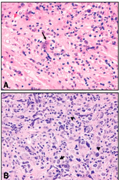
variants (
Figure 1A
), are collectively called
Hodgkin-Reed-Sternberg (HRS) cells in memory
of the three pioneers in the description and
diagnosis of cHL [3-5]. Recent investigation
suggests that HRS cells are
bona fide B-cells
that are crippled in the production of
immuno-globulin [6, 7], as a result of aberrant
expres-sion of transcription regulators [8]. Because of
this, HRS cells are weakly PAX5+, occasionally
express CD20, and have
IGH
rearrangement
[9]. However, cHL is usually CD30+ and CD15+,
but is negative for CD45, CD3, and CD79a.
Since the HRS cells secret many cytokines such
as PDGF, TGF-
β
and IL-6 etc. [10, 11], cHL is
often associated with a background of reactive
lymphocytes, plasma cells, eosinophils and
fibrosis (
Figure 1A
). The reactive background is
so characteristic that some hematopathologists
believe that a diagnosis of cHL cannot be made
without the associated reactive background.
However, similar features can also be seen in
the PMLBL, which is characterized by proliferation
of large atypical lymphoid cells in a background
of compartmentalizing fibrosis, or delicate
mesh-like sclerosis. Sometimes individual neoplastic
cells are scattered in a background of fibrosis,
resembling shells in the sand (
Figure 1B
). These
fibrotic backgrounds can also be seen in DLBCL,
particularly in DLBCL of retroperitoneal lymph
nodes. In contrast, the cHL fibrosis is usually
band-like, with neoplastic cells forming clusters
surrounded by fibrotic bands. These patterns
are particularly useful in evaluating needle core
biopsy specimens that have limited amount
of tissue and usually lack a histological
architecture. Since both cHL and PMLBL are
lymphomas of the mediastinum, the
overlap-ping features of cHL and PMLBL cause a major
challenge in lymphoma diagnosis.
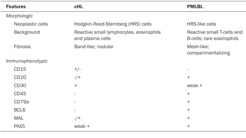
[image:2.612.323.523.69.372.2]To distinguish these two entities, several features
(
Table 2
) are useful: 1) cHL usually have classical
RS cells; while neoplastic cells in PMLBL resemble
RS cells; 2) cHL fibrosis is usually band-like and
surrounding the aggregates of neoplastic cells;
while fibrosis in PMLBL is compartmentalizing
and mesh-like; 3) CD45 and CD79a are positive
in PMLBL and negative in cHL; 4) PAX5 is weakly
positive in cHL, but strongly positive in PMLBL; 5)
CD15 is usually negative in PMLBL; only rarely,
large B-cell lymphoma could express CD15
[13]. MAL is usually expressed in PMLBL [14],
but not in cHL. When MAL is indeed expressed,
Table 1.
Common lymphomas with overlapping
or “grey zone” features
Classical Hodgkin lymphoma
Primary mediastinal large B-cell lymphoma
Burkitt lymphoma
Diffuse large B-cell lymphoma
Nodular lymphocyte predominant Hodgkin lymphoma
T-cell/histiocyte rich large B-cell lymphoma
Classical Hodgkin lymphoma
ALK-negative anaplastic cell lymphoma (ALCL) Peripheral T-cell lymphoma
Classical Hodgkin lymphoma T-cell classical Hodgkin lymphoma ALCL-like Hodgkin lymphoma
Table 2.
Features to differentiate classical Hodgkin lymphoma (cHL) and Primary mediastinal large
B-cell lymphoma (PMLBL)
Features cHL PMLBL
Morphologic
Neoplastic cells Hodgkin-Reed-Sternberg (HRS) cells HRS-like cells Background Reactive small lymphocytes, eosinophils
and plasma cells Reactive small T-cells and B-cells; rare eosinophils
Fibrosis Band-like; nodular Mesh-like;
compartmentalizing
Immunophenotypic
CD15 +/-
-CD20 -/+ +
CD30 + weak +
CD45 - +
CD79a - +
BCL6 - +
MAL -/+ +
PAX5 weak + +
the cHL is usually associated with an adverse
prognosis [15], and may be transforming into
large B-cell lymphoma. Although recent work
showed that these two lymphomas had
simi-larities in molecular signature [16], the clinical
management differs [17, 18].
Simply, when the large neoplastic cells are
CD45-negative, diagnosis favors cHL. When
the large neoplastic cells are CD45+/CD79+,
but are negative for CD15, diagnosis favors
large B-cell lymphoma. When lymphoma cells
express CD15, CD20, CD30, CD45, CD79a,
PAX5 and/or MAL, they represent a “grey zone
lymphoma”. The “grey zone lymphoma” is
cur-rently defined by the 2008 WHO Classification
as “B-cell lymphoma, unclassifiable, with
fea-tures intermediate between DLBCL and cHL” [2].
It is reasonable to believe that “grey zone
lym-phoma” and lymphocyte-depleted cHL may be
the intermediate between these two lymphomas.
The cHL to DLBCL transformation may represent
an example of lymphoma evolution (
Figure 2
).
Currently, “grey zone lymphoma” is managed as
DLBCL by using REPOCH regimen [19].
Burkitt lymphoma versus diffuse large B-cell
lymphoma
Burkitt lymphoma (BL) is a well established
patho-logic and clinical entity, which is featured by the
characteristic “starry-sky” histology, monotonous
medium-sized cytology with vacuolated
baso-philic cytoplasm and multiple small nucleoli,
extremely high proliferation index and presence
of c-
MYC
rearrangement [20]. Due to its
germi-nal center origin, the cells are CD10+ and
BCL6+, and negative for BCL2. Morphologic
variants that have prominent nucleoli and c-
MYC
they have a similar immunophenotype as BL.
In contrast, approximately 30% of DLBCL have
a t(14;18)(q32;q21) chromosomal
abnor-mality and typically no c-
MYC
rearrangement.
Morphologically, DLBCL cells are large and
pleomorphic, and they frequently also have
a high proliferation index. It is generally
not difficult to differentiate classical BL
from DLBCL. However, due to their similar
origin, overlaps between atypical BL and
DLBCL do exist and impose a challenge for the
hematopathologists.
Lymphomas with overlapping features of DLBCL
and BL have long been recognized [22].
Lymphomas with DLBCL morphology and c-
MYC
rearrangement were identified and diagnosed
as DLBCL with c-
MYC
rearrangement [23].
DLBCL with both t(14;18)(q32;q21) and c-
MYC
rearrangement were found to have a more
aggressive clinical course than DLBCL without
c-
MYC
rearrangement [24]. A group of
aggres-sive B-cell lymphomas with atypical morphology
(atypical for either BL or DLBCL) have been
identified [25], which could have BCL2
expres-sion, lower (than classical BL) proliferation rate
and presence of c-
MYC
rearrangement. These
lymphomas could also have “starry-sky” histology,
extremely high proliferation rate, and absence
of c-
MYC
rearrangement. Clinically, these
lym-phomas behaved aggressively and responded
favorably to high-intensive chemotherapy for BL.
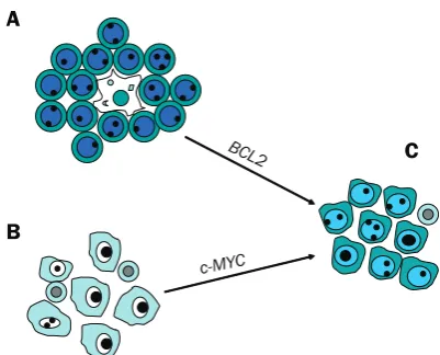
The atypical morphology of these lymphomas is
determined by their genetic abnormalities, such
as the coexistence of t(14;18)(q32;q21) and
c-
MYC
rearrangement, trisomy 8, or other yet
unidentified abnormalities that have a similar
effect as c-
MYC
(
Figure 3
). In 2005, an American
group and a European group independently
identified the BL molecular signature in both
classical BL and some DLBCL [26, 27]. Based
on accumulating information of these
lympho-mas, the 2008 WHO Classification listed them
as a separate entity [28]. It is generally agreed
that these lymphomas should be managed
aggressively as BL.
Since c-
MYC
rearrangement is a hallmark of BL,
detection of c-
MYC
rearrangement is required
for the diagnosis of BL. Although t(2;8)(p12;q24),
t(8;14)(q24;q32) and t(8;22)(q24;q11) could
cover up to 90% of the c-
MYC
rearrangements,
other c-
MYC
rearrangement was also detected
[29]. Whether c-
MYC
rearrangement is specific
for BL remains controversial. Besides BL, c-
MYC
rearrangement has been identified in chronic
lymphocytic leukemia [30], follicular lymphoma
[31], and DLBCL [32]. Presence of c-
MYC
rear-rangement is often associated with an
aggres-sive clinical course and poor prognosis in all
those lymphomas.
Nodular lymphocyte predominant Hodgkin
lymphoma versus T-cell/histiocyte rich large
B-cell lymphoma
It is currently believed that progressively
transformed germinal center (PTGC), NLPHL
and T/HRLBL are related and represent different
stages of a progressive disease [33, 34]. PTGC
is a benign disease, whereas NLPHL is a mature
B-cell lymphoma with indolent clinical course.
T/HRLBL has an aggressive clinical course and
should be treated as DLBCL. Because of this
clinical implication, distinguishing NLPHL from
T/HRLBL is critically important.
NLPHL is limited to lymph nodes of a young
patient, while T/HRLBL often involves the
extra-nodal sites, such as bone and soft tissue, in
addition to lymph node. Several arbitrary
fea-tures were proposed to differentiate NLPHL and
T/HRLBL (
Table 3
). It is generally believed that
NLPHL is almost always associated with a
back-ground of reactive small B-cells. Previous
stud-ies used <10% of the background small B-cells
to favor T/HRLBL. In my experience, classical T/
HRLBL has hardly any small B-cells in the
Figure 3. A model to explain the occurrence [image:4.612.87.288.67.229.2]background. Background small lymphocytes are
almost entirely CD3+ T-cells. Reactive histiocytes
can be prominent (
Figure 4
). Some believe that
vague nodularity favors NLPHL; however nodular
large cell transformation has been reported [35].
In these cases, “NLPHL with focal large cell
trans-formation” is preferred for the diagnosis.
It is rare that NLPHL involves bone marrow [36].
When marrow or other organs are involved, it is
usually diagnosed as T/HRLBL [37]. NLPHL may
be regarded as an “in situ” T/HRLBL – when it
goes to an extranodal site, it becomes T/HRLBL.
Even if there are rare cases of true NLPHL
involving bone marrow, it should be managed
as DLBCL or T/HRLBL [38].
Classical Hodgkin lymphoma versus
anaplastic large cell lymphoma versus
peripheral T-cell lymphoma
ALCL is another CD30+ lymphoma that should be
differentiated from cHL [39]. Morphologically,
ALCL cells can resemble HRS cells and often show
“hall-mark” cells. ALK-positivity distinguishes
ALK+ ALCL from cHL [40, 41]. However,
differenti-ating ALK-negative ALCL from cHL can be a
chal-lenge. Since cHL is of a B-cell origin [6, 7], it is
often weakly positive for PAX5. Conversely,
ALK-negative ALCL is a T-cell lymphoma, and is almost
never positive for PAX5. When peripheral T-cell
lymphoma (PTCL) expresses CD30, it becomes
another differential entity in this complicated
“trio”. As in ALK-negative ALCL, PTCL is also
nega-tive for PAX5. A consensus is still lacking among
experts on the definition of ALK-negative ALCL
[42]. Some believe that ALK-negative ALCL should
be included in PTCL, unspecified. Others argue
that ALK-negative ALCL has distinctive
morpho-logical features. A recent large study from the
Internaltional Peripheral T-cell Lymphoma Project
suggested that ALK-negative ALCL was clinically
and immunophenotypically different from PTCL
[43]. This study also showed that patients with
ALK-negative ALCL had a superior failure free
sur-vival (FFS) and overall sursur-vival (OS) rate than those
with PTCL (36% vs 20%; and 49% vs 32%,
respectively).
Classical Hodgkin lymphoma, B-cell or T-cell
origin?
[image:5.612.90.516.100.256.2]T-cell cHL has been reported [44, 45]. The
neoplastic cells in those reported cases were
even weakly positive for PAX5 [45], a B-cell
specific activator protein (BSAP). There was an
Figure 4. Classical T/HRLBL. Scattered large neoplastic B-cells (indicated by arrows) are in a background of numerous reactive histiocytes and small numbers of reactive small T-cells. Reactive small B-cells are extremely rare.Table 3.
Features to distinguish nodular lymphocyte predominant Hodgkin lymphoma (NLPHL) and
T-cell/histiocyte rich large B-cell lymphoma (T/HRLBL)
Features NLPHL T/HRLBL
Morphologic
Architecture Nodular Diffuse
Neoplastic cells “Popcorn”-like L&H cells Centroblastic, immunoblastic or RS-like Background Mixed populations of small T- cells and
B-cells; rosette small T-cells; aggregate small B-cells; nodular follicular dendritic cell meshwork
Scattered small T-cells and numerous reactive histiocytes; rare small B-cells
Immunophenotypic
Neoplastic cells CD20+/CD45+/CD79a-/BCL2- CD20+/CD45+/CD79a+/BCL2+ Background cells CD3+/CD57+ (rosette T-cells); CD3+ (small T-cells); CD68+
[image:5.612.325.526.258.422.2]ongoing debate over the existence of a “T-cell
cHL”. A recent study analyzed 259 cases of cHL
and identified that 5% of the HRS cells expressed
at least one T-cell markers [46], indicating that
T-cell antigens could be aberrantly expressed in
HRS cells. The REAL classification also
pro-posed an ALCL-like HL (ALCL-HL) as a provisional
entity [47] based on its RS-like morphology and
CD30+/CD15+ immunophenotype. However,
some PTCL can also be CD30+/CD15+ [48].
Although a randomized clinical trial concluded
that ALCL-HL had an equal response to
regi-mens for high-grade lymphomas and regiregi-mens
for cHL [49], microarray studies showed that
this lymphoma clustered with T-cell lymphomas
rather than cHL [50], suggesting that it is a
bona
fide T-cell lymphoma. Although some
patholo-gists believe T-cell cHL does exist [51], almost all
the microdissected single cell analyses of cHL
pointed to a B-cell origin [52, 53]. Currently,
“T-cell cHL” is not recognized by the WHO
Classification [2].
Summary
In summary, overlapping features are common
between malignant lymphomas, reflecting a
continuum of lymphocyte development and
lymphoma evolution. Awareness of these
fea-tures will help us to deal with atypical lymphomas
with a prepared mind. Recognizing the
chal-lenges in diagnosis of lymphomas with “grey
zone” features will increase our confidence in
committing to an accurate diagnosis.
Please address all correspondences to: X. Frank Zhao, MD, PhD, Department of Pathology, University of Maryland School of Medicine, 10 S. Pine Street, MSTF 611B, Baltimore, MD 21201; Tel: 410-328-2146, Fax: 410-328-5508; E-mail: [email protected]
References
Rudiger T, Jaffe ES, Delsol G, deWolf-Peeters C, [1]
Gascoyne RD, Georgii A, Harris NL, Kadin ME, MacLennan KA, Poppema S, Stein H, Weiss LE, Muller-Hermelink HK. Workshop report on Hodgkin’s disease and related diseases (‘grey zone’ lymphoma). Ann Oncol. 1998; 9 Suppl 5: S31-38.
Swerdlow SH, Campo E, Harris NL, Jaffe ES, [2]
Pileri SA, Stein H, Thiele J, Vardiman JW (Eds.): WHO Classification of Tumors of Hematopoietic and Lymphoid Tissues. IARC: Lyon 2008.
Watkins SM. The legacy of Thomas Hodgkin [3]
(1798-1866). J R Coll Physicians Lond.
2000; 34: 100-104.
Reed D. On the pathological changes in Hodg-[4]
kin’s disease, with special reference to its re-lation to tuberculosis. Johns Hopkins Hosp Rep. 1902; 10: 133-196.
Sternberg C. Uber eine eigenartige unter dem [5]
Bilde der Pseudoleukamie verlaufende Tubercu-lose des lymphatischen Apparates. Ztschr Heilk. 1898; 19: 21-90.
Küppers R, Hansmann ML, Rajewsky K. Clonal-[6]
ity and germinal centre B-cell derivation of Hodg-kin/Reed-Sternberg cells in Hodgkin’s dis-ease. Ann Oncol. 1998; 9 Suppl 5: S17-20. Marafioti T, Hummel M, Foss HD, Laumen H, [7]
Korbjuhn P, Anagnostopoulos I, Lammert H, Demel G, Theil J, Wirth T, Stein H. Hodgkin and reed-sternberg cells represent an expansion of a single clone originating from a germinal center B-cell with functional immunoglobulin gene rearrangements but defective immunoglobulin transcription. Blood. 2000; 95: 1443-1450. Mathas S, Janz M, Hummel F, Hummel M, [8]
Wollert-Wulf B, Lusatis S, Anagnostopoulos I, Lietz A, Sigvardsson M, Jundt F, Jöhrens K, Bommert K, Stein H, Dörken B. Intrinsic inhibi-tion of transcripinhibi-tion factor E2A by HLH proteins ABF-1 and Id2 mediates reprogramming of neo-plastic B cells in Hodgkin lymphoma. Nat Immu-nol. 2006; 7: 207-215.
Hummel M, Ziemann K, Lammert H, Pileri S, [9]
Sabattini E, Stein H. Hodgkin’s disease with monoclonal and polyclonal populations of Reed-Sternberg cells. N Engl J Med. 1995; 333: 901-906.
Hsu SM, Waldron JW Jr, Hsu PL, Hough AJ Jr. [10]
Cytokines in malignant lymphomas: review and prospective evaluation. Hum Pathol.
1993; 24: 1040-1057.
Skinnider BF, Elia AJ, Gascoyne RD, Trümper [11]
LH, von Bonin F, Kapp U, Patterson B, Snow BE, Mak TW. Interleukin 13 and interleukin 13 re-ceptor are frequently expressed by Hodgkin and Reed-Sternberg cells of Hodgkin lympho-ma. Blood. 2001; 97: 250-255.
Traverse-Glehen A, Pittaluga S, Gaulard P, [12]
Sorbara L, Alonso MA, Raffeld M, Jaffe ES. Mediastinal gray zone lymphoma: the missing link between classic Hodgkin’s lymphoma and mediastinal large B-cell lymphoma. Am J Surg Pathol. 2005; 29: 1411-1421.
García JF, Mollejo M, Fraga M, Forteza J, [13]
Muniesa JA, Pérez-Guillermo M, Pérez-Seoane C, Rivera T, Ortega P, Piris MA. Large B-cell lymphoma with Hodgkin’s features. Histopa-thology. 2005; 47: 101-110.
Copie-Bergman C, Plonquet A, Alonso MA, [14]
Leroy K, Gaulard P. MAL expression in lym-phoid cells: further evidence for MAL as a distinct molecular marker of primary medias-tinal large B-cell lymphomas. Mod Pathol.
2002; 15: 1172-1180.
Hsi ED, Sup SJ, Alemany C, Tso E, Skacel M, El-[15]
son P, Alonso MA, Pohlman B. MAL is expressed in a subset of Hodgkin lymphoma and identifies a population of patients with poor prognosis. Am J Clin Pathol. 2006; 125: 776-782.
Rosenwald A, Wright G, Leroy K, Yu X, Gaulard [16]
P, Gascoyne RD, Chan WC, Zhao T, Haioun C, Greiner TC, Weisenburger DD, Lynch JC, Vose J, Armitage JO, Smeland EB, Kvaloy S, Holte H, Delabie J, Campo E, Montserrat E, Lopez-Guill-ermo A, Ott G, Muller-Hermelink HK, Con-nors JM, Braziel R, Grogan TM, Fisher RI, Miller TP, LeBlanc M, Chiorazzi M, Zhao H, Yang L, Powell J, Wilson WH, Jaffe ES, Simon R, Klaus-ner RD, Staudt LM. Molecular diagnosis of pri-mary mediastinal B cell lymphoma identifies a clinically favorable subgroup of diffuse large B cell lymphoma related to Hodgkin lympho-ma. J Exp Med. 2003; 198: 851-862. Federico M, Luminari S, Iannitto E, Polimeno G, [17]
Marcheselli L, Montanini A, La Sala A, Merli F, Stelitano C, Pozzi S, Scalone R, Di Renzo N, Musto P, Baldini L, Cervetti G, Angrilli F, Mazza P, Brugiatelli M, Gobbi PG; HD2000 Gruppo Italiano per lo Studio dei Linfomi Trial. ABVD compared with BEACOPP compared with CEC for the initial treatment of patients with ad-vanced Hodgkin’s lymphoma: results from the HD2000 Gruppo Italiano per lo Studio dei Lin-fomi Trial. J Clin Oncol. 2009; 27: 805-811. Johnson PW, Davies AJ. Primary mediastinal [18]
B-cell lymphoma. Hematology Am Soc Hema-tol Educ Program. 2008; 2008: 349-358. Dunleavy K, Pittaluga S, Grant N, Steinberg S, [19]
Shovlin M, Tay K, Janik JE, Jaffe ES, Wilson WH. Gray Zone Lymphomas: Clinical and Histologi-cal Characteristics and Treatment with Dose-Adjusted-EPOCH-R. American Society of Hematology 2008, San Francisco (Moscone Center) 12/8/2008, Abstract 3590.
Leoncini L, Raphael M, Stein H, Harris NL, Jaffe ES, [20]
Kluin PM. Burkitt lymphoma. In Swerdlow SH, Campo E, Harris NL, Jaffe ES, Pileri SA, Stein H, Thiele J, Vardiman JW (Eds.): WHO Classification
of Tumors of Hematopoietic and Lymphoid Tissues. IARC: Lyon 2008, pp262-264. Ziegler JL, Cohen MH, Vogel CL, Sheagren JN, [21]
Carbone PP. Immunologic studies in American patients with Burkitt-like lymphoma. Cancer Res. 1967; 27: 2527-2531.
Ladanyi M, Offit K, Jhanwar SC, Filippa DA, [22]
Chaganti RS. MYC rearrangement and translo-cations involving band 8q24 in diffuse large cell lymphomas. Blood. 1991; 77: 1057-1063.
Steven Le Gouill, Pascaline Talmant, Cyrille [23]
Touzeau, Anne Moreau, Richard Garand, Na-dine Juge-Morineau, Fanny Gaillard, Thomas Gastinne, Noël Milpied, Philippe Moreau, Jean Luc Harousseau, Hervé Avet-Loiseau. The clini-cal presentation and prognosis of diffuse large B-cell lymphoma with t(14;18) and 8q24/c-MYC rearrangement. Haematologica. 2007; 92: 1335-1342.
Kanungo A, Medeiros LJ, Abruzzo LV, Lin P. [24]
Lymphoid neoplasms associated with concur-rent t(14;18) and 8q24/c-MYC translocation generally have a poor prognosis. Mod Pathol.
2006; 19: 25-33.
Zhao XF, Hassan A, Perry A, Ning Y, Stass SA, [25]
Dehner LP. C-MYC Rearrangements are Fre-quent in Aggressive Mature B-Cell Lymphoma with Atypical Morphology. Int J Clin Exp Pathol.
2008; 1: 65-74.
Hummel M, Bentink S, Berger H, et al. A bio-[26]
logic definition of Burkitt’s lymphoma from transcriptional and genomic profiling. N Engl J Med. 2006; 354: 2419-2430.
Dave SS, Fu K, Wright GW, et al. Molecular di-[27]
agnosis of Burkitt’s lymphoma. N Engl J Med. 2006; 354: 2431-2442.
Kluin PM, Raphael M, Harris NL, Campo E, [28]
Stein H, Jaffe ES, Leoncini L. B-cell lymphoma, unclassifiable, with features intermediate be-tween diffuse large B-cell lymphoma and Burkitt lymphoma. In Swerdlow SH, Campo E, Harris NL, Jaffe ES, Pileri SA, Stein H, Thiele J, Vardiman JW (Eds.): WHO Classification of Tu -mors of Hematopoietic and Lymphoid Tissues.
IARC: Lyon 2008: pp265-266.
Sandlund JT, Kastan MB, Kennedy W, Behm F, [29]
Entrekin E, Pui CH, Kalwinsky DT, Raimondi SC. A subtle t(3;8) results in plausible juxtaposition of MYC and BCL6 in a child with Burkitt lym-phoma/leukemia and ataxia-telangiectasia.
Cancer Genet Cytogenet. 2006; 168: 69-72. Huh YO, Lin KI, Vega F, Schlette E, Yin CC, Ke-[30]
ating MJ, Luthra R, Medeiros LJ, Abruzzo LV. MYC translocation in chronic lymphocytic leu-kaemia is associated with increased prolym-phocytes and a poor prognosis. Br J Haematol.
2008; 142: 36-44.
Lee JT, Innes DJ Jr, Williams ME. Sequential [31]
bcl-2 and c-myc oncogene rearrangements associated with the clinical transformation of non-Hodgkin’s lymphoma. J Clin Invest. 1989; 84: 1454-1459.
Niitsu N, Okamoto M, Miura I, Hirano M. [32]
Clinical features and prognosis of de novo diffuse large B-cell lymphoma with t(14;18) and 8q24/c-MYC translocations. Leukemia.
2009; 23: 777-783.
Chan WC. Cellular origin of nodular [33]
immunophenotypic and molecular studies. Semin Hematol. 1999; 36: 242-252.
Zhao FX. Nodular Lymphocyte-Predominant [34]
Hodgkin Lymphoma or T-cell/Histiocyte Rich Large B-cell Lymphoma: The Problem in “Grey Zone” Lymphomas. Int J Clin Exp Pathol. 2008; 1: 300-305.
Boudová L, Torlakovic E, Delabie J, Reimer P, [35]
Pfistner B, Wiedenmann S, Diehl V, Müller-Hermelink HK, Rüdiger T. Nodular lymphocyte-predominant Hodgkin lymphoma with nodules resembling T-cell/histiocyte-rich B-cell lympho-ma: differential diagnosis between nodular lym-phocyte-predominant Hodgkin lymphoma and T-cell/histiocyte-rich B-cell lymphoma.
Blood. 2003; 102: 3753-3758.
Pappa VI, Norton AJ, Gupta RK, Wilson AM, Ro-[36]
hatiner AZ, Lister TA. Nodular type of lymphocyte predominant Hodgkin’s disease. A clinical study of 50 cases. Ann Oncol. 1995; 6: 559-565. Couronné L, Schneider P, Picquenot JM, La-[37]
berge S, Bastard C, Vannier JP. Refractory nod-ular lymphocyte predominant Hodgkin lympho-ma transformed to T-cell/histiocyte-rich B-cell lymphoma in an adolescent: salvage therapy with allogeneic bone marrow transplantation. J Pediatr Hematol Oncol. 2008; 30: 959-962. Khoury JD, Jones D, Yared MA, Manning JT Jr, [38]
Abruzzo LV, Hagemeister FB, Medeiros LJ. Bone marrow involvement in patients with nodular lymphocyte predominant Hodgkin lymphoma.
Am J Surg Pathol. 2004; 28: 489-495. Perkins PL, Ross CW, Schnitzer B. CD30-posi-[39]
tive, anaplastic large-cell lymphomas that ex-press CD15 but lack CD45. A possible diagnos-tic pitfall. Arch Pathol Lab Med. 1992; 116: 1192-1196.
Herling M, Rassidakis GZ, Viviani S, Bonfante V, [40]
Giardini R, Gianni M, Morris SW, Cabanillas F, Me-deiros LJ, Sarris AH. Anaplastic lymphoma kinase (ALK) is not expressed in Hodgkin’s disease: re-sults with ALK-11 antibody in 327 untreated pa-tients. Leuk Lymphoma. 2001; 42: 969-979. Vassallo J, Lamant L, Brugieres L, Gaillard F, [41]
Campo E, Brousset P, Delsol G. ALK-positive anaplastic large cell lymphoma mimicking nod-ular sclerosis Hodgkin’s lymphoma: report of 10 cases. Am J Surg Pathol. 2006; 30: 223-9. Medeiros LJ, Elenitoba-Johnson KS. Anaplastic [42]
Large Cell Lymphoma. Am J Clin Pathol. 2007; 127: 707-722.
Savage KJ, Harris NL, Vose JM, Ullrich F, Jaffe [43]
ES, Connors JM, Rimsza L, Pileri SA, Chhanab-hai M, Gascoyne RD, Armitage JO, Weisenburg-er DD; IntWeisenburg-ernational PWeisenburg-eriphWeisenburg-eral T-Cell Lympho-ma Project. ALK- anaplastic large-cell lymphoma is clinically and immunophenotypi-cally different from both ALK+ ALCL and pe-ripheral T-cell lymphoma, not otherwise speci-fied: report from the International Peripheral
T-Cell Lymphoma Project. Blood. 2008; 111: 5496-5504.
Müschen M, Rajewsky K, Bräuninger A, Baur [44]
AS, Oudejans JJ, Roers A, Hansmann ML, Küp-pers R. Rare occurrence of classical Hodgkin’s disease as a T cell lymphoma. J Exp Med.
2000; 191: 387-394.
Willenbrock K, Ichinohasama R, Kadin ME, Miura I, [45]
Terui T, Meguro K, Fukuhara O, DeCoteau JF, Hansmann ML. T-cell variant of classical Hodgkin’s lymphoma with nodal and cutaneous manifesta-tions demonstrated by single-cell polymerase chain reaction. Lab Invest. 2002; 82: 1103-1109. Tzankov A, Bourgau C, Kaiser A, Zimpfer A, Mau-[46]
rer R, Pileri SA, Went P, Dirnhofer S. Rare expres-sion of T-cell markers in classical Hodgkin’s lym-phoma. Mod Pathol. 2005; 18: 1542-1549. Harris NL, Jaffe ES, Stein H, Banks PM, Chan JK, [47]
Cleary ML, Delsol G, De Wolf-Peeters C, Falini B, Gatter KC, Grogan TM, Isaacson PG, Knowles DM, Mason DY, Muller-Hermelink HK, Pileri SA, Piris MA, Ralfkiaer E, Warnke RA. A revised European-American classification of lymphoid neoplasms: a proposal from the International Lymphoma Study Group. Blood. 1994; 84: 1361-1392. Barry TS, Jaffe ES, Sorbara L, Raffeld M, [48]
Pittaluga S. Peripheral T-cell lymphomas expressing CD30 and CD15. Am J Surg Pathol.
2003; 27: 1513-1522.
Zinzani PL, Martelli M, Magagnoli M, Zaccaria [49]
A, Ronconi F, Cantonetti M, Bocchia M, Marra R, Gobbi M, Falini B, Gherlinzoni F, Moretti L, De Renzo A, Mazza P, Pavone E, Sabattini E, Amendola A, Bendandi M, Pileri SA, Mandelli F, Tura S. Anaplastic large cell lymphoma Hodg-kin’s-like: a randomized trial of ABVD versus MACOP-B with and without radiation therapy.
Blood. 1998; 92: 790-794.
Vanhentenrijk V, Vanden Bempt I, Dierickx D, [50]
Verhoef G, Wlodarska I, De Wolf-Peeters C. Re-lationship between classic Hodgkin lymphoma and overlapping large cell lymphoma investi-gated by comparative expressed sequence hy-bridization expression profiling. J Pathol. 2006; 210: 155-162.
Küppers R. The biology of Hodgkin’s lympho-[51]
ma. Nat Rev Cancer. 2009; 9: 15-27.
Tamaru J, Hummel M, Zemlin M, Kalvelage B, [52]
Stein H.Hodgkin’s disease with a B-cell pheno-type often shows a VDJ rearrangement and somatic mutations in the VH genes. Blood.
1994; 84: 708-715.
Küppers R, Rajewsky K, Zhao M, Simons G, [53]