Original Article
TLR2-deficiency of cKit
+bone marrow cells is associated
with augmented potency to stimulate angiogenic
processes
Nana-Maria Wagner1, Laura Bierhansl1, Antje Butschkau², Gabriele Noeldge-Schomburg1, Jan Patrick
Roesner1, Brigitte Vollmar2
1Clinic for Anesthesiology and Critical Care Medicine, University Hospital Rostock, Rostock, Germany; 2Institute for
Experimental Surgery, University Hospital Rostock, Rostock, Germany
Received September 11, 2013; Accepted October 21, 2013; Epub November 15, 2013; Published December 1, 2013
Abstract: Objective: Toll-like receptor 2 (TLR2)-deficiency is associated with the preservation of vascular function
and TLR2-deficient (TLR2-/-) mice exhibit increased neovascularization following induction of hindlimb ischemia. He
-matopoietic stem cells play an important role in ischemia-induced angiogenesis and we now investigated whether the effects observed in TLR2-/- mice may be attributed to TLR2 deficiency on bone marrow-derived stem cells. Ap -proach and Results: cKit-positive (cKit+) bone marrow cells (BMC) were isolated from wild type (WT) and TLR2-/- mice
employing MACS-bead technology. Co-incubation of TLR2-/-cKit+ BMC with mature endothelial cells (ECs) resulted in
increased tube formation of ECs on matrigel, augmented sprouting in a 3D-collagen matrix and increased migratory capacity compared to co-incubation with WT cKit+ BMC. In an in vivo matrigel plug assay, TLR2-/-cKit+ BMC exhibited
enhanced formation of capillary-like networks. In a murine model of hindlimb ischemia, administration of TLR2-/- cKit+ BMC to WT mice augmented capillary density and reperfusion of ischemic M. gastrocnemius muscle tissue to
the level of TLR2-/- mice. Western Blot analysis revealed comparable expression of CXCR4 on TLR2-/-cKit+ BMC but
increased activation of the PI3K downstream signaling molecule protein kinase B (PKB/AKT) compared to WT cKit+
cells. Conclusions: The absence of TLR2 on cKit+ BMC is associated with augmented potency to support angiogenic processes in vitro and in vivo. Functional inhibition of TLR2 may therefore provide a novel tool to enhance stem cell function for the treatment of vascular diseases.
Keywords: TLR2, cKit, hematopoietic stem cells, angiogenesis, ischemia
Introduction
Toll-like receptors (TLRs) are pattern recogni-tion receptors that can induce innate immune
responses. Activation of TLRs by exogenous
ligands such as pathogens or endogenous dan-ger-associated molecular patterns (DAMPs)
results in the activation of nuclear factor kap
-pa-light-chain-enhancer of activated B cells (NFκB) and the promotion of a pro-inflammato
-ry milieu [1]. Recently, Toll-like receptor 2 (TLR2) has been shown to be involved not only in the activation of inflammatory cells at sites of tis
-sue injury or local infection, but also in mediat
-ing increased inflammatory cell egress from the bone marrow [2]. During infection or applica
-tion of TLR2 ligands, activa-tion of TLR2 on
hematopoietic stem cells triggers their differen
-tiation towards the myeloid cell lineage [3], resulting in increased numbers of circulating immune cells such as macrophages [4]. Alth-ough augmented generation of cells belonging to first line host defense is mandatory to fight acute infection, TLR2-mediated modulation of stem- and progenitor cell fate inside or outside of the bone marrow may imbalance the provi
-sion of the number or potency of regular stem cell progeny. For example, TLR2 agonists have been shown to impair proliferation and matura
-tion of neuronal and glial progenitor cells [5, 6]. In turn, blockade of TLR2 reduced the dental pulp stem cell suppressing effect of pathogens in the oral cavity [7]. In the bone marrow,
2814 Int J Clin Exp Pathol 2013;6(12):2813-2823
result in the depletion of endothelial progenitor cell pools required for vascular regeneration and these effects were absent in TLR2-deficient
(TLR2-/-) mice [8].
TLR2 deficiency and functional blockade have been associated with the preservation of vas
-cular function. Prevention from excess TLR2-mediated activation of inflammatory cascades protected the vasculature from ischemia and reperfusion injury [9], atherosclerosis lesion progression [10, 11] and endothelial injury in response to cerebral [12, 13], myocardial [14] and skeletal muscle ischemia [15]. Although
evidence points towards endothelial TLR2 in
mediating at least part of these vasculoprotec
-tive effects, the contribution of the absence of
TLR2 signaling in stem- and progenitor cells that can participate in vascular regenerative
processes has not been determined so far. In the present study, we compared the potency of cKit-positive (cKit+) bone marrow-derived
stem cells (BMCs) from TLR2-/- mice to those
isolated from WT mice to augment endothelial cell functions relevant to angiogenic processes.
cKit+ cells were co-incubated with mature
endo-thelial cells and investigated for their capacity to modulate endothelial tube formation, endo -thelial sprouting and migration in vitro.
Furth-ermore, the capillary-like tube formation activi
-ty of TLR2-/-cKit+ BMC was compared to WT
cKit+ BMC in vivo and their potency to modulate
neovascularization processes in response to
ischemia was analyzed employing a second in vivo mouse model, the murine model of
hindlimb ischemia. Moreover, both TLR2-/-cKit+
and WT cKit+ BMC were analyzed for their
expression of CXCR4 and activation of the CXCR4 downstream signaling molecule protein
kinase B (PKB)/AKT relevant to progenitor cell
homing [16].
Materials and methods
Isolation of cKit+ bone marrow-derived cells
cKit+ cells were isolated from 8-10 week old WT
(C57BL/6J) or TLR2-/- mice (B6.129-Tlr2tm1Kir/J)
as described previously [17]. Mouse bone mar
-row was isolated from femur and tibia and cell
suspensions were incubated with magnetic microbeads coated with anti-cKit monoclonal
antibody (Miltenyi Biotec, Bergisch Gladbach, Germany). MS columns® and the MiniMacs® cell separator system were employed to obtain
cKit+ cell fractions. For fluorescence labeling of
cKit+ cells, cells were incubated with 2.5 µg/mL
CellTracker™ CM-DiI (Invitrogen, Karlsruhe,
Germany).
Cell culture of endothelial cells
Human Umbilical Vein Endothelial Cells (HUVECs) were purchased from PromoCell, Germany, and cultivated in endothelial growth medium (EndoPrime Kit, PAA, United Kingdom) supplemented with 10% fetal calf serum on
gelatin-coated dishes (Attachment Factor,
Gibco, Germany). Cells were harvested by tryp
-sinization (0.05%, Gibco, Germany) and used from passage 2 to 5.
Matrigel angiogenesis assay
As described previously [18], 1x10⁴ HUVECs
were incubated alone or in the presence of
either 3x10³ WT or TLR2-/-cKit+ BMC in
dupli-cate in 100 µL endothelial growth medium
including reagents or vehicle for 8 hours in 96-well plates precoated with 70 µL Matrigel
Basement Membrane Matrix (BD Bioscience,
USA). Tubular HUVEC structures and cKit+ cells
were photographed using a fluorescence micro
-scope (Leica, Germany) employing 100x magni
-fication at 8 random high power fields (HPF) per
variant. Tubular length was assessed per
high-power field employing ImageProPlus Software, CA, USA. Per independent experiment, mean values of all variants were expressed as rela -tive to control set to 1.0.
Spheroid angiogenesis assay
3.2x10⁴ HUVECs were suspended either alone
or together with 8x10³ WT or TLR2-/-cKit+ cells
in 10 mL endothelial basal medium containing
20% methylcellulose solution (dissolved in M199 medium; Sigma, Germany) and incubat -ed in round-bottom 96-well plates (100 µL per
well) for 24 hours to form spheroids as previ
-ously described [18]. Type I rat tail collagen (BD Biosciences, MA, USA) was diluted 1:1 with 0.1% acetic acid, mixed with 10X M199 medi
-um, and neutralized with 0.2 N NaOH immedi
-ately before use. Spheroids were harvested, centrifuged and resuspended in methylcellu -lose solution supplemented with 5% FCS and mixed (1:1) with collagen working solution. Spheroid suspensions were then distributed
2815 Int J Clin Exp Pathol 2013;6(12):2813-2823
min. After solidification of the collagen, 300 µL of medium were added to each well and incu
-bated for 24 hours at 37°C. Pictures of 10 spheroids at random fields were taken employ
-ing a fluorescence microscope and the mean cumulative sprout length and the number of
labeled cKit+ cells was determined by
Image-ProPlus software.
Migration assay
HUVECs were grown on 6-well plates until con
-fluence. A 10 µL pipette tip was employed to scratch over well plates twice vertically and horizontally for obtaining four 90° crosses on each well. Wells were gently washed and 2 mL of medium containing 5x10⁴ WT or TLR2-/-cKit+
cells were added in duplicate per variant. All
scratched-crosses were photographed every other hour until a total of 6 hours of incubation and scratch wounds were analyzed employing ImageProPlus software.
Matrigel plug assay
Animal experiments were approved by the gov
-ernmental ethical board for animal research in Mecklenburg-Vorpommern (7221.3-1.1-108/ 12 and 7221.3-1.2-038/12) and are in accor -dance with German law on animal protection
and the Guide for the Care and Use of Laboratory Animals. Animals were bred and housed at the Institute for Experimental Surgery, Central Animal Care Facility, Rostock University, Rostock, Germany. Eight to ten week-old male wild-type (WT; C57BL/6J) and
TLR2 knock-out (TLR2-/-; B6.129-Tlr2tm1Kir/J)
mice were subjected to intraperitoneal
anes-thesia by injection of 8 mg/kg xylazine hydro
-chloride (cp-pharma; Burgdorf, Germany) and 12 mg/kg ketamine hydrochloride (Pharmanovo GmbH; Hannover, Germany). 2x10⁶ WT or TLR2 -/-cKit+ cells were resuspended in 500 µL of
Matrigel and subjected to subcutaneous
injec-tion. Plugs were harvested following three weeks of incubation, fixed and counterstained with DAPI for visualization of cell nuclei. In some mice, 100 µL of fluorescein griffonia (bandei
-raea) simplicifolia lectin I (Vector Laboratories, Burlingame, CA, USA) were applied by left-ven
-tricular injection in anesthetized mice for analy
-sis of plug perfusion. 10 min later, mice were
euthanized, plugs were harvested and
investi-gated employing a fluorescence microscope and lengths of capillary networks were quanti
-fied per HPF employing ImageProPlus
soft-ware.
Murine hindlimb ischemia model
Unilateral hindlimb ischemia was induced as previously described [18-20]. In brief, the right femoral artery (immediately distal to the branch of the deep femoral artery) as well as the distal portion of the saphenous artery were perma
-nently ligated employing a 7-0 polypropylene suture (Prolene™, Ethicon, Germany) and the ligated femoral artery was then removed. Wounds were carefully sutured using 6-0 sutures (Prolene™). 24 hours after the induc
-tion of ischemia, mice were subjected to intra
-cardiac injection of 3x10⁶ labeled WT or TLR2
-/-cKit+ cells or vehicle.
Infrared thermal imaging (thermography)
Thermal imaging was performed as previously described [20]. In brief, before and immediately after surgical ligation of the femoral artery and
duringfollow-up on post-operative day (POD) 1,
5, 10 and 21, mice were anesthetized as described above and placed ona 37°C heating pad for 6 min following 3 min on a table surface at room temperature and infrared imaging was performed employing a ThermaCAM B20HS camera, FLIR Systems, Wilsonville, OR, USA. Images were analyzed using FLIR QuickReport 1.2 software by determination of the tempera
-ture at the middle of the pad of both the oper
-ated and non-oper-ated hindlimb. The differ
-ence in temperature (°C) between both pads of each animal was analyzed using GraphPadPrism software.
Immunohistochemistry
For harvest of M. gastrocnemius muscle tissue on POD 21, mice were euthanized and capillary density in the gastrocnemius muscle was assessed on 5 µm-thick, acetone-fixed frozen sections after staining with ABs against CD31 (1:50 dilution; SantaCruz, USA) followed by Cy3-labeled secondary ABs (Molecular Probes, USA). Cell nuclei were counterstained with DAPI. The number of CD31-immunopositive cells per muscle fiber was manually counted on 8 random microscope fields per section (200x magnification).
Western blot analysis
cKit+ cells were resuspended in lysis buffer con
-taining fresh protease and phosphatase inhibi
2816 Int J Clin Exp Pathol 2013;6(12):XXX-XXX
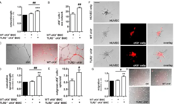
Figure 1. Bone marrow-derived cKit+ cells from TLR2-/- mice more potently promote the angiogenic function of endothelial cells in vitro. (A) HUVEC showed increased
capillary-like tube formation in the presence of TLR2-/-cKit+ cells compared to WT cKit+ cells or at basal level (ctrl). ***P<0.001 vs. HUVECs alone, ##P<0.01 vs. WT cKit+ cells. Quantitative summary of n=6 independent experiments. (B) Increased numbers of TLR2-/-cKit+ cells integrated into endothelial cell tubules provided by
HUVECs compared to WT cKit+ cells. ##P<0.01, n=6. (C) Representative tubular networks provided by HUVECs (translucent) after 8 hours of incubation in the pres
-ence of either WT or TLR2-/-cKit+ cells (labeled in red). (D) In the spheroid angiogenesis-assay, co-incubation of HUVECs with TLR2-/-cKit+ cells resulted in increased
outgrowth of endothelial cells from collagen-embedded HUVEC spheroids and (E) enhanced migration of TLR2-/-cKit+ cells along HUVEC sprouts in comparison to co-incubation with WT cKit+ cells. **P<0.01 vs. HUVECs at basal level, #P<0.05 and ##P<0.01, quantitative summary of n=6 independent experiments. (F) Repre
-sentative spheroids exhibiting HUVEC tubular sprouts (translucent) and migrated cKit+ cells (red) following 24 h of incubation in a collagen matrix. (G) TLR2-/-cKit+
cells more potently promoted the migration of HUVECs in a scratch wound assay compared to WT cKit+ cells or HUVECs at basal level (ctrl). Quantitative summary of
n=5 independent experiments. **P<0.01 vs. ctrl, #P<0.05 vs. WT cKit+ cells. (H) Representative pictures of scratch-wound closures following 4 hours of incubation.
2817 Int J Clin Exp Pathol 2013;6(12):2813-2823
lysates were cleared by centrifugation and equal amounts of protein were loaded and frac
-tionated by electrophoresis on 10-12% SDS polyacrylamide gels together with molecular weight standards and then transferred to nitro
-cellulose membranes (Immobilon transfer membranes, Millipore Corporation, USA).
Membranes were blocked in 2.5% BSA (in
TBS/0.1% Tween-20) for 2 hours at RT prior to incubation with primary ABs overnight at 4°C. Visualization of protein bands was achieved using a HRP-conjugated secondary donkey anti-rabbit (1:1000; Cell Signaling Technology, USA) or anti-mouse IgG AB (1:2500; Sigma, USA) for 1 hour at RT, followed by detection of HRP with enhanced chemiluminescent sub
-strate (Pierve ECL2, Thermo Scientific, USA) and autoradiography. Densitometry was per
-formed employing Quantity One 4.6.6.0 soft
-ware (Bio-Rad, USA). Antibodies against PKB/ AKT and phospho-PKB/AKT (S473) were pur
-chased from R&D Systems, USA, against CXCR4 from BD Biosciences and against beta-actin from Sigma, Germany.
Statistical analysis
Results are presented as mean ± SEM. All
sta-tistical analyses were performed employing One-way ANOVA followed by Bonferroni’s com
-parison for three or more variables, Students t-test was applied for the comparison of two
variables. A P value of less than 0.05 was con
-sidered statistically significant. All statistical analyses were performed using GraphPad Prism software 4.01 (GraphPad Software Inc., San Diego, CA, USA).
Results
TLR2-/-cKit+ BMC more potently support
endo-thelial cell angiogenesis in vitro compared to WT cKit+ BMC
In order to compare the potency of TLR2-/-cKit+
BMC to stimulate mature endothelial cell
angio-genic function to the effects exerted by WT
cKit+ BMC, we first co-incubated either TLR2
-/-cKit+ BMC or WT cKit+ BMC with endothelial
cells on matrigel. Following 8 hours of incuba -tion, endothelial cells co-incubated with TLR2
-/-cKit+ BMC exhibited increased tubular network
formation compared to those co-incubated
with WT cKit+ BMC (Figure 1A, 1C). Analysis of
the number of cKit+ cells incorporated into the
capillary-like network formations of HUVECs
revealed that more TLR2-/-cKit+ BMC had
inte-grated into HUVEC networks compared to WT
cKit+ cells (Figure 1B). Investigations employing
the spheroid angiogenesis assay revealed that the presence of TLR2-/-cKit+ BMC resulted in
increased sprouting activity of HUVECs in a 3D-collagen matrix compared to HUVECs incu
-bated in the presence of WT cKit+ BMC (Figure
1D, 1F). Moreover, TLR2-/-cKit+ BMC revealed
enhanced activity to migrate along outgrown HUVEC sprouts compared to WT cKit+ BMC
(Figure 1E). In a scratch wound assay, the pres
-ence of TLR2-/-cKit+ BMC stimulated HUVECs to
faster close a scratch wound in an otherwise confluent HUVEC cell layer in contrast to the presence of WT cKit+ BMC (Figure 1G, 1H), that
did not significantly alter the migration process of HUVECs. Hence, we gathered evidence that
TLR2-/-cKit+ BMC exert beneficial influence on
the angiogenic potency of mature endothelial
cells and stimulated angiogenic processes
more potently than WT cKit+ BMC.
In vivo, TLR2-/-cKit+ BMC exhibit increased
capacity to form capillary-like networks
The potency of TLR2-/-cKit+ BMC to form capil
-lary-like tubular networks themselves was next investigated employing the in vivo matrigel plug
assay. Three weeks following subcutaneous injection and incubation of either TLR2-/-cKit+
BMC or WT cKit+ BMC suspended in matrigel,
longer coherent capillary-like network struc -tures were detected in matrigel plugs in mice that had received TLR2-/-cKit+ BMC compared
to WT cKit+ BMC (Figure 2A, 2B). Tubular
struc-tures formed by both TLR2-/-cKit+ and WT cKit+
cells were not functional blood vessels as they did not stain positive for lectin upon systemic perfusion of animals prior to plug harvest (not
shown).
Ischemia-induced neovascularization is aug-mented in TLR2-/- and WT mice that received
TLR2-/-cKit+ BMC
Ischemia is one of the most potent inductors of angiogenic processes [21] and bone
marrow-derived stem- and progenitor cells participate
in the process of neovascularization in isch
-emic tissues [22]. We have previously shown
that TLR2-/- mice exhibit an increased
angiogen-ic response in a mouse model of hindlimb isch
2818 Int J Clin Exp Pathol 2013;6(12):2813-2823
order to address whether these effects are exerted by TLR2-deficiency on the endothelium itself or can be, at least in part, also attributed
to missing TLR2 signaling in hematopoietic stem- and progenitor cells, WT mice were
sub-jected to intracardiac delivery of either TLR2
-/-cKit+ BMC or WT cKit+ BMC and compared to
both WT and TLR2-/- mice receiving only vehicle
24 hours after the induction of ischemia. Evaluation of the capillary density in all treat
-ment groups 21 days after surgery revealed that the injection of TLR2-/-cKit+ BMC into WT
mice resulted in significantly augmented den
-sity of CD31/DAPI double-positive endothelial
cells in M. gastrocnemius muscle tissue
com-pared to the delivery of WT cKit+ cells to WT
mice (Figure 3A, 3B). These effects obtained by treatment of WT mice with TLR2-/-cKit+ BMC
were comparable in magnitude to those
obtained in mice exhibiting a whole-body knock-out of TLR2. Both TLR2-/-cKit+
BMC-treated WT and TLR2-/- mice exhibited signifi
-cantly more pronounced angiogenic response
to ischemia compared to WT mice treated with
vehicle alone. The recovery of blood flow was furthermore monitored employing thermal
imaging. Analogous to increased vascular
den-sity, we observed an increase in temperature of
formerly ischemic, i.e. chilled hindlimbs in both
TLR2-/-cKit+ BMC-treated WT and TLR2-/- mice
on post operative day (POD) 21 displayed as a decrease in temperature difference between both pads of the same animal (Figure 3C, 3D). In contrast, both WT cKit+ BMC- and
vehicle-treated WT mice did not exhibit a relevant change in temperature on POD 21 compared to
immediately following induction of ischemia. Thus, TLR2-deficiency was associated with increased neovascularization capacity and augmented restoration of blood flow in isch
-emic hindlimbs and these beneficial effects could be encompassed by application of TLR2 -/-cKit+ BMC to WT mice.
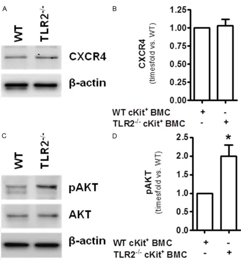
TLR2-/-cKit+ BMC exhibit equal protein
expres-sion of CXCR4 but increased basal activation of the PI3K downstream signaling molecule AKT
Homing of hematopoietic cells to sites of isch -emia is an important mechanism that
contrib-utes to neovascularization processes [22]. During ischemia, increased levels of the cyto
[image:6.612.97.521.74.365.2]-kine stromal-cell derived factor-1 (SDF-1) are expressed by tissues and SDF-1 augments the recruitment of CXCR4-positive BMC from the
Figure 2. cKit+ cells from TLR2-/- mice exhibit
in-creased capillary formation in vivo. A: Quantita
-tive summary of the mean cumula-tive capillary length per high power field (HPF) of n=3 mice
per group in Matrigel plugs with incorporated cKit+ cells three weeks after subcutaneous in
-jection. ##P<0.01. B: Representative HPFs of
Matrigel plugs exhibiting either WT or TLR2 -/-cKit+ cells, respectively (red); blue shows stain
-ing of cell nuclei with DAPI. Nuclei exhibit-ing no red fluorescence equal host-derived cells. Bars
2819 Int J Clin Exp Pathol 2013;6(12):2813-2823
circulation [16]. Comparing the amount of CXCR4 protein expression in WT cKit+ and TLR2 -/-cKit+ BMC however revealed no differences
(Figure 4A, 4B). Instead, increased activation
(i.e. phosphorylation) of the CXCR4 downstream
signaling molecule protein kinase B (PKB)/AKT
[23] was detected in TLR2-/-cKit+ BMC and may
thus account for the increased potency of these
cells to support angiogenic processes com-pared to WT cKit+ cells (Figure 4C, 4D) [24].
Discussion
Endothelial tube formation, migration and the sprouting of endothelial cells into the extracel
-lular matrix are the major constituents of the angiogenic process [21]. Hematopoietic cells home to sites of neovascularization where they exert stimulatory effects on the pre-existing vasculature, thereby augmenting the formation
of new capillaries [22]. TLR2 deficiency has been associated with various protective effects on the vasculature and the preservation of vas
-cular function [25, 26]. Studies employing mod
-els of transplanting TLR2-deficient bone mar -row into WT mice observed a reduction in tissue
injury for example after myocardial infarction that was mainly attributed to TLR2 deficiency on leukocytes [27]. In the present study, evi
-dence is provided that extends the spectrum of the beneficial effects of TLR2-deficiency on
hematopoietic stem cells to subsets that express cKit and augment the angiogenic
capacity of endothelial cells. cKit+ cells isolated
from the bone marrow of TLR2-deficient mice
promoted in vitro capillary tube formation, sprouting as well as the migratory capacity of mature endothelial cells more potently com -pared to WT cKit+ cells. Since the employment
[image:7.612.90.519.73.352.2]of anti-TLR2 antibodies is currently discussed
Figure 3. Bone marrow-derived cKit+ cells from TLR2-/- mice exhibit increased capacity to promote neovasculariza
-tion in vivo. A: 21 days following ischemia induction and cKit+ cell administration, WT mice that had received TLR2 -/-cKit+ bone-marrow derived cells (BMC) exhibited increased vascular density (CD31/DAPI double-positive cells) in M. Gastrocnemius tissue similar to TLR2-/- mice and in contrast to both WT and mice treated with cKit+ WT
BMCs. B: Quantitative summary of the CD31/DAPI double-positive cells per mm² of n=5-6 mice/group. #P<0.05 and ##P<0.01 vs. Non-ischemic control leg of the same animal, ***P<0.001. C: Thermal imaging of mice hindlimbs revealed augmented perfusion in both TLR2-/- and WT mice that had received TLR2-/-cKit+ cells. D: Quantitative sum
2820 Int J Clin Exp Pathol 2013;6(12):2813-2823
in the context of the treatment of a variety of vascular diseases [25], these results may point towards a potential therapeutical use of hema -topoietic stem cells pre-treated with anti-TLR2
antibodies for therapeutical
neovasculariz-ation.
We have recently shown that incubation of endothelial cells with mono- or polyclonal anti-TLR2 antibodies resulted in the activation of CXCR4 canonical signaling [20]. CXCR4 is
expressed on bone marrow-derived precursor
cells and mediates the effects of its ligand stro
-mal cell-derived factor-1 (SDF-1), one of the key players in progenitor cell homing [16]. In the present study, CXCR4 was verified to be
expressed on cKit+ cells but the amount of
CXCR4 expression in TLR2-/-cKit+ was equal to
those detected in WT cKit+ BMC. However, an
increased basal level of activation of the key dow-nstream signal transduction molecule of the SDF-1/CXCR4 system, namely AKT, has been
observed in cKit+ BMC isolated from TLR2
-/-mice compared to those derived from WT mice.
would challenge the notion that a reduction in
TLR signaling accompanied by a diminished activation of inflammatory signaling cascades may represent the underlying mechanism of the beneficial effects of TLR2 functional defi
-ciency for vascular homeostasis. In this con -text, TLR2 has also been reported to be an
important mediator of pro-angiogenic effects in the context of inflammatory processes [32, 33] and disruption of TLR2 signaling may impair tis -sue regenerative processes such as wound
healing due to impaired angiogenic events [34]. Thus, further studies are needed to decipher the exact phenotype of hematopoietic stem and progenitor as well as of cells of the vascu
-lature and the role of inflammatory processes
in correspondence to the vasculoprotective
phenotype of TLR2-deficient mice.
The stem cell factor receptor cKit is expressed on murine hemangioblasts during embryonic
development and on hematopoietic stem cells and endothelial progenitor cells during
[image:8.612.92.340.70.337.2]adult-hood [35]. In recent years, the characterization
Figure 4. TLR2-/-cKit+ cells exhibit similar expression of CXCR4 but
increased basal phosphorylation of AKT compared to WT cKit+ cells.
A: Western Blot analysis of both TLR2-/- and WT cKit+ cells for CXCR4
protein expression. β-actin indicates equal loading. B: Quantitative densitometric analysis of CXCR4 protein in cells isolated from n=3 mice per group. C: Western Blot analysis for phospho-AKT and AKT. D: Results of the densitometric analysis, *P<0.05, n=3 mice per
group.
Increased expression and activation
of AKT in TLR2-/- mice has been
observed previously and is associat
-ed with the cardio-protective effects of TLR2-deficiency in the context of myocardial ischemia [28]. Moreover,
AKT has been shown to be an
essen-tial mediator of homing of hematopoi -etic stem cells that contribute to
angiogenic processes and the level of
AKT activation correlates with the
quality of entrapment of BMC at sites of neovascularization [24]. In this regard, increased basal levels of AKT activation together with our finding that intracardiac delivery of TLR2
-/-cKit+ cells into mice resulted in
incr-eased capillary density following hindlimb ischemia may suggest that
TLR2-/-cKit+ BMC exhibit an increased
responsiveness towards
chemoat-tractants throughout the process of homing. However, the upstream acti
-vators of AKT in TLR2-/-cKit+ BMC
remain unclear. TLR2-deficient mice
have been reported to express higher
levels of TLR4 and TLR9 [29] and both TLR4 and TLR9 result in down
2821 Int J Clin Exp Pathol 2013;6(12):2813-2823
and potency of hematopoietic progenitors that specifically contribute to vascular regenerative processes have been redefined [36]. Specific combinations of surface antigens may identify different types of progenitor subsets with vari
-able potential to either differentiate into endo
-thelial cells themselves or serve as sources of angiogenic cytokines [37]. In the present study,
cKit+ BMC were employed irrespective of fur
-ther characterization for the expression of spe
-cific endothelial progenitor cell marker subsets. In the matrigel plug assay, cKit+ cells formed
capillary-like structures and this finding was found to be more pronounced when cKit+ cells
isolated from TLR2-/- mice were employed.
However, the tubular structures formed by both
TLR2-/- and WT cKit+ cells were not associated
with functionality as they were not connected to the circulation as indicated by negative stain
-ing for lectin upon systemic lectin perfusion. These findings are in line with previous studies reporting that the vast majority of bone marrow
derived precursor cells do not contribute to
neovascularization by structural contribution to new vessel formation [38]. On the other hand, the supportive role of cKit+ cells in
neovascular-ization processes is undisputed and supported
in the present study by the finding of augment -ed neovascularization in WT mice treat-ed with WT cKit+ BMC in contrast to WT mice having
received vehicle alone 24 hours after the induc
-tion of hindlimb ischemia.
Further studies are nevertheless required
addressing the exact identity and potency of hematopoietic cells that mediate the beneficial effects of TLR2-deficiency on vascular regener
-ative processes. However, the employment of hematopoietic cells selected by the expression of the marker for differentiation potential cKit may result in the utilization of a cell population
that exerts ubiquitous potential. Thus, the
pres-ent study employing cKit+ cells may also extend
the implications of TLR2-deficiency to other
implications involving hematopoietic stem cells such as liver, muscle or cardiac tissue regen- eration.
Acknowledgements
The authors would like to thank Berit Blendow, Dorothea Frenz, Maren Nerowski and Eva
Lorbeer-Rehfeldt (Institute for Experimental Surgery, University of Rostock, Germany) for
excellent technical assistance.
Disclosure of conflict of interest
The authors confirm that there are no conflicts of interest.
Address correspondence to: Dr. Nana-Maria
Wagner, Clinic for Anesthesiology and Critical Care Medicine, University Hospital Rostock, Schillingallee 35, D-18057 Rostock, Germany. Tel: +49 381 494 6401; Fax: +49 381 494 6402; E-mail: nana-maria.
References
[1] Kawai T, Akira S. Signaling to Nf-Kappab by Toll-Like Receptors. Trends Mol Med 2007; 13: 460-469.
[2] King KY, Goodell MA. Inflammatory Modulation of Hscs: Viewing the Hsc as a Foundation for
the Immune Response. Nat Rev Immunol 2011; 11: 685-692.
[3] De Luca K, Frances-Duvert V, Asensio MJ,
Ih-sani R, Debien E, Taillardet M, Verhoeyen E, Bella C, Lantheaume S, Genestier L, Defrance T. The Tlr1/2 Agonist Pam(3)Csk(4) Instructs Commitment of Human Hematopoietic Stem Cells to a Myeloid Cell Fate. Leukemia 2009; 23: 2063-2074.
[4] Megias J, Yanez A, Moriano S, O’Connor JE, Go -zalbo D, Gil ML. Direct Toll-Like
Receptor-Medi-ated Stimulation of Hematopoietic Stem and
Progenitor Cells Occurs in Vivo and Promotes
Differentiation toward Macrophages. Stem Cells 2012; 30: 1486-1495.
[5] Okun E, Griffioen KJ, Son TG, Lee JH, Roberts NJ, Mughal MR, Hutchison E, Cheng A, Arumu
-gam TV, Lathia JD, van Praag H, Mattson MP. Tlr2 Activation Inhibits Embryonic Neural Pro
-genitor Cell Proliferation. J Neurochem 2010; 114: 462-474.
[6] Sloane JA, Batt C, Ma Y, Harris ZM, Trapp B, Vartanian T. Hyaluronan Blocks Oligodendro
-cyte Progenitor Maturation and Remyelination through Tlr2. Proc Natl Acad Sci U S A 2010; 107: 11555-11560.
[7] Yamagishi VT, Torneck CD, Friedman S, Huang GT, Glogauer M. Blockade of Tlr2 Inhibits Por
-phyromonas Gingivalis Suppression of Miner
-alized Matrix Formation by Human Dental Pulp Stem Cells. J Endod 2011; 37: 812-818. [8] Kebschull M, Haupt M, Jepsen S, Deschner J,
Nickenig G, Werner N. Mobilization of Endothe
-lial Progenitors by Recurrent Bacteremias with
a Periodontal Pathogen. PLoS One 2013; 8:
e54860.
2822 Int J Clin Exp Pathol 2013;6(12):2813-2823
[10] Mullick AE, Soldau K, Kiosses WB, Bell TA 3rd, Tobias PS, Curtiss LK. Increased Endothelial
Expression of Toll-Like Receptor 2 at Sites of Disturbed Blood Flow Exacerbates Early Ath
-erogenic Events. J Exp Med 2008; 205:
373-383.
[11] Mullick AE, Tobias PS, Curtiss LK. Modulation
of Atherosclerosis in Mice by Toll-Like Receptor 2. J Clin Invest 2005; 115: 3149-3156. [12] Ziegler G, Freyer D, Harhausen D, Khojasteh U,
Nietfeld W, Trendelenburg G. Blocking Tlr2 in Vivo Protects against Accumulation of Inflam
-matory Cells and Neuronal Injury in Experimen -tal Stroke. J Cereb Blood Flow Metab 2011; 31:
757-766.
[13] Ziegler G, Harhausen D, Schepers C, Hoffmann O, Rohr C, Prinz V, Konig J, Lehrach H, Nietfeld W, Trendelenburg G. Tlr2 Has a Detrimental
Role in Mouse Transient Focal Cerebral
Isch-emia. Biochem Biophys Res Commun 2007; 359: 574-579.
[14] Favre J, Musette P, Douin-Echinard V, Laude K,
Henry JP, Arnal JF, Thuillez C, Richard V. Toll-Like Receptors 2-Deficient Mice Are Protected against Postischemic Coronary Endothelial Dysfunction. Arterioscler Thromb Vasc Biol 2007; 27: 1064-1071.
[15] Patel H, Shaw SG, Shi-Wen X, Abraham D, Bak -er DM, Tsui JC. Toll-Like Receptors in
Isch-aemia and Its Potential Role in the Pathophysi
-ology of Muscle Damage in Critical Limb
Ischaemia. Cardiol Res Pract 2012; 2012:
121237.
[16] Ceradini DJ, Kulkarni AR, Callaghan MJ, Tepper OM, Bastidas N, Kleinman ME, Capla JM, Galiano RD, Levine JP, Gurtner GC. Progenitor
Cell Trafficking Is Regulated by Hypoxic Gradi
-ents through Hif-1 Induction of Sdf-1. Nat Med 2004; 10: 858-864.
[17] Furlani D, Donndorf P, Westien I, Ugurlucan M,
Pittermann E, Wang W, Li W, Vollmar B,
Stein-hoff G, Kaminski A, Ma N. Hmgb-1 Induces
C-Kit+ Cell Microvascular Rolling and Adhesion Via Both Toll-Like Receptor-2 and Toll-Like
Re-ceptor-4 of Endothelial Cells. J Cell Mol Med 2012; 16: 1094-1105.
[18] Heida NM, Leifheit-Nestler M, Schroeter MR, Muller JP, Cheng IF, Henkel S, Limbourg A, Lim
-bourg FP, Alves F, Quigley JP, Ruggeri ZM, Hasenfuss G, Konstantinides S, Schafer K. Leptin Enhances the Potency of Circulating An -giogenic Cells Via Src Kinase and Integrin
(Al-pha)Vbeta5: Implications for Angiogenesis in Human Obesity. Arterioscler Thromb Vasc Biol
2010; 30: 200-206.
[19] Limbourg A, Korff T, Napp LC, Schaper W, Drex
-ler H, Limbourg FP. Evaluation of Postnatal Ar -teriogenesis and Angiogenesis in a Mouse
Model of Hind-Limb Ischemia. Nat Protoc 2009; 4: 1737-1746.
[20] Wagner NM, Bierhansl L, Noldge-Schomburg G, Vollmar B, Roesner JP. Toll-Like Receptor 2-Blocking Antibodies Promote Angiogenesis
and Induce Erk1/2 and Akt Signaling Via Cxcr4
in Endothelial Cells. Arterioscler Thromb Vasc
Biol 2013; 33: 1943-1951.
[21] Carmeliet P. Mechanisms of Angiogenesis and
Arteriogenesis. Nat Med 2000; 6: 389-395.
[22] Rafii S, Lyden D. Therapeutic Stem and Pro
-genitor Cell Transplantation for Organ Vascu -larization and Regeneration. Nat Med 2003; 9:
702-712.
[23] Ho TK, Tsui J, Xu S, Leoni P, Abraham DJ, Baker DM. Angiogenic Effects of Stromal Cell-Derived Factor-1 (Sdf-1/Cxcl12) Variants in Vitro and the in Vivo Expressions of Cxcl12 Variants and Cxcr4 in Human Critical Leg Ischemia. J Vasc
Surg 2010; 51: 689-699.
[24] Hur J, Yoon CH, Lee CS, Kim TY, Oh IY, Park KW, Kim JH, Lee HS, Kang HJ, Chae IH, Oh BH, Park YB, Kim HS. Akt Is a Key Modulator of Endothe
-lial Progenitor Cell Trafficking in Ischemic Mus
-cle. Stem Cells 2007; 25: 1769-1778. [25] Lin E, Freedman JE, Beaulieu LM. Innate
Im-munity and Toll-Like Receptor Antagonists: A Potential Role in the Treatment of Cardiovascu
-lar Diseases. Cardiovasc Ther 2009; 27:
117-123.
[26] Frantz S, Vincent KA, Feron O, Kelly RA. Innate Immunity and Angiogenesis. Circ Res 2005;
96: 15-26.
[27] Arslan F, Smeets MB, O’Neill LA, Keogh B, Mc
-Guirk P, Timmers L, Tersteeg C, Hoefer IE, Doe
-vendans PA, Pasterkamp G, de Kleijn DP. Myo
-cardial Ischemia/Reperfusion Injury Is Mediated by Leukocytic Toll-Like Receptor-2 and Reduced by Systemic Administration of a Novel Anti-Toll-Like Receptor-2 Antibody. Circu -lation 2010; 121: 80-90.
[28] Mersmann J, Tran N, Latsch K, Habeck K, Is
-kandar F, Zimmermann R, Zacharowski K. Akt
or Phosphoinositide-3-Kinase Inhibition Re-verses Cardio-Protection in Toll-Like Receptor
2 Deficient Mice. Resuscitation 2012; 83: 1404-1410.
[29] Ehrentraut SF, Dorr A, Ehrentraut H, Lohner R, Lee SH, Hoeft A, Baumgarten G, Knuefermann P, Boehm O, Meyer R. Vascular Dysfunction Following Polymicrobial Sepsis: Role of Pattern Recognition Receptors. PLoS One 2012; 7: e44531.
[30] Sester DP, Brion K, Trieu A, Goodridge HS, Rob
-erts TL, Dunn J, Hume DA, Stacey KJ, Sweet
MJ. Cpg DNA Activates Survival in Murine Mac-rophages through Tlr9 and the
Phosphati-dylinositol 3-Kinase-Akt Pathway. J Immunol 2006; 177: 4473-4480.
[31] Dauphinee SM, Voelcker V, Tebaykina Z, Wong F, Karsan A. Heterotrimeric Gi/Go Proteins
Indepen-2823 Int J Clin Exp Pathol 2013;6(12):2813-2823
dent of the Myd88-Dependent Pathway. Am J Physiol Heart Circ Physiol 2011; 301:
H2246-2253.
[32] Grote K, Schuett H, Salguero G, Grothusen C, Jagielska J, Drexler H, Muhlradt PF, Schieffer B.
Toll-Like Receptor 2/6 Stimulation Promotes
Angiogenesis Via Gm-Csf as a Potential Strate
-gy for Immune Defense and Tissue Regenera
-tion. Blood 2010; 115: 2543-2552.
[33] Saber T, Veale DJ, Balogh E, McCormick J,
NicAnUltaigh S, Connolly M, Fearon U. Toll-Like
Receptor 2 Induced Angiogenesis and Invasion Is Mediated through the Tie2 Signalling
Path-way in Rheumatoid Arthritis. PLoS One 2011; 6: e23540.
[34] West XZ, Malinin NL, Merkulova AA, Tischenko
M, Kerr BA, Borden EC, Podrez EA, Salomon
RG, Byzova TV. Oxidative Stress Induces Angio
-genesis by Activating Tlr2 with Novel Endoge
-nous Ligands. Nature 2010; 467: 972-976.
[35] Kabrun N, Buhring HJ, Choi K, Ullrich A, Risau W, Keller G. Flk-1 Expression Defines a Popula
-tion of Early Embryonic Hematopoietic Precur
-sors. Development 1997; 124: 2039-2048. [36] Fadini GP, Losordo D, Dimmeler S. Critical
Re-evaluation of Endothelial Progenitor Cell Phe
-notypes for Therapeutic and Diagnostic Use. Circ Res 2012; 110: 624-637.
[37] Yoder MC, Mead LE, Prater D, Krier TR, Mroueh KN, Li F, Krasich R, Temm CJ, Prchal JT, Ingram
DA. Redefining Endothelial Progenitor Cells Via Clonal Analysis and Hematopoietic Stem/Pro
-genitor Cell Principals. Blood 2007; 109:
1801-1809.
[38] Ziegelhoeffer T, Fernandez B, Kostin S, Heil M, Voswinckel R, Helisch A, Schaper W. Bone Mar -row-Derived Cells Do Not Incorporate into the
Adult Growing Vasculature. Circ Res 2004; 94: