Original Article
Pathogenesis and significance of glomerular C4d
deposition in lupus nephritis: activation
of classical and lectin pathways
Min-Kyung Kim1, Young-In Maeng1, Sun-Jae Lee1,In Hee Lee2, Jisuk Bae3, Yu-Na Kang4, Byung-Tae Park5, Kwan-Kyu Park1
1Department of Pathology, Catholic University of Daegu School of Medicine, Daegu, Republic of Korea; 2
Depart-ment of Internal Medicine, Catholic University of Daegu School of Medicine, Daegu, Republic of Korea; 3
Depart-ment of Preventive Medicine, Catholic University of Daegu School of Medicine, Daegu, Republic of Korea; 4
Depart-ment of Pathology, Keimyung University School of Medicine, Republic of Korea; 5Department of Microbiology,
Catholic University of Daegu School of Medicine, Daegu, Republic of Korea
Received August 13, 2013; Accepted August 28, 2013; Epub September 15, 2013; Published October 1, 2013
Abstract: Immune complex-mediated complement activation through the classic pathway plays a key role in the pathogenesis of lupus nephritis (LN). C4d deposition in renal tissue reflects the prognosis of systemic lupus erythe -matosus (SLE). The aim of the current study is to investigate the pathogenesis and clinicopathologic significance of glomerular C4d deposition in LN. We retrospectively analyzed clinical and histopathological data of 20 SLE patients with renal biopsy-proven LN and 10 non-SLE renal biopsy samples as control. LN biopsies showed varying degrees of glomerular C4d staining associated with immune complex deposits, IgG (p = 0.015), C1q (p = 0.032) and C3 (p
= 0.049). 7 LN biopsies had all of C4d, C1q and C3 deposits in their glomeruli, indicative of the activation of the classical pathway, whereas 2 LN biopsies had C4d and C3 deposits without accompanying C1q deposits, indicating the activation of the lectin pathway. Glomerular C4d deposition was correlated with the LN subtype (p < 0.001). In particular, a diffusely intense and coarsely granular pattern of C4d deposition in all glomeruli was detected in class V membranous LN. However, glomerular C4d deposition was correlated with neither disease activity of SLE nor histological activity and chronicity of LN. In conclusion, the activation of the lectin pathway as well as the clas-sical pathway seems to play a crucial role in the pathogenesis of LN. Glomerular C4d staining could be helpful for diagnosing class V membranous LN, although glomerular C4d deposition does not reflect SLE disease activity and histological activity and chronicity.
Keywords: Lupus nephritis, C4d, classical pathway, lectin pathway, disease activity
Introduction
SLE is a prototypic autoimmune disease char-acterized by loss of self-tolerance and the pro-duction of autoantibodies to double-stranded DNA and nucleosomes [1]. Immune complex-mediated complement activation via the classi-cal pathway plays a key role in the tissue injury leading to LN, a serious complication of SLE [1]. However, the pathogenesis of LN remains unclear.
A complement split product C4d is produced by breakdown of C4b into C4c and C4d through the classical pathway or lectin pathway after being activated by immune complexes [2]. C4d
stud-ies on the association of C4d deposition with disease activity of SLE. C4d deposition in the peritubular capillaries is correlated with dis-ease activity of SLE [10], whereas C4d deposi-tion in the glomeruli is not correlated with dis-ease activity of SLE [5-11].
Therefore, the aims of this study are to (1) determine what complement pathway is involved in the pathogenesis of LN, to (2) exam-ine whether glomerular C4d deposition is help-ful for classifying LN subtypes through evalua-tion of the pattern of glomerular C4d deposievalua-tion, and to (3) identify whether glomerular C4d deposition is correlated with disease activity of SLE and histological activity and chronicity of LN.
Materials and methods
Patients
We retrospectively studied 20 SLE patients with biopsy-proven LN at Daegu Catholic Medical Center from January 2010 to March 2013. All patients met the diagnostic criteria for SLE according to the American College of
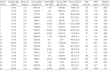
[image:2.612.95.521.83.353.2]Rheumatology. The medical records of all patients were reviewed independently of the analysis of pathologic features. The demo-graphic and clinical data are summarized in Table 1.
The control sample was composed of negative and positive control groups. The negative con-trol group included 3 renal biopsy specimens with minimal change disease and 1 renal biop-sy specimen with thin basement membrane disease. The positive control group included 3 renal biopsy specimens with grade III IgA nephropathy and 3 renal biopsy specimens with membranous glomerulopathy.
Renal tissue and histological evaluation
Percutaneous renal biopsies were performed in 20 SLE and 10 non-SLE patients under ultraso-nographic guidance. 10% formalin-fixed tissue was embedded in paraffin. Four μm-thick sec-tions were stained with hematoxylin and eosin (H&E), periodic acid-Schiff (PAS), periodic acid methenamine silver (PAM), masson-trichrome and polyclonal rabbit anti-human C4d antibody (Biomedia, Austria) using an autostainer (Bond-Table 1. Overview of clinical and laboratory data of 20 patients with lupus nephritis
Patient
with LN Gender/Age (years) Serum Cr (mg/dL) Urine protein (mg/24 hr) Urine RBCs (per HPF) Anti-ds DNA IgG (IU/mL) Serum C3/C4 (mg/dL) (mm/hr)ESR (mg/L)CRP (x10Platelet 3/μL)
1 F/41 1.3 6873.6 > 30 424.00 49.6/7.8 43 3.4 200
2 F/10 0.6 1124.2 1-3 486.70 24.8/1.6 62 5.5 145
3 F/50 2.2 2775.6 5-10 1.09 83.2/24.4 13 4.8 48
4 F/58 0.6 1445.4 20-30 21.06 74.0/12.1 23 4.8 129
5 F/32 1.2 286.2 > 30 46.56 37.1/2.3 15 4.8 197
6 F/14 1.0 180.0 10-20 258.20 116.1/28.5 101 4.8 164
7 F/34 0.7 7386.6 > 30 499.10 21.6/5.5 19 3.8 120
8 F/18 0.7 3504.0 20-30 > 500.00 67.8/11.1 45 5.8 190
9 F/24 0.8 1638.0 10-20 122.70 72.0/8.4 47 4.8 186
10 F/25 0.7 4956.0 > 30 > 500.00 18.3/4.1 3 4.5 159
11 F/26 0.6 1537.8 0-1 3.07 49.9/11.6 10 4.8 154
12 M/39 0.9 2337.0 1-3 > 300.00 29.8/3.1 2 54.4 141
13 F/52 0.8 1203.6 0-1 3.82 91.2/34.7 24 8.4 212
14 F/27 0.7 2540.4 10-20 < 1.00 36.7/5.8 6 0.1 225
15 F/29 0.7 291.0 > 30 43.00 34.7/17.3 2 0.5 239
16 F/28 0.7 922.6 3-5 150.00 24.5/1.0 39 0.3 167
17 F/28 0.6 880.1 10-20 > 300.00 16.1/2.1 84 17.5 259
18 F/54 0.6 716.1 > 30 5.00 20.4/2.4 105 4.8 207
19 F/37 0.6 4330.0 > 30 > 379 40.2/4.1 53 8.7 79
20 M/29 1.5 1431.4 10-20 197.00 7.5/3.2 90 79.6 75
Max, Leica, USA). Renal biopsy specimens of SLE patients were assessed using the most recent modification of the World Health Organization (WHO) classification proposed by a joint committee of the International Society of Nephrology and the Renal Pathology Society (2003 ISN/RPS) [12, 13]. Activity and chronicity indexes of renal lesions were evaluated accord-ing to the NIH scoraccord-ing system [14, 15].
C4d immunohistochemical staining
As in the previously described method, renal C4d staining was performed with polyclonal rabbit anti-human C4d antibody (Biomedia, Austria) using an autostainer (Bond-Max, Leica, USA). Glomerular C4d deposition was defined as glomerular capillary wall and/or mesangial C4d staining. The pattern of glomerular C4d staining was documented, and C4d positivity was semi-quantitatively graded as follows: -, no glomerular staining; +, mild glomerular stain-ing; ++, moderate glomerular stainstain-ing; and
+++, intense glomerular staining. Images were captured by a digital camera with OLYMPUS (Tokyo, Japan) BX51 microscopy.
Immunofluorescence evaluation
Fresh frozen tissue sections were stained with fluorescein isothiocyanate-conjugated poly-clonal antibodies directed against human IgG, IgA, IgM, C3, C1q, kappa and lambda, respec-tively (Dako, Denmark). The pattern of immuno-fluorescent staining in the glomeruli was evalu-ated. Intensity of glomerular staining for IgG was also semi-quantitatively graded as +, ++ or +++, and for IgA and C3 as -, + or ++. Staining intensity for IgM and C1q was assessed as - or +, where - represented the absence of immuno-fluorescent staining, and + represented the presence of immunofluorescent staining.
Electron microscopic evaluation
[image:3.612.92.523.97.385.2]Renal biopsy tissues was fixed in 2.5% glutaral-dehyde in 0.1 M sodium phosphate buffer, Table 2. Overview of C4d and immune complex deposition in the glomeruli with possible complement pathway related to the pathogenesis of lupus nephritis
Patients
with LN ISN/RPS Class Immunohistochemical C4d staining
Immunofluorescent staining
Complement pathway
IgG IgA IgM C1q C3
1 IV ++ +++ + + + ++ Classical
2 II - ++ + + + + Classical
3 V +++ + - + - - Lectin
4 V +++ +++ + - - - Lectin
5 IV ++ ++ + + + + Classical
6 IV + + + - - - Lectin
7 IV + + + - - + Lectin
8 IV ++ ++ + - + + Classical
9 IV + + - - - + Lectin
10 IV + ++ + + + + Classical
11 II + + - - - - Lectin
12 III - ++ ++ + + ++ Classical
13 II - + - - - - Unknown
14 V ++ +++ + - + ++ Classical
15 IV ++ ++ + + + + Classical
16 III - ++ ++ - + ++ Classical
17 III - ++ + - + ++ Classical
18 II - ++ ++ + - - Unknown
19 IV + + - - + + Classical
20 III - + + - + ++ Classical
post-fixed in 1% osmium tetroxide (OsO4) in 0.1 M sodium phosphate buffer, and embedded in epoxy-resin. Ultrathin sections using an Ultramicrotome were stained with uranyl ace-tate and lead citrate, and examined with a transmission electron microscope (H-7000, HITACHI Japan). The presence and location of electron dense deposits in the glomeruli were examined.
Statistical analysis
The data were expressed as mean ± standard deviation for continuous variables and frequen-cy and percentage for categorical variables. Mann-Whitney tests were used to compare the mean differences between patient groups. The
differences in proportions between various groups were assessed by Fisher’s exact tests. A value of P < 0.05 was considered statistically significant. All statistical analyses were per-formed by software IBM SPSS Statistics 19.0 (IBM Corp., Armonk, NY, USA).
Results
Relationship between glomerular C4d deposits and immune complex deposits
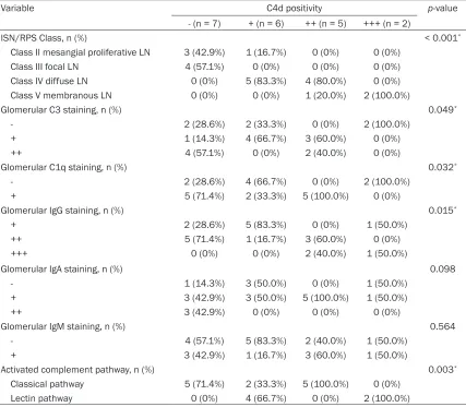
[image:4.612.92.519.95.468.2]C4d deposit was absent in the negative control group, whereas C4d deposits in the mesangi-um or along the pericapillary loops were detect-ed in the positive control group. In LN, glomeru-lar deposition of immune complex was present Table 3. Relationship between glomerular C4d deposition and the pathologic classes and immune complex deposition in 20 patients with lupus nephritis
Variable C4d positivity p-value
- (n = 7) + (n = 6) ++ (n = 5) +++ (n = 2)
ISN/RPS Class, n (%) < 0.001*
Class II mesangial proliferative LN 3 (42.9%) 1 (16.7%) 0 (0%) 0 (0%) Class III focal LN 4 (57.1%) 0 (0%) 0 (0%) 0 (0%) Class IV diffuse LN 0 (0%) 5 (83.3%) 4 (80.0%) 0 (0%) Class V membranous LN 0 (0%) 0 (0%) 1 (20.0%) 2 (100.0%)
Glomerular C3 staining, n (%) 0.049*
- 2 (28.6%) 2 (33.3%) 0 (0%) 2 (100.0%)
+ 1 (14.3%) 4 (66.7%) 3 (60.0%) 0 (0%)
++ 4 (57.1%) 0 (0%) 2 (40.0%) 0 (0%)
Glomerular C1q staining, n (%) 0.032*
- 2 (28.6%) 4 (66.7%) 0 (0%) 2 (100.0%)
+ 5 (71.4%) 2 (33.3%) 5 (100.0%) 0 (0%)
Glomerular IgG staining, n (%) 0.015*
+ 2 (28.6%) 5 (83.3%) 0 (0%) 1 (50.0%)
++ 5 (71.4%) 1 (16.7%) 3 (60.0%) 0 (0%)
+++ 0 (0%) 0 (0%) 2 (40.0%) 1 (50.0%)
Glomerular IgA staining, n (%) 0.098
- 1 (14.3%) 3 (50.0%) 0 (0%) 1 (50.0%)
+ 3 (42.9%) 3 (50.0%) 5 (100.0%) 1 (50.0%)
++ 3 (42.9%) 0 (0%) 0 (0%) 0 (0%)
Glomerular IgM staining, n (%) 0.564
- 4 (57.1%) 5 (83.3%) 2 (40.0%) 1 (50.0%)
+ 3 (42.9%) 1 (16.7%) 3 (60.0%) 1 (50.0%)
Activated complement pathway, n (%) 0.003*
Classical pathway 5 (71.4%) 2 (33.3%) 5 (100.0%) 0 (0%)
Lectin pathway 0 (0%) 4 (66.7%) 0 (0%) 2 (100.0%)
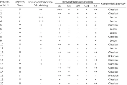
with varying degrees of staining intensity. The staining intensities of glomerular C4d and immune complex in LN are summarized in Table 2. Immunofluorescent staining of glomerular IgG was detected in 100%, IgA in 75.0%, IgM in 40.0%, C1q in 60.0% and C3 in 70.0% of the 20 cases. Table 2 shows possible complement pathways related to the pathogenesis of LN, based on the relative deposition of C4d, C1q and C3. Glomerular C4d deposition was corre-lated with deposition of immune complexes, especially IgG (p = 0.015), C1q (p = 0.032) and C3 (p = 0.049) (Table 3).
Relationship between pattern of glomerular C4d staining and LN subtype
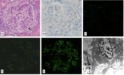
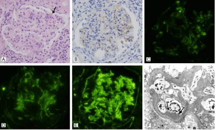
Out of the 20 cases of LN, glomerular C4d deposits were detected in 13 cases. 1 case of class II LN and 5 cases of class IV LN showed mild C4d deposition along the glomerular
[image:5.612.91.524.72.336.2]capil-lary loops and in the mesangium (Figure 1). 4 cases of class IV LN and 1 case of class V LN showed moderate C4d deposition along the glomerular capillary loops (Figure 2). 2 cases of class V LN showed a diffusely intense and coarsely granular pattern of C4d deposition along the glomerular capillary loops, involving the entire capillary circumference (Figure 3). As well as glomerular staining of C4d, peritubular capillary staining was observed in 4 cases (case No. 3, 9, 15 and 16) and tubular base-ment membrane staining in 2 cases (case No. 3 and 18). Although glomerular C4d deposits were detected in various subtypes of LN, a uni-formly strong and diffusely granular pattern of C4d deposition in all glomeruli was found exclu-sively in class V membranous LN (Figure 3). Moreover, glomerular C4d deposition was sig-nificantly correlated with ISN/RPS Class (p < 0.001) (Table 3).
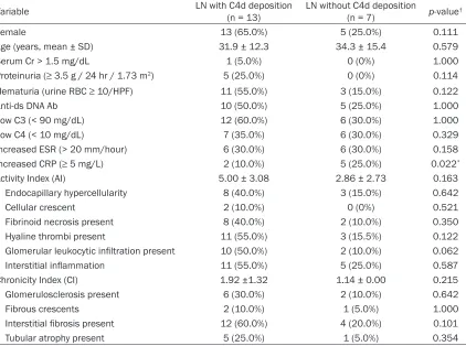
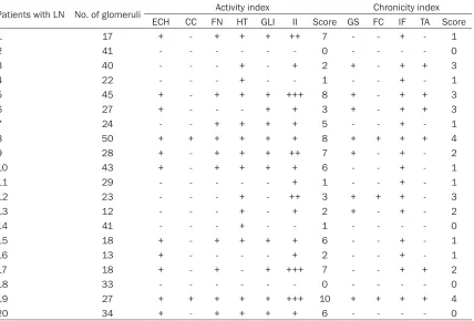
the two groups. The histological features indi-cating the activity and chronicity of LN are sum-marized in Table 5.
Discussion
Immune complex-mediated complement acti-vation via the classical pathway plays a key role in the pathogenesis of LN [1]. The classical pathway is initiated by binding of the comple-ment C1 complex to the antigen-antibody com-plex or CRP, which is followed by the cleavage of the complement component C4 into C4a and C4b, and then C4b is cleaved into C4c and C4d [2]. The presence of C4d and C1q deposits with C3 deposits in the glomerulus is considered to be the evidence for the activation of the classi-cal pathway. In the present study, glomerular C4d deposition, accompanied by C1q deposits, was detected in 7 of 20 LN patients (case No. 1, 5, 8, 10, 14, 15 and 19). Glomerular C3 deposition was also observed in all of them
Relationship between glomerular C4d staining and clinical features of SLE
We divided LN patients into two groups based on the glomerular C4d deposition, and com-pared the various indexes affecting disease activity of SLE between the two groups. However, there was no significant difference between the two groups, except that LN patients with glomerular C4d deposition showed a lower frequency of increased C-reactive protein (CRP) (p = 0.022). That is, the presence of glomerular C4d deposition was not correlated with the disease activity of SLE (Table 4).
Relationship between the glomerular C4d staining and histological features of LN
[image:6.612.91.525.72.335.2]We compared parameters of activity and chro-nicity of LN between the two groups (Table 4). There was no significant difference between
with glomerular C4d deposition showed C3 deposits without C1q deposits (case No. 7 and 9), which indicates the possibility of the activa-tion of the lectin pathway in the pathogenesis of LN. In addition, glomerular C4d deposition without accompanying C1q deposition was found in another 4 LN patients. However, this was not accompanied by C3 deposition (case No. 3, 4, 6 and 11), which indicates there is needed for further evaluation using MBL or L-ficolin staining to check for activation of the lectin pathway in these patients.
Glomerular C3 deposition without C4d and C1q deposits could be considered as a marker for the activation of the alternative pathway. In the present study, we could not find any case show-ing activation of an alternative pathway. Moreover, no deposits for C1q, C4d and C3 were detected in 2 LN patients, who were clas-sified as class II mesangial proliferative LN (Table 2), indicating that activation of the
clas-sical pathway might be involved in the progres-sion of LN.
[image:7.612.90.523.71.335.2]C4d is also produced by activation of the lectin pathway [2]. The lectin pathway is activated by binding of mannose-binding lectin (MBL) or fico-lins to the carbohydrate ligand on the microbial surface, leading to activation of MBL-associated serine protease (MASP) and cleavage of C4 [2, 16]. In addition, the microbial surface directly cleaves C3, leading to activation of the alterna-tive pathway, and factor B, factor D and proper-din are involved in the cascade event [2, 16]. Recently, Sato et al. [3] reported that activation of the lectin and alternative pathways was involved in the glomerular injury of LN. Glomerular deposition of C4d and C3 without accompanying C1q deposits can be considered to be the evidence for the activation of the lec-tin pathway. In the present study, 2 LN patients
accompanying C4d deposits. Among these patients, 4 are classified as having class III focal LN (case No. 12, 16, 17 and 20) except for one who is having class II mesangial prolifera-tive LN (case No. 2). The activation of the clas-sical pathway could be also involved in the pro-gression of LN in these patients, considering the lower sensitivity of immunohistochemical staining for C4d on paraffin-embedded tissue compared to immunofluorescent staining for C1q and C3 on fresh frozen tissue.
Glomerular C4d deposition was associated with immune complex deposition detected by immunofluorescence or electron microscopy, and significantly correlated with immune com-plexes, especially IgG (p = 0.015), C1q (p = 0.032) and C3 (p = 0.049) deposits (Table 3). In addition, the pattern of glomerular C4d depo-sition varied with the LN subtype (p < 0.001) (Table 3): a uniformly strong and granular pat-(case No. 13 and 18). We thought the reason
[image:8.612.97.519.98.412.2]for the absence of deposition of these comple-ments in class II mesangial proliferative LN is that deposition of these complements is asso-ciated with deposition of immune complexes: the relatively small numbers of stable immune complexes prevents the mesangial clearing system from becoming overloaded and allows the complexes to be sequestered in the mesan-gium [17]. The sequestered immune complexes are apt to be degraded and removed rather than remaining at the sites where they could initiate complement activation, resulting in the absence of deposits of C1q, C4d and C3 in the glomeruli of class II mesangial proliferative LN. Immunohistochemical staining for C4d on par-affin-embedded tissue is less sensitive than immunofluorescent staining on frozen tissue [18]. In the present study, 5 LN patients had glomerular C1q and C3 deposits without
Table 4. Relationship between glomerular C4d deposition and clinicopathological parameters in 20 patients with lupus nephritis
Variable LN with C4d deposition (n = 13) LN without C4d deposition (n = 7) p-value†
Female 13 (65.0%) 5 (25.0%) 0.111
Age (years, mean ± SD) 31.9 ± 12.3 34.3 ± 15.4 0.579
Serum Cr > 1.5 mg/dL 1 (5.0%) 0 (0%) 1.000
Proteinuria (≥ 3.5 g / 24 hr / 1.73 m2) 5 (25.0%) 0 (0%) 0.114
Hematuria (urine RBC ≥ 10/HPF) 11 (55.0%) 3 (15.0%) 0.122
Anti-ds DNA Ab 10 (50.0%) 5 (25.0%) 1.000
Low C3 (< 90 mg/dL) 12 (60.0%) 6 (30.0%) 1.000
Low C4 (< 10 mg/dL) 7 (35.0%) 6 (30.0%) 0.329
Increased ESR (> 20 mm/hour) 6 (30.0%) 6 (30.0%) 0.158
Increased CRP (≥ 5 mg/L) 2 (10.0%) 5 (25.0%) 0.022*
Activity Index (AI) 5.00 ± 3.08 2.86 ± 2.73 0.163
Endocapillary hypercellularity 8 (40.0%) 3 (15.0%) 0.642
Cellular crescent 2 (10.0%) 0 (0%) 0.521
Fibrinoid necrosis present 8 (40.0%) 2 (10.0%) 0.350
Hyaline thrombi present 11 (55.0%) 3 (15.5%) 0.122
Glomerular leukocytic infiltration present 10 (50.0%) 2 (10.0%) 0.062
Interstitial inflammation 11 (55.0%) 5 (25.0%) 0.587
Chronicity Index (CI) 1.92 ±1.32 1.14 ± 0.00 0.215
Glomerulosclerosis present 6 (30.0%) 2 (10.0%) 0.642
Fibrous crescents 2 (10.0%) 1 (5.0%) 1.000
Interstitial fibrosis present 12 (60.0%) 4 (20.0%) 0.101
Tubular atrophy present 5 (25.0%) 1 (5.0%) 0.354
Data were expressed as mean ± SD for continuous variables and n (%) for categorical variables; LN, lupus nephritis; † Calcu-lated by Mann-Whitney tests for continuous variables and Fisher’s exact tests for categorical variables; Cr, creatinine; RBC, red blood cell; HPF, high power field; Anti-ds DNA, anti-double stranded DNA antibodies; C3, complement C3; C4, complement C4;
diffuse LN, tend to overcome the mesangial ability to clear these macromolecules, leading to their accumulation in a paramesangial sub-endothelial location, and then ultimately in the peripheral capillary loops [17]. Immune com-plexes localized to the subendothelial region have easy access to plasma inflammatory mediators, causing inflammation and injury of the endothelium, thus allowing the permeability of the capillary wall to increase, resulting in deposition of immune complexes in the suben-dothelial area [17] and immune complex-medi-ated complement activation.
In contrast, small, unstable, and circulating immune complexes formed by low-avidity and low-affinity antibodies, which are characteristic of class V membranous LN, may dissociate with the antigen or antibody lodging in the glomeru-lar capilglomeru-laries [17]. Subsequently, complexes may be formed by in situ attachment to the tar-get protein in the outer aspect of the GBM [17], where the complexes may induce activation and deposition of complements. Thus, the dif-ferent pathogenesis of each LN class seems to tern of C4d deposition in the glomeruli was
found exclusively in class V membranous LN (Figure 3), but this pattern was less strong in some class IV diffuse LN (Figures 1 and 2) and class III focal LN. In addition, the pattern of glo-merular C4d staining with thickened gloglo-merular basement membrane and spikes in class V membranous LN, was reminiscent of the find-ings in membranous nephropathy. Thus, glo-merular C4d immunohistochemical staining can be a useful method for confirmation of class V membranous LN when there is absence of glomeruli in the frozen renal section.
[image:9.612.96.524.85.375.2]The pathogenesis of LN varies with the class of the lesion [17]. The pattern of glomerular C4d deposition also varied with the LN subtype. The reason that the pattern of glomerular C4d deposition varies with the LN subtype may be due to the size, quantity or stability of the immune complexes and location of the depos-ited immune complexes. Large numbers of intermediate-size complexes or large complex-es formed by high-affinity antibodicomplex-es, which are characteristic of class III focal LN and class IV
Table 5. Summary of histological features of activity and chronicity in lupus nephritis
Patients with LN No. of glomeruli Activity index Chronicity index ECH CC FN HT GLI II Score GS FC IF TA Score
1 17 + - + + + ++ 7 - - + - 1
2 41 - - - 0 - - - - 0
3 40 - - - + - + 2 + - + + 3
4 22 - - - + - - 1 - - + - 1
5 45 + - + + + +++ 8 + - + + 3
6 27 + - - - + + 3 + - + + 3
7 24 - - + + + + 5 - - + - 1
8 50 + + + + + + 8 + + + + 4
9 28 + - + + + ++ 7 + - + - 2
10 43 + - + + + + 6 - - + - 1
11 29 - - - + 1 - - + - 1
12 23 - - - + - ++ 3 + + + - 3
13 12 - - - + - + 2 + - + - 2
14 41 - - - + - - 1 - - - - 0
15 18 + - + + + + 6 - - + - 1
16 13 + - - - - + 2 - - + - 1
17 18 + - + - + +++ 7 - - + + 2
18 33 - - - 0 - - - - 0
19 27 + + + + + +++ 10 + + + + 4
20 34 + - + + + + 6 - - - - 0
merular C4d deposition does not reflect dis-ease activity of SLE and histological activity and chronicity of LN.
Acknowledgements
This work was supported by the grant of Research Institute of Medical Science, Catholic University of Daegu, Republic of Korea (2012).
Address correspondence to: Dr. Kwan-Kyu Park, Department of Pathology, Catholic University of Daegu School of Medicine, 3056-6 Daemyung-4 Dong, Nam-gu, Daegu, 705-718, Republic of Korea. Tel: 82-53-650-4149; Fax: 82-53-650-3050; E-mail: [email protected]; Dr. Byung-Tae Park, Depart- ment of Microbiology, Catholic University of Daegu School of Medicine, 3056-6 Daemyung-4 Dong, Nam-gu, Daegu, 705-718, Republic of Korea. Tel: 82-53-650-4476; Fax: 82-53-621-4106; E-mail: [email protected]
References
[1] Tang S, Lui SL and Lai KN. Pathogenesis of lu-pus nephritis: an update. Nephrology (Carlton) 2005; 10: 174-179.
[2] Murata K and Baldwin WM 3rd. Mechanisms of complement activation, C4d deposition, and their contribution to the pathogenesis of anti-body-mediated rejection. Transplant Rev (Or-lando) 2009; 23: 139-150.
[3] Sato N, Ohsawa I, Nagamachi S, Ishii M, Kusa-ba G, Inoshita H, Toki A, Horikoshi S, Ohi H, Matsushita M and Tomino Y. Significance of glomerular activation of the alternative path-way and lectin pathpath-way in lupus nephritis. Lu-pus 2011; 20: 1378-1386.
[4] Manzi S, Ahearn JM and Salmon J. New in-sights into complement: a mediator of injury and marker of disease activity in systemic lu-pus erythematosus. Lulu-pus 2004; 13: 298-303.
[5] Batal I, Liang K, Bastacky S, Kiss LP, McHale T, Wilson NL, Paul B, Lertratanakul A, Ahearn JM, Manzi SM and Kao AH. Prospective assess-ment of C4d deposits on circulating cells and renal tissues in lupus nephritis: a pilot study. Lupus 2012; 21: 13-26.
[6] Manzi S, Rairie JE, Carpenter AB, Kelly RH, Jag-arlapudi SP, Sereika SM, Medsger TA Jr and Ramsey-Goldman R. Sensitivity and specificity of plasma and urine complement split prod-ucts as indicators of lupus disease activity. Ar-thritis Rheum 1996; 39: 1178-1188.
[7] Liu CC, Manzi S, Kao AH, Navratil JS, Ruffing MJ and Ahearn JM. Reticulocytes bearing C4d as biomarkers of disease activity for systemic result in varying patterns of glomerular C4d
deposition according to the LN subtype. The complement split product C4d can cova-lently bind to endothelial surfaces and base-ment membranes via a thioester moiety [19]. C4d deposition on endothelial cells induces insertion of sublytic levels of membrane attack complex (MAC), which is followed by von Willebrand factor release from endothelial cells, and then platelet aggregation [2]. This event might bring about a decrease in platelet levels and renal microthrombi. In the present study, intensity of glomerular C4d deposition was marginally significant by platelet levels (p = 0.056, data not shown). However, only 1 of 13 LN patients with glomerular C4d deposition had microthrombi (case No. 19), and another 1 LN patient had microthrombi without glomeru-lar C4d deposition (case No. 17). Thus we thought that glomerular C4d deposition was not an indicator of thrombotic microangiopathy in LN, which was different the results of Cohen
et al. [20]. Furthermore, we divided the LN patients into two groups based on glomerular C4d deposition and compared parameters of SLE disease activity and histological features of activity and chronicity between the two groups. However, glomerular C4d deposition showed a poor correlation with SLE disease activity and histological activity and chronicity, as reported by Batal et al. [5] and Kim et al. [11] (Table 4). In addition, regardless of which com-plement pathway was activated, there was no significant difference in SLE disease activity and histological activity and chronicity (data not shown).
[15] Austin HA 3rd, Muenz LR, Joyce KM, An-tonovych TT and Balow JE. Diffuse proliferative lupus nephritis: identification of specific patho -logic features affecting renal outcome. Kidney Int 1984; 25: 689-695.
[16] Monticielo OA, Mucenic T, Xavier RM, Brenol JC and Chies JA. The role of mannose-binding lec-tin in systemic lupus erythematosus. Clin Rheumatol 2008; 27: 413-419.
[17] Fogo AB, Kashgarian M. Lupus nephritis. In: Diagnostic Atlas of Renal Pathology. Second edition. Elsevier Saunders, 2012; pp: 222-223.
[18] Chantranuwat C, Qiao JH, Kobashigawa J, Hong L, Shintaku P and Fishbein MC. Immuno -peroxidase staining for C4d on paraffin-em -bedded tissue in cardiac allograft endomyo-cardial biopsies: comparison to frozen tissue immunofluorescence. Appl Immunohistochem Mol Morphol 2004; 12: 166-171.
[19] Feucht HE and Opelz G. The humoral immune response towards HLA class II determinants in renal transplantation. Kidney Int 1996; 50: 1464-1475.
[20] Cohen D, Koopmans M, Kremer Hovinga IC, Berger SP, Roos van Groningen M, Steup-Beek-man GM, de Heer E, Bruijn JA and Bajema IM. Potential for glomerular C4d as an indicator of thrombotic microangiopathy in lupus nephritis. Arthritis Rheum 2008; 58: 2460-2469. lupus erythematosus. Arthritis Rheum 2005;
52: 3087-3099.
[8] Muso E, Sekita K, Doi T, Kuwahara T, Yoshida H, Tamura T, Kawai C and Hamashima Y. Im-munopathological correlation between mesan-gial C3d-deposition and C3d-fixing circulating immune complexes in lupus nephritis. Clin Im-munol Immunopathol 1984; 32: 351-358. [9] Kao AH, Navratil JS, Ruffing MJ, Liu CC,
Hawkins D, McKinnon KM, Danchenko N, Ahearn JM and Manzi S. Erythrocyte C3d and C4d for monitoring disease activity in systemic lupus erythematosus. Arthritis Rheum 2012; 62: 837-844.
[10] Li SJ, Liu ZH, Zen CH, Wang QW, Wang Y and Li LS. Peritubular capillary C4d deposition in lu-pus nephritis different from antibody-mediated renal rejection. Lupus 2007; 16: 875-880. [11] Kim SH and Jeong HJ. Glomerular C4d
deposi-tion indicates in situ classic complement path-way activation, but is not a marker for lupus nephritis activity. Yonsei Med J 2003; 44: 75-80.
[12] Weening JJ, D’Agati VD, Schwartz MM, Seshan SV, Alpers CE, Appel GB, Balow JE, Bruijn JA, Cook T, Ferrario F, Fogo AB, Ginzler EM, Hebert L, Hill G, Hill P, Jennette JC, Kong NC, Lesavre P, Lockshin M, Looi LM, Makino H, Moura LA and Nagata M. The classification of glomerulo -nephritis in systemic lupus erythematosus re-visited. J Am Soc Nephrol 2004; 15: 241-250. [13] Markowitz GS and D’Agati VD. Classification of
lupus nephritis. Curr Opin Nephrol Hypertens 2009; 18: 220-225.