Introduction
Enchondromas are common, benign, and
usu-ally asymptomatic hyaline cartilage forming
neo-plasms in the metaphyses and diaphyses of the
short and long tubular bones of the limbs,
espe-cially the hands and feet [1,2]. They usually
oc-cur as a single lesion (solitary enchondroma)
which is most often found incidentally when
radiographic studies are performed for other
reasons.
Occasionally patients present with multiple
en-chondromas causing severe deformity of the
affected bones, generally defined as
matosis [2,3]. The distribution of the
enchondro-mas, and other accompanying symptoms as
well as the mode of inheritance define the
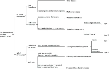
dif-ferent subtypes of enchondromatosis (Figure 1),
which mainly includes Ollier disease, Maffucci
syndrome, metachondromatosis,
genochondro-matosis, spondyloenchondrodysplasia,
cheiro-spondyloenchondromatosis and
dysspondyloen-chondromatosis. These subtypes should be
dis-tinguished and adequately diagnosed, not only
to guide therapeutic decision and genetic
coun-seling, but also to enable future studies to shed
light on whether these are different ends of a
spectrum caused by a single gene, or that they
represent true different diseases. We therefore
review the available clinical information for all
enchondromatosis subtypes and discuss the
little molecular data available hinting towards
their cause.
Enchondroma
On conventional radiographs enchondromas
Review Article
Enchondromatosis: insights on the different subtypes
Twinkal C. Pansuriya
1, Herman M. Kroon
2, Judith V.M.G. Bovée
11Department of Pathology, Leiden University Medical Center, Leiden, The Netherlands. 2Department of Radiology,
Leiden University Medical Center, Leiden, The Netherlands.
Received May 31, 2010, accepted June 18, 2010, available online June 26, 2010
Abstract: Enchondromatosis is a rare, heterogeneous skeletal disorder in which patients have multiple enchondro-mas. Enchondromas are benign hyaline cartilage forming tumors in the medulla of metaphyseal bone. The disorder manifests itself early in childhood without any significant gender bias. Enchondromatosis encompasses several differ-ent subtypes of which Ollier disease and Maffucci syndrome are most common, while the other subtypes (metachondromatosis, genochondromatosis, spondyloenchondrodysplasia, dysspondyloenchondromatosis and cheirospondyloenchondromatosis) are extremely rare. Most subtypes are non-hereditary, while some are autosomal dominant or recessive. The gene(s) causing the different enchondromatosis syndromes are largely unknown. They should be distinguished and adequately diagnosed, not only to guide therapeutic decisions and genetic counseling, but also with respect to research into their etiology. For a long time enchondromas have been considered a develop-mental disorder caused by the failure of normal endochondral bone formation. With the identification of genetic ab-normalities in enchondromas however, they were being thought of as neoplasms. Active hedgehog signaling is re-ported to be important for enchondroma development and PTH1R mutations have been identified in ~10% of Ollier patients. One can therefore speculate that the gene(s) causing the different enchondromatosis subtypes are involved in hedgehog/PTH1R growth plate signaling. Adequate distinction within future studies will shed light on whether these subtypes are different ends of a spectrum caused by a single gene, or that they represent truely different dis-eases. We therefore review the available clinical information for all enchondromatosis subtypes and discuss the little molecular data available hinting towards their cause.
present as multiple, oval-shaped, linear and/or
pyramidal osteolytic lesions with well-defined
margins in the metaphysis and/or diaphysis of
the long tubular and in the flat bones [4].
Mag-netic resonance (MR) studies demonstrate
lobu-lated lesions with intermediate signal intensity
on T1-weighted images and predominantly high
signal intensity on T2-weighted sequences [5].
Histologically enchondromas show low
cellular-ity with an abundant hyaline cartilage matrix
sometimes with extensive calcifications
[1].
When enchondromas are located in the
pha-langeal bones or when they occur in
enchondro-matosis patients, cellularity is increased and
more worrisome histological features are
toler-ated, since they are not correlated with
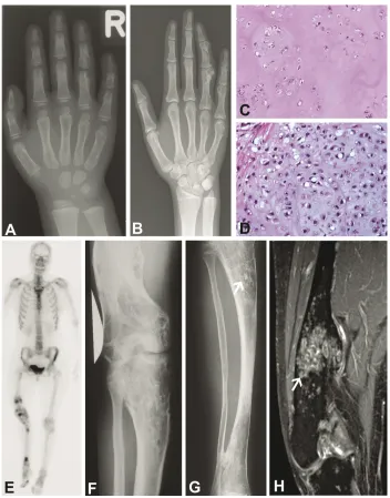
malig-nant behavior in this specific context (Figure 2)
[1].
Treatment of solitary enchondroma is surgical
but only in case of complaints or cosmetic
de-formity [6]. In case of enchondromatosis, the
deformities as well as malignant progression of
enchondromas may require multiple surgical
interventions [7-12].
Secondary central chondrosarcoma
While solitary enchondromas almost never
pro-gress to secondary central chondrosarcoma,
malignant transformation in enchondromatosis
is estimated to occur in 25-30% of the patients
[1]. Central chondrosarcoma is a malignant
bone tumor forming hyaline cartilage and
aris-ing centrally in the medullary cavity of bone
[13]. The distinction between enchondroma and
low grade chondrosarcoma is difficult on
con-ventional radiographs [14]. Fast
contrast-enhanced dynamic MRI is more helpful in this
regard [15]. At the histological level the
distinc-tion can also be very difficult and is subject to
interobserver variability [16] [17]. Low grade
chondrosarcomas (grade I) can be treated
surgi-cally with local curettage combined with
cryosur-gery or phenol treatment while resection and
reconstruction is obligatory in case of grade II or
III chondrosarcoma [18].
Enchondromagenesis
[image:2.612.84.529.85.366.2]The underlying mechanism for enchondroma
development is largely unknown. Several
cyto-genetic/genetic reports are present in the
litera-ture using solitary enchondromas, suggesting
these lesions to be neoplastic (http://
atlasgeneticsoncology.org//Tumors/
EnchondromaID5333.html). Enchondromas in
Ollier disease are comparable to solitary
en-chondromas at m-RNA expression level [19].
Since enchondromas arise in the metaphysis in
close proximity to the growth plate, they may
result from failure of terminal differentiation of
growth plate chondrocytes. In support of this,
transgenic mice expressing the hedgehog (Hh)
regulated transcription factor
Gli2
in
chondro-cytes, which mimics activated Hh signaling,
de-velop lesions similar to human enchondromas
[20]. Hedgehog signaling is a crucial regulator
of normal chondrocytes proliferation and
differ-entiation within the normal growth plate.
En-chondromas indeed demonstrate levels of
hedgehog signaling that are comparable to
nor-mal growth plate [20-23].
Additionally, ten percent of patients with
en-chondromatosis harbour a mutation in the
PTHLH receptor,
PTH1R,
in their tumor tissue
[20,22,23]. The mutations were shown to
de-crease the function of the PTHLH receptor with
~30% [22]. PTH1R is a receptor for parathyroid
hormone and for parathyroid hormone-related
peptide which acts in a negative feedback loop
with Indian Hedgehog (IHH) regulating normal
endochondral bone formation [24-26]. PTHLH
signaling is active in solitary enchondromas and
in chondrosarcomas [27].
In parallel, multiple osteochondromas (MO)
syn-drome (multiple benign cartilaginous tumors
arising from the surface of bone) is an
auto-somal dominant disorder caused by mutations
in the
EXT1
and
EXT2
genes, leading to
dis-turbed hedgehog signaling based on their
in-volvement in heparan sulphate (HS)
biosynthe-sis [28-30]. The
EXT
genes are not affected in
central chondrosarcomas and their m-RNA
ex-pression is normal [21]. It may be hypothesized
that the genes causing the different
enchondro-matosis subtypes also affect the HS dependent
signaling pathways.
Ollier disease
Ollier disease (also known as dyschondroplasia,
multiple cartilaginous enchondromatosis,
en-chondromatosis Spranger type I), is the most
common subtype, first described in 1889. It is
defined by the presence of multiple
enchondro-mas with asymmetric distribution (Figure 2)
[4,31-34]. Ollier disease is non-familial and
mostly encountered early in childhood, affecting
both sexes equally. The estimated prevalence of
Ollier disease is 1/100,000 [2]. The true
inci-dence can be higher since mild phenotypes
without skeletal deformities can go undetected.
Few cases of familial occurrence have been
reported (OMIM 166000) [35-37] There is large
clinical variability with respect to size, number,
location, age of onset, and requirement of
sur-gery [1,2,4,34]. Lesions are usually distributed
unilaterally and may involve the entire skeleton,
although the skull and vertebral bodies are very
rarely involved. Sometimes lesions are bilateral
or present in only one extremity [4].
Malignant transformation of one or more
en-chondromas towards secondary central
chon-drosarcoma is estimated to occur in 5-50% of
the patients and can be life threatening [38-42].
Malignant transformation most frequently
oc-curs in long tubular and flat bones while this is
far less common in the small bones of hands
and feet (Figure 2). This is interesting since
en-chondromas preferentially occur at the hands
and feet.
In addition to the risk of developing
chondrosarcoma, Ollier patients also seem to
have an increased risk for the development of
non-skeletal malignancies as reported in Table
1, especially intracranial tumors of glial origin
[43]. There is at present no curative or
preven-tive treatment option for patients with Ollier
disease.
The underlying cause of Ollier disease is so far
unknown. The non-hereditary asymmetrical
polyostotic distribution of the lesions might
sug-gest a somatic mosaic mutation [14]. This is
similar to McCune-Albright syndrome /
polyostotic fibrous dysplasia in which an
activat-ing mutation in
GNAS1
occurs during early
em-bryogenesis leading to a somatic mosaic state
resulting in fibrous dysplasia affecting several
bones [44,45].
patients (~10%). Thus,
PTH1R
mutations may
contribute to the disease in a small subset of
Ollier patients but is probably not causative for
the disease [22].
Maffucci syndrome
Maffucci syndrome (also known as
dyschondro-dysplasia with haemangiomas,
enchondromato-sis with multiple cavernous haemangiomas,
Kast syndrome, haemangiomatosis
chondrodys-trophica, enchondromatosis Spranger type II)
was first described in 1881[32,46,47]. It is
non-hereditary and characterized by the presence of
multiple enchondromas combined with multiple
haemangiomas of soft tissue or, less commonly,
lymphangiomas (Figure 3) [34,48]. Lesions are
asymmetrically distributed and there is no
gen-der discrimination. The disease appears to
de-velop in 25% of cases from the time of birth or
during the first year of life, in 45% of cases
symptoms start before the age of 6 and in 78%
of cases symptoms developed before puberty
[49,50]. Lewis et al reviewed ninety-eight cases
[image:5.612.78.537.100.155.2] [image:5.612.86.532.190.348.2]and showed that hand, foot, femur, tibia, and
Table 1. Non-cartilaginous malignancies associated with Ollier disease
Table 2. Genetic findings in Ollier enchondromas and chondrosarcoma
Associated tumors No. of patients References
Glioma 17 [38,43,90-99]
Juvenile granulosa cell tumor 7 [38,100-105]
Non-small cell lung cancer 1 [106]
Tumor per patient Technique used Results of chromosomal abnormality References
low grade CS cytogenetics deletion at 1p [108]
high grade CS microsatellite marker,
SSCP, IHC LOH at 13q14 (of TP53 RB1), 9p21 and over expression [109]
Enchondroma array-CGH no alteration [110]
Enchondroma array-CGH deletion of 6
CS II array-CGH gain at 1, 2, 5, 7, 8, 9, 15, 16, 17, 18, 19, 20,
21 and 22 [110]
CS II array-CGH gain at 6, 7, 12, 14, 15, 16, 17, 18, 19 and loss
at 1, 3, 4, 6, 9, 10, 13, 15, 16 and 22
[110]
[image:5.612.322.533.401.651.2]fibula were most frequently affected by
enchon-dromas [50].
Haemangiomas are benign vascular tumors
which often protrude as bluish or reddish soft
nodules. They can be found anywhere in the
body. In addition to the enchondromas,
radio-graphs can show phleboliths, associated with
soft tissue calcifications in haemangiomas.
His-tologically, haemangiomas can be of the
capil-lary or cavernous subtype. Spindle cell
haeman-gioendothelioma is more specific for Maffucci
syndrome [51,52]. Both the enchondromas and
the vascular lesions may progress to
malig-nancy. The risk of malignancy is higher than
in
Ollier disease [2,49]. When considering
intracra-nial tumors, the majority is of mesenchymal
origin and includes secondary central
chon-drosarcoma and angiosarcoma associated with
Maffucci syndrome [43]. Non-mesodermal
tu-mors associated with Maffucci syndrome are
summarized in Table 3. While in Ollier disease
intracranial tumors other than
chondrosarco-mas of the cranium are exclusively of glial
ori-gin, in Maffucci syndrome different tumor types
are encountered [43]. Also, patients with
Maf-fucci syndrome are almost 10 years older when
developing intracranial tumors, and are more
likely to live in Asia or South America as
com-pared to Ollier disease [43].
Genetic studies on Maffucci syndrome are
sparse. An inversion of chromosome bands p11
and q21 of chromosome 1 were reported in one
patient with Maffucci syndrome [53]. Robinson
et al showed an increased number of nerve
fi-bers in tumors as well as in normal tissue of
Maffucci patients, while in enchondroma and
haemangioma tissue numerous methionine
enkephalin positive nerves were detected,
serv-ing as a growth factor for cartilage proliferation
[54]. In total, 26 patients with Maffucci
syn-drome were screened for mutations in
PTH1R
and revealed absence of mutation [23,55].
Metachondromatosis
[image:6.612.82.533.99.372.2]This rare hereditary condition displays a
combi-nation of multiple enchondromas with multiple
osteochondroma-like lesions [32,56,57] (MIM
156250, enchondromatosis Spranger type III).
The enchondromas mainly involve the iliac
crests and metaphyses of the long bones of the
lower extremities while the osteochondroma-like
Table 3. Non-cartilaginous and non-vascular neoplasms associated with Maffucci syndrome
Associated tumors No. of patients References
Astrocytoma 6 [38,50,111-113]
Pituitary adenoma 4 [50,114-116]
Juvenile granulosa cell tumor 6 [50,117-120]
Pancreatic adenocarcinoma 3 [38,50,121]
Adrenal adenoma 1 [50]
Intracranial chordoma 1 [122]
Biliary adenocarcinoma 1 [38]
Olfactory neuroblastoma 1 [123]
Paraganglioma 1 [124]
Fibrosarcoma 1 [50]
Thyroid adenoma 1 [51]
Hepatic adenocarcinoma 1 [121]
Fibro adenoma breast 2 [125]
Breast carcinoma 1 [114]
Squamous cell carcinoma 1 [126]
Fibroadenoma of thorax and canalicular adenoma 1 [127]
Acute myeloblastic leukemia 1 [51]
Lymphoid leukemia 1 [128]
lesions are mainly located in hands and feet
[58]. The syndrome manifests early in childhood
[59,60]. In contrast to conventional
osteochon-dromas, the osteochondroma-like lesions in
metachondromatosis point towards the joint, do
not cause bone deformities, and may regress
spontaneously [3,59] (Figure 4). Importantly,
malignant transformation has not been
re-ported. Avascular necrosis of the femoral
epiphysis can be found due to interference with
the integrity of the lateral epiphyseal vessels by
enchondromas [59,61-63].
The mode of inheritance is autosomal dominant
but the underlying gene has not been identified
so far, due to the extreme rarity of the disease.
In a single case, mutations in the
EXT
genes
causing multiple osteochondromas were absent
and EXT mRNA expression levels were normal.
IHH/PTHLH signaling was normally active in two
cases. These data suggest that EXT related
pathways are not involved in the pathogenesis
of metachondromatosis [64].
Genochondromatosis
[image:7.612.118.498.84.372.2]Genochondromatosis is an extremely rare
auto-somal dominant disorder manifesting itself in
childhood [65](MIM 137360). Patients have
normal stature and enchondromas are
distrib-uted symmetrically with characteristic
localiza-tion in the metaphyses of the proximal humerus
and distal femur. Two subtypes are
distin-guished: genochondromatosis type I includes
the presence of a chondroma on the medial
side of the clavicle while in type II the short
tu-bular bones of the hand, wrist and feet are
af-fected while the clavicle is normal [65-68]. The
enchondroma-like lesions will not lead to any
bone deformities, may be discovered
acciden-tally, and tend to regress in adulthood [65] in
which they differ from the enchondromas in the
other subtypes. Moreover, no malignancies
as-sociated with genochondromatosis have been
described in the literature. No spinal
involve-ment is reported which emphasizes it being
dif-ferent from spondyloenchondrodysplasia,
cheirospondyloenchondromatosis and
dyss-pondyloenchondromatosis [3]. No genetic
stud-ies have been reported for this rare subtype.
Spondyloenchondrodysplasia
Spondyloenchondrodysplasia (SPENCD,
enchon-dromatosis Spranger type IV) is an autosomal
recessive inherited disorder characterized by
vertebral dysplasia combined with
enchon-droma like lesions in the pelvis or long tubular
bones (MIM 271550) [32,69-81]. Estimated
prevalence is higher in Israel [77]. The spinal
aberrations include platyspondyly; flat, often
rectangular vertebral bodies are seen at
radiog-raphy with irregular areas of increased and
de-creased mineralization, and short broad ilia.
Spondyloenchondrodysplasia can manifest itself
from birth to later infancy [71]. Patients usually
have a short stature (short limbs), with
in-creased lumbar lordosis, barrel chest and
ky-phoscoliosis, genua valga or vara, facial
anoma-lies, and may show clumsy movements [3]. Type
I is classic, and type II also affects the central
nervous system including spasticity,
develop-mental delay, and late-onset cerebral
calcifica-tions [76,81]. In addition, clinical manifestation
of autoimmunity can be seen. The spine is less
severely affected as compared with
dysspondy-loenchondromatosis and
cheirospondyloen-chondromatosis, in which the vertebral lesions
are less uniform and the ilia are not short [3].
The clinical features of
spondyloenchondrodys-plasia are highly variable within and between
the families; neurological and autoimmune
manifestations were seen in different
combina-tions within one single family suggesting
re-markable pleiotropic manifestations of a single
disease [81]. In addition, there are two reports
suggesting an association of
spondyloenchon-dromatosis with D-2-hydroxyglutaric aciduria, a
neurometabolic disorder [82,83]. Although the
disease is thought to be autosomal recessive,
an autosomal dominant inheritance pattern has
also been described [77,80].
Cheirospondyloenchondromatosis
Cheirospondyloenchondromatosis (generalized
enchondromatosis with platyspondyly,
enchon-dromatosis Spranger type VI) combines
symmet-rically distributed multiple enchondromas with
marked involvement of metacarpals and
pha-langes resulting in short hands and feet with
mild platyspondyly [3,32]. It occurs at very early
age. There is mild to moderate dwarfism and
joints, especially of the fingers, become
enlarged. Mental retardation is frequently seen.
Genetic transmission is unknown [3].
Dysspondyloenchondromatosis
Dysspondyloenchondromatosis
(enchondroma-tosis with irregular vertebral lesions,
enchondro-matosis type V) is a non-hereditary disorder
characterized by spondyloenchondromatosis
combined with malformation of the spine
[3,32,70,80,84]. The irregularity of the vertebral
anomalies differentiates
dysspondyloenchon-dromatosis from other enchondysspondyloenchon-dromatosis with
spinal involvement. Severe segmentation
abnor-malities and secondary deformities of the
verte-bral column can be seen [3]. In addition,
neona-tal dwarfism, unequal limb length or asymmetric
limb shortening, a flat midface with a frontal
prominence, and progressive kyphoscoliosis can
be found [3,80,85]. Multiple enchondromas are
present in the long tubular and flat bones while
the bones of hands and feet are not or only
mildly affected. The disease manifests itself at
birth. Haga et al reported a case with
dyss-pondyloenchondromatosis along with Maffucci
syndrome in one patient [86] which suggests
that these two different syndromes may be part
of one spectrum caused by a pleiotropic gene.
Malignant transformation is not yet reported in
the literature.
Other less well delineated subtypes
epiphy-seal-metahyseal enchondromatosis as
pro-posed by the authors [88]. In addition,
metaphy-seal chondrodysplasia, Vandraager-Pena type
may also be considered an enchondromatosis
subtype (MIM 250300) [89] although it is more
often considered a subtype of metaphyseal
dys-plasia. It is an autosomal recessive disorder in
which the metaphyses of the long bones have
an extensive sponge-like appearance at
radio-graphs while histologically numerous islands of
cartilage reminiscent of enchondromatosis are
seen [89].
Conclusions
Many different syndromes are present with
mul-tiple enchondromas, and most of them are
ex-tremely rare. None of these syndromes seem to
be determined by a simple mendelian manner,
and it is unclear whether they represent
sepa-rate entities, or that they are manifestations of
a single causal process. Since they all share the
occurrence of multiple enchondromas, this may
suggest that the same gene or gene family may
be involved in at least a proportion of the
differ-ent types of enchondromatosis. It can be
ex-pected that within the upcoming era of next
generation sequencing approaches, elucidating
the genes causing rare genetic disorders, also
the gene(s) that causes the different
enchon-dromatosis subtypes may be identified.
Ade-quate classification of the different
enchondro-matosis subtypes as reviewed here is not only
relevant for clinical management with regards
to genetic counseling and the risk of malignant
transformation, but also to allow future
molecu-lar studies to reveal whether (a proportion of)
the different subtypes are different ends of a
spectrum caused by a pleiotropic single gene, or
that they truly represent separate disease
enti-ties.
Note added in proof
Sobreira et al recently reported mutations in the
PTPN11 gene in two metachondromatosis
fami-lies (PLOS Genet, 2010;6;6). Future studies
should reveal whether this gene is also involved
in other enchondromatosis subtypes.
Acknowledgements
We are thankful to S.H.M Verdegaal and R. J.
Grimer for kindly providing Figure 3A and
Henk-Jan van der Woude for Figure 3B. Our work on
enchondromatosis is supported by The
Nether-lands Organization for Scientific Research
(917-76-315) and is performed within EuroBoNet, a
European Commission granted Network of
Ex-cellence for studying the pathology and genetics
of bone tumors (018814).
Please address correspondence to: Judith V.M.G. Bovée, MD, PhD, Department of Pathology, Leiden University Medical Center , Albinusdreef 2, 2333 ZA Leiden , The Netherlands. Tel: +31715266617. Fax: +31715266952. Email: [email protected]
References
[1] Lucas DR, Bridge JA: Chondromas: enchon-droma,periosteal chondroma,and enchondro-matosis. World Health Organization classifica-tion of tumours. Pathology and genetics of tu-mours of soft tissue and bone. Edited by Fletcher CDM, Unni KK, Mertens F. Lyon: IARC Press, 2002, pp. 237-40.
[2] Silve C, Juppner H. Ollier disease. Orphanet J Rare Dis 2006; 1: 37.
[3] Spranger JW, Brill P.W., Poznanski AK: Bone Dysplasias, An Atlas of Genetic Disorders of Skeletal Development. second ed. New York: Oxford University Press; 2002, pp. 554-70. [4] Unni KK. Cartilaginous lesions of bone. J Orthop
Sci 2001; 6: 457-72.
[5] Loder RT, Sundberg S, Gabriel K, Mehbod A, Meyer C. Determination of bone age in children with cartilaginous dysplasia (multiple hereditary osteochondromatosis and Ollier's enchondro-matosis). J Pediatr Orthop 2004; 24: 102-8. [6] Shapiro F. Ollier's Disease. An assessment of
angular deformity, shortening, and pathological fracture in twenty-one patients. J Bone Joint Surg Am 1982; 64: 95-103.
[7] Watanabe K, Tsuchiya H, Sakurakichi K, Yama-shiro T, Matsubara H, Tomita K. Treatment of lower limb deformities and limb-length discrep-ancies with the external fixator in Ollier's dis-ease. J Orthop Sci 2007; 12: 471-5.
[8] Baumgart R, Burklein D, Hinterwimmer S, Thaller P, Mutschler W. The management of leg -length discrepancy in Ollier's disease with a fully implantable lengthening nail. J Bone Joint Surg Br 2005; 87: 1000-4.
[9] Van LP, Lammens J. Malformation of the hume-rus in a patient with Ollier disease treated with the Ilizarov technique. J Shoulder Elbow Surg 2008; 17: e9-11.
[10] Urist MR. A 37-year follow-up evaluation of mul-tiple-stage femur and tibia lengthening in dyschondroplasia (enchondromatosis) with a net gain of 23.3 centimeters. Clin Orthop Relat Res 1989; 137-57.
[12] Martson A, Haviko T, Kirjanen K. Extensive limb lengthening in Ollier's disease: 25-year follow-up. Medicina (Kaunas ) 2005; 41: 861-6. [13] Bertoni F, Bacchini P, Hogendoorn PCW:
Chon-drosarcoma. World Health Organisation classifi-cation of tumours. Pathology and genetics of tumours of soft tissue and bone. Edited by-Fletcher CDM, Unni KK, Mertens F. Lyon: IARC Press, 2002, pp. 247-51.
[14] D'Angelo L, Massimi L, Narducci A, Di RC. Ollier disease. Childs Nerv Syst 2009; 25: 647-53. [15] Geirnaerdt MJ, Hogendoorn PCW, Bloem JL,
Taminiau AHM, Van der Woude HJ. Cartilagi-nous tumors: fast contrast-enhanced MR imag-ing. Radiology 2000; 214: 539-46.
[16] Eefting D, Schrage YM, Geirnaerdt MJ, Le Ces-sie S, Taminiau AH, Bovee JVMG, Hogendoorn PCW. Assessment of interobserver variability and histologic parameters to improve reliability in classification and grading of central cartilagi-nous tumors. Am J Surg Pathol 2009; 33: 50-7. [17] Reliability of Histopathologic and Radiologic
Grading of Cartilaginous Neoplasms in Long Bones. J Bone Joint Surg Am 2007; 89-A: 2113-23.
[18] Veth R, Schreuder B, van Beem H, Pruszczynski M, de Rooy J. Cryosurgery in aggressive, benign, and low-grade malignant bone tumours. Lancet Oncol 2005; 6: 25-34.
[19] Rozeman LB, Hameetman L, van Wezel T, Taminiau AHM, Cleton-Jansen AM, Hogendoorn PCW, Bovée JVMG. cDNA expression profiling of central chondrosarcomas: Ollier disease resem-bles solitary tumors and alteration in genes coding for energy metabolism with increasing grade. J Pathol 2005; 207: 61-71.
[20] Hopyan S, Gokgoz N, Poon R, Gensure RC, Yu C, Cole WG, Bell RS, Juppner H, Andrulis IL, Wunder JS, Alman BA. A mutant PTH/PTHrP type I receptor in enchondromatosis. Nat Genet 2002; 30: 306-10.
[21] Schrage YM, Hameetman L, Szuhai K, Cleton-Jansen AM, Taminiau AHM, Hogendoorn PCW, Bovée JVMG. Aberrant heparan sulfate pro-teoglycan localization, despite normal exosto-sin, in central chondrosarcoma. Am J Pathol 2009; 174: 979-88.
[22] Couvineau A, Wouters V, Bertrand G, Rouyer C, Gerard B, Boon LM, Grandchamp B, Vikkula M, Silve C. PTHR1 mutations associated with Ollier disease result in receptor loss of function. Hum Mol Genet 2008; 17: 2766-75.
[23] Rozeman LB, Sangiorgi L, Bruijn IH, Mainil-Varlet P, Bertoni F, Cleton-Jansen AM, Hogen-doorn PC, Bovée JVMG. Enchondromatosis (Ollier disease, Maffucci syndrome) is not caused by the PTHR1 mutation p.R150C. Hum Mutat 2004; 24: 466-73.
[24] Alexandre C, Jacinto A, Ingham PW. Transcrip-tional activation of hedgehog target genes in Drosophila is mediated directly by the cubitus interruptus protein, a member of the GLI family
of zinc finger DNA-binding proteins. Genes Dev 1996; 10: 2003-13.
[25] Vortkamp A, Lee K, Lanske B, Segre GV, Kronenberg HM, Tabin CJ. Regulation of rate of cartilage differentiation by indian hedgehog and PTH-related protein. Science 1996; 273: 613-22.
[26] Amling M, Neff L, Tanaka S, Inoue D, Kuida K, Weir E, Philbrick WM, Broadus AE, Baron R. Bcl-2 lies downstream of parathyroid hormone related peptide in a signalling pathway that regulates chondrocyte maturation during skele-tal development. J Cell Biol 1997; 136: 205-13. [27] Bovée JVMG, Van den Broek LJCM, Cleton-Jansen AM, Hogendoorn PCW. Up-regulation of PTHrP and Bcl-2 expression characterizes the progression of osteochondroma towards pe-ripheral chondrosarcoma and is a late event in central chondrosarcoma. Lab Invest 2000; 80: 1925-33.
[28] Ahn J, Ludecke H-J, Lindow S, Horton WA, Lee B, Wagner MJ, Horsthemke B, Wells DE. Cloning of the putative tumour suppressor gene for hereditary multiple exostoses (EXT1). Nature Genet 1995; 11: 137-43.
[29] Stickens D, Clines G, Burbee D, Ramos P, Tho-mas S, Hogue D, Hecht JT, Lovett M, Evans GA. The EXT2 multiple exostoses gene defines a family of putative tumour suppressor genes. Nature Genet 1996; 14: 25-32.
[30] Wuyts W, Van Hul W, De Boulle K, Hendrickx J, Bakker E, Vanhoenacker F, Mollica F, Ludecke H-J, Sitki Sayli B, Pazzaglia UE, Mortier G, Hamel B, Conrad EU, Matsushita M, Raskind WH, Willems PJ. Mutations in the EXT1 and EXT2 genes in hereditary multiple exostoses. Am J Hum Genet 1998; 62: 346-54.
[31] Ollier M. De la dyschondroplasia. Bull Soc Chir Lyon 1899; 3: 22-3.
[32] Spranger J, Kemperdieck H, Bakowski H, Opitz JM. Two peculiar types of enchondromatosis. Pediatr Radiol 1978; 7: 215-9.
[33] Flemming DJ, Murphey MD. Enchondroma and chondrosarcoma. Semin Musculoskelet Radiol 2000; 4: 59-71.
[34] Mertens F, Unni KK: Enchondromatosis: Ollier disease and Maffucci syndrome. World Health Organization Classification of Tumours. Pathol-ogy and genetics of tumours of soft tissue and bone. Edited byFletcher CDM, Unni KK, Mertens F. Lyon: IARC Press, 2002, pp. 356-7. [35] ROSSBERG A. [Heredity of osteochondromas.].
Fortschr Geb Rontgenstr Nuklearmed 1959; 90: 138-9.
[36] CARBONELL JM, VINETA TJ. [A further case of congenital generalized dyschondrotheosis, Ollier type.]. Rev Esp Pediatr 1962; 18: 91-9. [37] Lamy M, Aussannaire M, Jammet ml, Nezelof C.
[Three cases of Ollier's disease in one family.]. Bull Mem Soc Med Hop Paris 1954; 70: 62-70. [38] Schwartz HS, Zimmerman NB, Simon MA,
malig-nant potential of enchondromatosis. J Bone Joint Surg Am 1987; 69: 269-74.
[39] Schaison F, Anract P, Coste F, De PG, Forest M, Tomeno B. [Chondrosarcoma secondary to multiple cartilage diseases. Study of 29 clinical cases and review of the literature]. Rev Chir Orthop Reparatrice Appar Mot 1999; 85: 834-45.
[40] Rozeman LB, Hogendoorn PCW, Bovée JVMG. Diagnosis and prognosis of chondrosarcoma of bone. Expert Rev Mol Diagn 2002; 2: 461-72. [41] Liu J, Hudkins PJ, Swee RG, Unni KK. Bone
sarcomas associated with Ollier's disease. Can-cer 1987; 59: 1376-85.
[42] Bukte Y, Necmioglu S, Nazaroglu H, Kilinc N, Yilmaz F. A case of multiple chondrosarcomas secondary to severe multiple symmetrical en-chondromatosis (Ollier's disease) at an early age. Clin Radiol 2005; 60: 1306-10.
[43] Ranger A, Szymczak A. Do intracranial neo-plasms differ in Ollier disease and maffucci syndrome? An in-depth analysis of the litera-ture. Neurosurgery 2009; 65: 1106-13.
[44] Riminucci M, Saggio I, Robey PG, Bianco P. Fibrous dysplasia as a stem cell disease. J Bone Miner Res 2006; 21 Suppl 2: 125-31. [45] Cohen MM, Jr., Siegal GP: McCune-Albright
syndrome. World Health Organization Classifi-cation of Tumours. Pathology & Genetics. Tu-mours of Soft Tissue and Bone. Edited by-Fletcher C.D.M, Unni KK, Mertens F. Lyon: IARC Press, 2002, pp. 357-9.
[46] Maffucci A. Di un caso encondroma ed an-gioma multiplo. Movimento medico-chirurgico, Napoli 1881; 3: 399-412; 565-575.
[47] Mertens F, Unni KK: Congenital and inherited syndromes associated with bone and soft tis-sue tumours: Enchondromatosis. World Health Organisation classification of tumours. Pathol-ogy and genetics of tumours of soft tissue and bone. Edited by Fletcher CDM, Unni KK, Mertens F. Lyon: IARC Press, 2002, pp. 356-7. [48] Auyeung J, Mohanty K, Tayton K. Maffucci
lym-phangioma syndrome: an unusual variant of Ollier's. J Pediatr Orthop B 2003; 12: 147-50. [49] Zwenneke FH, Ginai AZ, Wolter OJ. Best cases
from the AFIP. Maffucci syndrome: radiologic and pathologic findings. Armed Forces Insti-tutes of Pathology. RadioGraphics 2001; 21: 1311-6.
[50] Lewis RJ, Ketcham AS. Maffucci's syndrome: functional and neoplastic significance. Case report and review of the literature. J Bone Joint Surg Am 1973; 55: 1465-79.
[51] Fanburg JC, Meis-Kindblom JM, Rosenberg AE. Multiple enchondromas associated with spindle -cell hemangioendotheliomas. An overlooked variant of Maffucci's syndrome. Am J Surg Pathol 1995; 19: 1029-38.
[52] Perkins P, Weiss SW. Spindle cell hemangioen-dothelioma. An analysis of 78 cases with reas-sessment of its pathogenesis and biologic
be-havior. Am J Surg Pathol 1996; 20: 1196-204. [53] Matsumoto N, Fukushima T, Tomonaga M,
Ima-mura M. [Maffucci's syndrome with intracranial manifestation and chromosome abnormalities--a cabnormalities--ase report]. No Shinkei Gekabnormalities--a 1986; 14: 403-10.
[54] Robinson D, Tieder M, Halperin N, Burshtein D, Nevo Z. Maffucci's syndrome--the result of neu-ral abnormalities? Evidence of mitogenic neuro-transmitters present in enchondromas and soft tissue hemangiomas. Cancer 1994; 74: 949-57.
[55] Couvineau A, Wouters V, Bertrand G, Rouyer C, Gerard B, Boon LM, Grandchamp B, Vikkula M, Silve C. PTHR1 mutations associated with Ollier disease result in receptor loss of function. Hum Mol Genet 2008; 17: 2766-75.
[56] Maroteaux P. [Metachondromatosis]. Z Kinder-heilkd 1971; 109: 246-61.
[57] Kennedy LA. Metachondromatosis. Radiology 1983; 148: 117-8.
[58] Wittram C, Carty H. Metachondromatosis. Pedi-atr Radiol 1995; 25 Suppl 1: S138-S139. [59] Bassett GS, Cowell HR. Metachondromatosis.
Report of four cases. J Bone Joint Surg Am 1985; 67: 811-4.
[60] Beals RK. Metachondromatosis. Clin Orthop Relat Res 1982; 167-70.
[61] Keret D, Bassett GS. Avascular necrosis of the capital femoral epiphysis in metachondromato-sis. J Pediatr Orthop 1990; 10: 658-61. [62] Wenger DR, Birch J, Rathjen K, Tobin R, Billman
G. Metachondromatosis and avascular necrosis of the femoral head: a radiographic and his-tologic correlation. J Pediatr Orthop 1991; 11: 294-300.
[63] Ikegawa S, Nagano A, Matsushita T, Nakamura K. Metachondromatosis: a report of two cases in a family. Nippon Seikeigeka Gakkai Zasshi 1992; 66: 460-6.
[64] Bovee JVMG, Hameetman L, Kroon HM, Aigner T, Hogendoorn PCW. EXT-related pathways are not involved in the pathogenesis of dysplasia epiphysealis hemimelica and metachondroma-tosis. J Pathol 2006; 209: 411-9.
[65] Le Merrer M, Fressinger P, Maroteaux P. Geno-chondromatosis. J Med Genet 1991; 28: 485-9.
[66] Kozlowski KS, Masel J. Distinctive enchondro-matosis with spine abnormality, regressive lesions, short stature, and coxa vara: impor-tance of long-term follow-up. Am J Med Genet 2002; 107: 227-32.
[67] Kozlowski K, Jarrett J. Genochondromatosis II. Pediatr Radiol 1992; 22: 593-5.
1976; 118: 133-9.
[70] Uhlmann D, Rupprecht E, Keller E, Hormann D. Spondyloenchondrodysplasia: several pheno-types--the same syndrome. Pediatr Radiol 1998; 28: 617-21.
[71] Menger H, Kruse K, Spranger J. Spondyloen-chondrodysplasia. J Med Genet 1989; 26: 93-9.
[72] Gustavson KH, Holmgren G, Probst F. Spondy-lometaphyseal dysplasia in two sibs of normal parents. Pediatr Radiol 1978; 7: 90-6.
[73] Sauvegrain J, Maroteaux P, Ribier J, Garel L, Tato L, Rochiccioli P, de Magalhaes J, Duhamel B. [Multiple chondroma affecting the spine: spondylo-enchondroplasia and other forms (author's trans)]. J Radiol 1980; 61: 495-501. [74] Chagnon S, Lacert P, Blery M.
[Spondylo-enchondrodysplasia]. J Radiol 1985; 66: 75-7. [75] Ziv N, Grunebaum M, Kornreich L, Mimouni M.
Case report 512: Spondyloenchondrodysplasia (SED) in two siblings. Skeletal Radiol 1989; 17: 598-600.
[76] Frydman M, Bar-Ziv J, Preminger-Shapiro R, Brezner A, Brand N, Ben-Ami T, Lachman RS, Gruber HE, Rimoin DL. Possible heterogeneity in spondyloenchondrodysplasia: quadriparesis, basal ganglia calcifications, and chondrocyte inclusions. Am J Med Genet 1990; 36: 279-84. [77] Robinson D, Tieder M, Copeliovitch L, Halperin
N. Spondyloenchondrodysplasia. A rare cause of short-trunk syndrome. Acta Orthop Scand 1991; 62: 375-8.
[78] Zack P, Beighton P. Spondyloenchondromato-sis: syndromic identity and evolution of the phenotype. Am J Med Genet 1995; 55: 478-82. [79] Tuysuz B, Arapoglu M, Ungur S. Spondyloen-chondrodysplasia: clinical variability in three cases. Am J Med Genet A 2004; 128A: 185-9. [80] Bhargava R, Leonard NJ, Chan AK, Spranger J.
Autosomal dominant inheritance of spondy-loenchondrodysplasia. Am J Med Genet A 2005; 135: 282-8.
[81] Renella R, Schaefer E, Lemerrer M, Alanay Y, Kandemir N, Eich G, Costa T, Ballhausen D, Boltshauser E, Bonafe L, Giedion A, Unger S, Superti-Furga A. Spondyloenchondrodysplasia with spasticity, cerebral calcifications, and im-mune dysregulation: clinical and radiographic delineation of a pleiotropic disorder. Am J Med Genet A 2006; 140: 541-50.
[82] Honey EM, van RM, Knoll DP, Mienie LJ, van dW, I, Beighton P. Spondyloenchondromatosis with D-2-hydroxyglutaric aciduria: a report of a second patient with this unusual combination. Clin Dysmorphol 2003; 12: 95-9.
[83] Talkhani IS, Saklatvala J, Dwyer J. D-2-hydroxyglutaric aciduria in association with spondyloenchondromatosis. Skeletal Radiol 2000; 29: 289-92.
[84] Freisinger P, Finidori G, Maroteaux P. Dyss-pondylochondromatosis. Am J Med Genet 1993; 45: 460-4.
[85] Kozlowski K, Brostrom K, Kennedy J, Lange H, Morris L. Dysspondyloenchondromatosis in the newborn. Report of four cases. Pediatr Radiol 1994; 24: 311-5.
[86] Haga N, Nakamura K, Taniguchi K, Nakamura S. Enchondromatosis with features of dyss-pondyloenchondromatosis and Maffucci syn-drome. Clin Dysmorphol 1998; 7: 65-8. [87] Halal F, Azouz EM. Generalized
enchondroma-tosis in a boy with only platyspondyly in the father. Am J Med Genet 1991; 38: 588-92. [88] Gabos PG, Bowen JR. Epiphyseal-metaphyseal
enchondromatosis. A new clinical entity. J Bone Joint Surg Am 1998; 80: 782-92.
[89] Spranger JW. Metaphyseal chondrodysplasia. Postgrad Med J 1977; 53: 480-7.
[90] Walid MS, Troup EC. Cerebellar anaplastic as-trocytoma in a teenager with Ollier Disease. J Neurooncol 2008; 89: 59-62.
[91] Patt S, Weigel K, Mayer HM. A case of dyschon-droplasia associated with brain stem glioma: diagnosis by stereotactic biopsy. Neurosurgery 1990; 27: 487-91.
[92] Rawlings CE, III, Bullard DE, Burger PC, Fried-man AH. A case of Ollier's disease associated with two intracranial gliomas. Neurosurgery 1987; 21: 400-3.
[93] Bendel CJ, Gelmers HJ. Multiple enchondroma-tosis (Ollier's disease) complicated by malig-nant astrocytoma. Eur J Radiol 1991; 12: 135-7.
[94] Mellon CD, Carter JE, Owen DB. Ollier's disease and Maffucci's syndrome: distinct entities or a continuum. Case report: enchondromatosis complicated by an intracranial glioma. J Neurol 1988; 235: 376-8.
[95] Mahafza WS. Multiple enchondromatosis Ollier's disease with two primary brain tumors. Saudi Med J 2004; 25: 1261-3.
[96] Hofman S, Heeg M, Klein JP, Krikke AP. Simul-taneous occurrence of a supra- and an infraten-torial glioma in a patient with Ollier's disease: more evidence for non-mesodermal tumor pre-disposition in multiple enchondromatosis. Skeletal Radiol 1998; 27: 688-91.
[97] Chang S, Prados MD. Identical twins with Ollier's disease and intracranial gliomas: case report. Neurosurgery 1994; 34: 903-6.
[98] van Nielen KM, de Jong BM. A case of Ollier's disease associated with two intracerebral low-grade gliomas. Clin Neurol Neurosurg 1999; 101: 106-10.
[99] Frappaz D, Ricci AC, Kohler R, Bret P, Mottolese C. Diffuse brain stem tumor in an adolescent with multiple enchondromatosis (Ollier's dis-ease). Childs Nerv Syst 1999; 15: 222-5. [100] Rietveld L, Nieboer TE, Kluivers KB, Schreuder
HW, Bulten J, Massuger LF. First case of juve-nile granulosa cell tumor in an adult with Ollier disease. Int J Gynecol Pathol 2009; 28: 464-7. [101] Leyva-Carmona M, Vazquez-Lopez MA,
tumors in infants. J Pediatr Hematol Oncol 2009; 31: 304-6.
[102] Vaz RM, Turner C. Ollier disease
(enchondromatosis) associated with ovarian juvenile granulosa cell tumor and precocious pseudopuberty. J Pediatr 1986; 108: 945-7. [103] Tamimi HK, Bolen JW. Enchondromatosis
(Ollier's disease) and ovarian juvenile granulosa cell tumor. Cancer 1984; 53: 1605-8.
[104] Le GC, Bouvier R, Chappuis JP, Hermier M. [Ollier's disease and juvenile ovarian granulosa tumor]. Arch Fr Pediatr 1991; 48: 115-8. [105] Gell JS, Stannard MW, Ramnani DM, Bradshaw
KD. Juvenile granulosa cell tumor in a 13-year-old girl with enchondromatosis (Ollier's dis-ease): a case report. J Pediatr Adolesc Gynecol 1998; 11: 147-50.
[106] Sendur OF, Turan Y, Odabasi BB, Berkit IK. A case of Ollier disease with non-small cell lung cancer and review of the literature. Rheumatol Int 2010; 30: 699-703.
[107] Balcer LJ, Galetta SL, Cornblath WT, Liu GT. Neuro-ophthalmologic manifestations of Maf-fucci's syndrome and Ollier's disease. J Neu-roophthalmol 1999; 19: 62-6.
[108] Ozisik YY, Meloni AM, Spanier SS, Bush CH, Kingsley KL, Sandberg AA. Deletion 1p in a low-grade chondrosarcoma in a patient with Ollier disease. Cancer Genet Cytogenet 1998; 105: 128-33.
[109] Bovee JV, van Roggen JF, Cleton-Jansen AM, Taminiau AH, Van der Woude HJ, Hogendoorn PC. Malignant progression in multiple enchon-dromatosis (Ollier's disease): an autopsy-based molecular genetic study. Hum Pathol 2000; 31: 1299-303.
[110] Rozeman LB, Szuhai K, Schrage YM, Rosenberg C, Tanke HJ, Taminiau AHM, Cleton-Jansen AM, Bovee JVMG, Hogendoorn PCW. Array-comparative genomic hybridization of central chondrosarcoma - Identification of ribosomal protein S6 and cyclin-dependent kinase 4 as candidate target genes for genomic aberra-tions. Cancer 2006; 107: 380-8.
[111] Jirarattanaphochai K, Jitpimolmard S, Jirarat-tanaphochai K. Maffucci's syndrome with fron-tal lobe astrocytoma. J Med Assoc Thai 1990; 73: 288-93.
[112] Goto H, Ito Y, Hirayama A, Sakamoto T, Kowada M. [Maffucci's syndrome associated with pri-mary brain tumor: report of a case]. No Shinkei Geka 1987; 15: 971-5.
[113] Cremer H, Gullotta F, Wolf L. The Mafucci-Kast Syndrome. Dyschondroplasia with hemangio-mas and frontal lobe astrocytoma. J Cancer Res Clin Oncol 1981; 101: 231-7.
[114] Marymont JV, Fisher RF, Emde GE, Limbird TJ. Maffucci's syndrome complicated by carcinoma of the breast, pituitary adenoma, and mediasti-nal hemangioma. South Med J 1987; 80: 1429 -31.
[115] Miki K, Kawamoto K, Kawamura Y, Matsumura
H, Asada Y, Hamada A. A rare case of Maf-fucci's syndrome combined with tuberculum sellae enchondroma, pituitary adenoma and thyroid adenoma. Acta Neurochir (Wien ) 1987; 87: 79-85.
[116] Ruivo J, Antunes JL. Maffucci syndrome associ-ated with a pituitary adenoma and a probable brainstem tumor. J Neurosurg 2009; 110: 363-8.
[117] Tanaka Y, Sasaki Y, Nishihira H, Izawa T, Nishi T. Ovarian juvenile granulosa cell tumor associ-ated with Maffucci's. Am J Clin Pathol 1992; 97: 523-7.
[118] Hamdoun L, Mouelhi C, Zhioua F, Jedoui A, Meriah S, Houet S. [Maffucci syndrome and ovarian tumor]. Bull Cancer 1993; 80: 816-9. [119] Hachi H, Othmany A, Douayri A, Bouchikhi C,
Tijami F, Laalou L, Chami M, Boughtab A, Jalil A, Benjelloun S, Ahyoud F, Kettani F, Souadka A. [Association of ovarian juvenile granulosa cell tumor with Maffucci's syndrome]. Gynecol Ob-stet Fertil 2002; 30: 692-5.
[120] Yuan JQ, Lin XN, Xu JY, Zhu J, Zheng WL. Ovar-ian juvenile granulosa cell tumor associated with Maffucci's syndrome: case report. Chin Med J (Engl ) 2004; 117: 1592-4.
[121] Sun TS, Swee RG, Shives TC, Unni KK. Chon-drosarcoma in Maffucci's syndrome. J Bone Joint Surg 1985; 67A: 1214-9.
[122] Nakayama Y, Takeno Y, Tsugu H, Tomonaga M. Maffucci's syndrome associated with intracra-nial chordoma: case. Neurosurgery 1994; 34: 907-9.
[123] Kurian S, Ertan E, Ducatman B, Crowell EB, Rassekh C. Esthesioneuroblastoma in Maf-fucci's syndrome. Skeletal Radiol 2004; 33: 609-12.
[124] Lamovec J, Frkovic-Grazio S, Bracko M. Nonsporadic cases and unusual morphological features in. Arch Pathol Lab Med 1998; 122: 63-8.
[125] Cheng FC, Tsang PH, Shum JDOGB. Maffucci's syndrome with fibroadenomas of the breast. J Roy Coll Surg Edinburgh 1981; 26: 181-3. [126] Yazidi A, Benzekri L, Senouci K, Bennouna-Biaz
F, Hassam B. [Maffucci syndrome associated with epidermoid carcinoma of the nasophar-ynx]. Ann Dermatol Venereol 1998; 125: 50-1. [127] Strzalka M, Drozdz W, Kulawik J. [Maffucci's
syndrome with giant tumor of the thoracic wall]. Przegl Lek 2003; 60 Suppl 7:77-80.: 77-80. [128] Rector JT, Gray CL, Sharpe RW, Hall FW,
Tho-mas W, Jones W. Acute lymphoid leukemia associated with Maffucci's syndrome. Am J Pediatr Hematol Oncol 1993; 15: 427-9. [129] Christman JE, Ballon SC. Ovarian fibrosarcoma