Original Article
Effects of bone marrow MSCs transfected with sRAGE
on the intervention of HMGB1 induced
immuno-inflammatory reaction
Jun Wang1*, Hao Wang2,3,4*, Jiong Shi5, Yitao Ding1,2,3,4
1Department of Hepatobiliary Surgery, Drum Tower Clinical Medical College of Nanjing Medical University, Nanjing, China; 2Department of Hepatobiliary Surgery, Affiliated Drum Tower Hospital of Nanjing University Medical School, Nanjing, China; 3Jiangsu Province’s Key Medical Center for Digestive Disease, Nanjing, China; 4Institute of Hepatobiliary Surgery, Nanjing University, Nanjing, China; 5Department of Pathology, Drum Tower Clinical Medical College of Nanjing Medical University, Nanjing, China. *Equal contributors.
Received August 19, 2015; Accepted September 24, 2015; Epub October 1, 2015; Published October 15, 2015
Abstract: Background: High mobility group box chromosomal protein 1 (HMGB1) is an important proinflammatory molecule in many inflammatory disorders, but little is known about its role in acute liver failure (ALF). In this study, we determined the plasma and hepatic tissue levels of HMGB1 in a d-galactosamine-induced rat ALF model and in
-vestigated the effect of soluble receptor for advanced glycation end products (sRAGE) on ALF successfully. Methods: Male Sprague-Dawley rats were divided into five groups randomly. Group A (Control group, n=20) received adminis
-trated saline via peritoneal cavity. Group B (ALF group, n=20) induced by d-galactosamine (0.6 g/kg) via peritoneal cavity. Group C (HMGB1 group, n=20) were treated with HMGB1 recombination protein (200 μg/kg) via penile vein after ALF model induced. Group D (sRAGE group, n=20) received administrated sRAGE recombination protein (400 μg/kg) via penile vein after ALF model induced. Group E (sRAGE-MSC group, n=20) received 3×10⁶ MSC
transplan-tation which could maintain a stable expression of sRAGE via penile vein after ALF model induced. Liver function, level of cytokines and liver pathological changes were measured. Results: We determined that the plasma levels and hepatic tissue levels of HMGB1 were significant increased in ALF model (P<0.05). SRAGE group and
sRAGE-MSC group could significantly prolong ALF rat survival time, as well as improve its liver functions, inflammatory cyto
-kines level and hepatocytes necrosis. Conclusion: SRAGE as a ligand decoy has illustrated largely beneficial effects on reducing immuno-inflammatory response, which holds promise for the identification of potential therapeutic targets and/or biomarkers of RAGE activity in ALF.
Keywords: Acute liver failure, bioartificial livers, high mobility group box chromosomal protein 1, soluble receptor for advanced glycation end products, Immuno-inflammatory reaction
Introduction
Acute liver failure (ALF) is a clinical syndrome characterized by progressive and massive hepatocellular necrosis [1]. The essence is that acute liver injury with hepatocellular necrosis was mainly caused by viral hepatitis or alcohol intake. Despite of the recent therapeutic advances, ALF remains a serious clinical condi-tion that is associated with a high mortality rate. Although liver transplantation is some-times the only effective treatment for ALF [2], the availability of both cadaveric and living donor organ is limited.
mation, facilitates gene tran-scription and regulates the activity of steroid hormone receptors [11]. Recently, HMG- B1 has been established as a late mediator of lethal systemic inflammatory disease. By itself or in conjunction with other pro-inflammatory cytokines (e.g., IL-1β, IFN-γ and TNF-α), HMGB1 amplifies an inflammatory re- sponse by stimulating the release of various proinflam-matory cytokines [12]. Mean- while, RAGE was initially identi-fied as a receptor for AGEs [13]. Since then, we have learned that this receptor has various binding partners. Rather than binding to a single specific ligand or even a group of close-ly related ligands, RAGE binds to several classes of molecules that lack sequence similarities. These ligands include HMGB1.
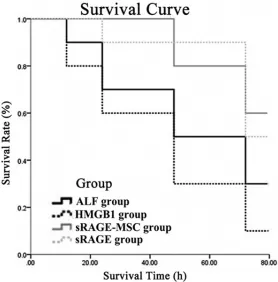
In light of the important role of HMGB1 in inflammatory in ALF and the exploration of the rela-Figure 1. Survival of each group was recorded every 12 h, For normal group,
ALF model group, rHMGB1 group, sRAGE group and GFP/sRAGE-transfect -ed MSCs group, overall survival are 100%, 30%, 10%, 60% and 50%, re-spectively (72 h).
primary hepatocytes gain access to environ-ment similar to that of normal human body, which help stabilize their structure and exert their biological function [7, 8]. This may provide insights into the current dilemma that bio-artifi-cial liver cells dedifferentiate rapidly and are of limited liver support function. Therefore, BAL would serve as a bridge to liver transplantation or regeneration, which is needed to reduce the morbidity and mortality caused by ALF [9]. Until now, BAL support system is still not routinely used in clinical treatment. One of the essential issues is that the BALs focus on liver function supporting, while ignore the immuno-inflamma-tory response caused by the inflammaimmuno-inflamma-tory cyto-kines “second attack” [10]. Furthermore, sev-eral differentially expressed functional proteins within co-cultured hepatocytes have been revealed by comparative proteomics such as high mobility group box chromosomal protein 1 (HMGB1).
HMGB1 is a ubiquitously expressed DNA-binding protein that stabilizes nucleosome
for-tionship between HMGB1 and RAGE, our pri-mary goals were (a) to determine the plasma and hepatic tissue levels of HMGB1 in a drug-induced model of ALF in rats and (b) to confirm a protective effect of specific HMGB1 anti-bodies and the HMGB1 antagonist nicotine in the ALF model.
Materials and methods
Animals
[image:2.612.92.370.72.354.2]Cell culture
MSCs were purchased from Invitrogen and cultured in Dulbecco’s modified Eagle’s medium (DMEM) containing 10% fetal bovine serum (FBS), 100 U/mL penicillin and 100 mg/mL streptomycin at 37°C with 5% CO2.
Establishment of GFP/sRAGE-MSC
MSC line stably expressing rat GFP and sRAGE was construct-ed by using the lentivirus sys-tem according to the manufac-turer’s instructions, and cul-tured cells mentioned above. GFP/sRAGE-MSCs were cul-Figure 2. Plasma NH3, AST, ALT and PT of each groups were measured at 24 h and 72 h after the model was estab
-lished. Compared to normal group, ALT, AST, PT and NH3 of model group significantly increased (12 h, P<0.05), and
aggravated over time (72 h, P<0.05), the same tend can be observed in rHMGB1 group. Giving recombined-sRAGE
and sRAGE-transfected MSCs reverse the aggravating tendency. ELISA was applied to determine the expression of HMGB1 and other cytokines in plasma collected at 24 h and 72 h. HMGB1 as well as TNF-α, IL-1β and IL-6 of each groups were measured and compared in histograms. Expression of HMGB1 as well as TNF-α, IL-1β and IL-6 are low in the plasma of normal group. The expressions were markedly raised after the model was established. For HMGB1 group at 24 h after administration, however, expression of HMGB1 is significantly higher than normal group, and the peak was reached at 72 h, this trend is also shown in TNF-α, IL-1β and IL-6, indicating severe inflammation due to ALF. The high expressions of indexes in HMGB1 group were markedly suppressed when recombinant-sRAGE or sRAGE-transfected MSCs was given. The suppression extend has no obviously difference for sRAGE group than for GFP/sRAGE-transfected MSCs group.
Reagents
L-DMEM medium, fetal bovine serum and tryp-sin were purchased from Gibco (Grand Island, NY). For in vitro experiments, recombinant HMGB1 proteins were obtained from Abcam (Cambridge, MA). Purified sRAGE and Srage ELASA Kit were purchased from ADIPOBIO- SCIENCE. RAGE was purchased from Abcam (Cambridge, MA). D-Gal was purchased from Sigma (St. Louis, MO). Rat HMGB1, IL-1, TNF-α, and IL-6 ELISA Kit were purchased from BD (Pharmingen, La Jolla, CA). TUNEL Kit was pur-chased from Thermo Fisher (Santa Cruz, CA).
Acute liver failure model
D-Gal was dissolved in 50 g/L glucose solution until a final concentration of 10% and pH was adjusted to 6.8. The animals were injected intraperitoneally with D-gal (0.6 g/kg) to induce Acute Liver Failure Model.
tured under routine conditions. Twenty days later, GFP expression was observed in up to 95.5% of the cultured MSCs and high fluores-cence intensity was sustained, suggesting that GFP and sRAGE were successfully transduced into MSCs and could maintain a stable expres-sion over a long time (Figure 7). Lentivirus infected in MSC cell line, the mRNA level of sRAGE genes has increased 6,000 times in len-tivirus infected MSC cell line than in the control cell line (Figure 8).
Experimental groups and treatments
Male Sprague-Dawley rats were divided into five groups randomly. Control group was inject-ed intraperitoneally with saline. ALF group was induced by d-galactosamine (0.6 g/kg) via intraperitoneal injection. HMGB1 group were treated with HMGB1 recombination protein (200 μg/kg) via penile vein right after ALF model induced. sRAGE group was injected Figure 3. Western Blot was used to determine the expression of HMGB1
and RAGE in liver. MGB1 expression of model group in liver is distinctly high -er at 12 h, even high-er at 72 h, and RAGE expression is in accordance with
[image:4.612.90.368.219.364.2]tify plasma levels of HM- GB1, IL-1, TNF-α, and IL-6 according to the manufac-turers’ instructions.
Expression of HMGB1 and RAGE in liver
Liver tissues were collect-ed at 72 h for each group. ALF group were collected at 12 h and 72 h. The lev-els of HMGB1 and RAGE in every 100 mg liver tissue were measured by Western blot. Tissues were homog-enized in lysate buffer con-taining protease and phos-phatase inhibitors. Equal amounts of proteins from each sample were resolved on 4% to 20% SDS-PAGE gels, transferred, and im- munoblotted onto nitrocel-lulose membrane. Antibo- dies for HMGB1, RAGE and β-actin antibodies were used.
Distribution of HMGB1 and RAGE in Liver
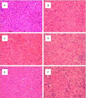
[image:5.612.90.390.71.410.2]Liver tissues were collect-ed at 72 h for each group. ALF group was collected at 12 h and 72 h. The liver tis-sues were used for histo-logical examination, and Figure 4. For HE staining, cytoplasm of liver tissue in control group was acidophil
-ia. Central vein was surrounded by hepatocytes radiated out in all directions (A).
24 h (B) and 72 h (C) after the D-gal was challenged; apoptotic bodies and acido
-philic change of hepatocytes can be observed, as well as infiltration of inflamma
-tion cell and spotty necrosis. In HMGB1 group, 2/3 of hepatocytes were observed necrosis and else phenomenon was in accordance with 72 h after modeling, but even severer (D). Administration of recombinant-Srage (E) or sRAGE-transfected MSCs (F) partly reversed these abnormalities in ALF group and HMGB1 group.
sRAGE recombination protein (400 μg/kg) via penile vein right after ALF model induced. sRAGE-MSC group received MSC transplanta-tion (3×10⁶) which could maintain a stable
expression of sRAGE via penile vein right after ALF model induced. Liver function, level of cyto-kines and liver pathological changes were mea-sured in the following days every 12 h. Overall survival was determined after 72 h for each group after the model was established.
Determination of plasma cytokines and HMGB1 level
Blood samples of each group were collected every 12 h after the model was established, centrifuged at 3000 rpm for 10 min at 4°C, and stored at -80°C. ELISA kits were used to
quan-immunohistochemical staining was applied to determine HMGB1 and RAGE distribution. The samples embedded in paraffin were rehydrated and treated with 3% H2O2. The HMGB1 antibody or RAGE antibody was added and preserved at 4°C overnight. Biotin-labeled secondary anti-body and streptavidin-biotin-peroxisome solu-tion were added, and the samples were stained by DAB, restained by hematoxylin, and sealed with neutral gum. Samples treated with phos-phate buffer served as the negative control.
Determination of liver cell apoptosis
POD were added. Each slice was stained by DAB, and liver cell apoptosis was observed under microscopy. The primary antibody was replaced by phosphate buffer on the posi-tive specimens in negaposi-tive control. Five fields were ran-domly selected in each slice under high-power field (400×). The number of positive liver cells and the total number of liver cells in each field were counted to calculate the per-centage of positive liver cells.
Statistical analysis
All statistical analyses were performed with SPSS v.21.0. Data were reported as mean ± SD. Statistical significance was analyzed by Student t-test;
P<0.05 was considered statis-tically significant.
Results
Survival rates
Survival of each group was recorded every 24 h, and the overall survival rate was deter-mined at 72 h after treatment (Figure 1). For control group, ALF group, HMGB1 group, sR- AGE-MSC group and sRAGE group, the overall survival rates were 100%, 30%, 10%, 60% and 50% respectively. Accor- ding to the survival curve, sur-vival rate of ALF model group significantly dropped over time than the control group, and the HMGB1 group presented a sharper drop than AFL model group. The dropping rate was much lower than sRAGE group, and similar as sRAGE-MSC group.
[image:6.612.93.372.71.526.2]Liver function and cytokines
Figure 5. Immunohistochemical staining was applied to determine HMGB1 and RAGE distribution in liver tissue. Nuclei are lightly stained with brown,
and hardly cytoplasm is stained in the control group (A). The number of
HMGB1-positive cells in the nucleus increased compared to the control
group at 24 h (B) and 72 h (C) after D-gal induction. After administration of
HMGB1 (D), nuclei were distinctly dense stained, and cytoplasm is stained
as well. sRAGE reversed the dense distribution of HMGB1 in nucleus (F).
RAGE is cytomembrane localized. Net-shaped structure already emerged at
24 h after the model was established (B). RAGE expression was greatly en
-hanced by the administration of HMGB1 (D), hepatocytes was surrounded by thicker net-shaped structure even than ALF group at 72 h. The enhanced expression was reversed by giving recombinant-RAGE (F) and sRAGE-trans
-fected MSCs (E).
After blocking endogenous peroxidase activity by methanol, permeability liquid (1 g/L TritonX-100 was dissolved in 0.1% sodium citrate), TUNEL reaction solution and
Converter-level in plasma
NH3 288.5±32.9 g/L, AST 721.8±79.8 u/L, ALT 1039.2± 25.3 u/L, PT 28.4±3.5, 72 h), as well as sRAGE-transfec- ted MSCs (sRAGE-MSC group, NH3 348.6±22.1, AST 195- 4.3±223.9 u/L, ALT 168- 65.3±206.3 u/L, PT 34.2± 0.3, 72 h). Reversing extent of sRAGE-MSC group is simi-lar as sRAGE group, which is also reflected in survival rate. ELISA was applied to deter-mine the expression of HM- GB1 and other cytokines in plasma collected at 24 h and 72 h. HMGB1 as well as TNF-α, IL-1β and IL-6 of each group were measured. As shown in
[image:7.612.89.379.71.397.2]Figure 2, expression of HMGB1, TNF-α, IL-1β and IL-6 are low in the plasma of con-trol group. However, after administrating the HMGB1 at 24 h, expression of HMGB1 was significantly higher than control group, and the peak reached at 72 h, this trend was also shown in TNF-α, IL- 1β and IL-6. The high expres-sions of the above indexes in HMGB1 group were markedly suppressed when recombi-nant-sRAGE or sRAGE-trans-fected MSCs was given. The suppression extension has Figure 6. TUNEL assay was used to determine apoptosis level of hepatocytes
of each group during ALF. There were hardly any apoptosis cells observed in the control group (A). The number of TUNEL stained cells was markedly increased in ALF group (B, C). The serious apoptosis level of HMGB1 group
(D) was reversed to a relatively low state by giving recombinant-sRAGE (E),
and the apoptosis level was closed to the normal state by sRAGE-transfected MSCs administration (F).
the model was established to determine liver function and compared in histograms (Figure 2). As shown in the figure, compared to control group (NH3 87.1±13.7g/L, AST 164.4±35.0 u/L, ALT 49.9±5.1 u/L, PT 13.1±2.6 s), ALT (440.1±32.9 u/L, 24 h), AST (871.7±94.2 u/L, 24 h) and NH3 (101.8±16.6 g/L) as well as PT (20.9±1.8 s, 24 h) of model group significantly increased (P<0.05), and aggravated over time (NH3 405.7±79.8 g/L, AST 4933.3±901.8 u/L, ALT 2466.6±378.5 u/L, PT 40.6±2.2 s, 72 h). The same tend can be observed in HMGB1 group, and each index is significantly higher than normal group (NH3 441.0±58.6 g/L, AST 5213.3±701.8 u/L, ALT 2870.3±344.9 u/L, PT 58.2±3.5, 72 h). The aggravating tendency was distinctly reversed by giving recombined-sRAGE (sRAGE group,
no obvious difference between sRAGE group and sRAGE-MSC group.
Expression of HMGB1 and RAGE in liver
Figure 7. The level of GFP expressed in GFP/sRA
-GEMSC been observed by fluorescence microscope (400×). GFP/sRAGE-MSC was cultured under routine conditions. Twenty days later, GFP expression was observed in up to 95.5% of the cultured MSCs and high fluorescence intensity was sustained, suggest
-ing that GFP and sRAGE were successfully trans -duced into MSCs and could maintain a stable expres-sion over a long time.
and 72 h was significantly higher than control group, and was similar to the level of HMGB1 group. The administration of recombinant-sRAGE as well as recombinant-sRAGE-MSC markedly reduced the RAGE expression compared with the HMGB1 group and the ALF group. However, there is no significant difference between the sRAGE and sRAGE-MSC group.
Effects of sRAGE-MSC on histopathological changes in D-gal challenged rats
According to Figure 4, cytoplasm of liver tissue in control group was acidophilia. Central vein surrounded by hepatocytes radiated out in all directions (Figure 4A). 24 h after the D-gal was challenged, apoptotic bodies and acidophilic change of hepatocytes can be observed, as well as infiltration of inflammation cell and spotty necrosis (Figure 4B). At 72 h, serious and extensive necrosis of hepatocytes occurred. More than 1/3 of hepatocytes were necrosis or bridging necrosis, disordered lob-ules of liver, fragmented hepatic cords, abnor-mally expanded bleeding sinus could be observed under microscopic examination, and a great amount of inflammation cells were infil-trated inside lobules, the other hepatocytes were distinctly steatosis (Figure 4C). In HMGB1
group, 2/3 of hepatocytes were observed necrosis and else phenomenon was in acc- ordance with 72 h after modeling, but ev- en severer (Figure 4D). Administration of recombinant-Srage (Figure 4E) or sRAGE- transfected MSCs (Figure 4F) partly reversed these abnormalities in ALF group and HMGB1 group, and no obvious differences were obser- ved between these two groups.
Distribution of HMGB1 and RAGE in liver
Distribution of HMGB1 and RAGE were ana-lyzed by immunohistochemical staining in the liver slices. HMGB1 was generally nucleus localized, and translocate to cytoplasm in inflammation, stress reaction and some other particular situation. D-gal and HMGB1 injection caused massive expression of HMGB1 in the liver. As shown in the Figure 5, nuclei were light-ly stained with brown, and hardlight-ly cytoplasm was stained in the control group (Figure 5A). The number of HMGB1-positive cells in the nucleus increased compared with the control group at 24 h after D-gal induction (Figure 5B), and at 72 h nearly all nuclei were densely stained, the shape of nucleus was abnormal (Figure 5C). Cytoplasm of cell in model group was hardly stained, which means the translo- cation of HMGB1 is not obvious. However, after administration of HMGB1 (Figure 5D), nuclei were distinctly densely stained, and cytoplasm was stained as well, which means the existence of HMGB1 translocation from nucleus to cyto-plasm. sRAGE reversed the dense distribution of HMGB1 in nucleus (Figure 5F) compared with HMGB1 group, and most nuclei were in their normal shape; Administration of sRAGE-transfected MSCs (Figure 5E) was in accor-dance with the effect of sRAGE group, and almost the same effect according to ob- servation.
RAGE was cytomembrane localized. As a recep-tor of HMGB1, amount of RAGE and HMGB1 was closely related. RAGE was represented by the brown stain on the edge of hepatocytes in
Figure 8. The mRNA level of sRAGE genes expressed in GFP/sRAGE-MSC. Lentivirus infected in MSC cell line, the mRNA level of sRAGE genes has increased 6,000 times in lentivirus infected MSC cell line than
in the control cell line.
at 72 h (Figure 5C), which indicated the increas-ing expression of RAGE over time. RAGE ex- pression was greatly enhanced by the admi- nistration of HMGB1 (Figure 5D), hepatocytes were surrounded by even thicker net-shaped structure than ALF group at 72 h. The enhanced expression was reversed by given recombinant-RAGE (Figure 5F) and sRAGE-transfected MSCs (Figure 5E). Net-shaped structure reduced on both treatments and the sharp edges of cells ree- merged. Overall, the change of RAGE has con-sistent tendency of HMGB1 due to their close relation.
Determination of hepatocytes apoptosis
Apoptosis is a direct result of ALF, and TUNEL assay was used to determine the apoptosis level of hepatocytes of each group during ALF. As shown in the Figure 6, solid red spots repre-sented non-specific erythrocyte stain, and the hollow circles marked by arrows were stained apoptosis nuclei. The result showed that there were hardly any apoptosis cells observed in the control group (Figure 6A). The number of TUNEL stained cells markedly increased in ALF group (Figure 6B), demonstrating that apoptosis greatly occurred in the D-gal challenged liver. The HMGB1 group, however, showed even more TUNEL positive cells than ALF group in the
Figure 6D, suggesting that apoptosis is a re- sult of HMGB1 mediated ALF. The serious apop-tosis level of HMGB1 group was reversed to a relatively low state by giving recombinant-sRAGE (Figure 6E), and the apoptosis level was closed to the normal state by sRAGE-transfect-ed MSCs administration (Figure 6F). The pro-tective effect of sRAGE on inhibiting apoptosis confirmed the effect of HMGB1 in ALF development.
Discussion
ALF has been currently recognized as a global problem to all clinicians. BAL is a safe and effective therapeutic modality for patients with end-stage liver diseases. It will serve as a bridge to liver transplantation or regeneration, which is needed to reduce the morbidity and mortality caused by ALF. Furthermore, several differentially expressed functional proteins within co-cultured hepatocytes in BAL have been revealed by comparative proteomics such as HMGB1. So the inevitable trend for BAL now-adays is to suppress systemic immuno-inflam-matory reaction provoking the conversion of BAL from passive support to active inter- vention.
Since the first report of HMGB1 as “death medi-ator” in 1999 [11] a growing number of articles have reported that HMGB1 plays a fundamen-tal role in various disease conditions, including acute pancreatitis [14], ARDS [15], sepsis [16] and other diseases [17-21]. In this study, we determined not only the plasma levels, but also the hepatic tissue levels of HMGB1 in a rat model of ALF induced by D-galactosamin and further investigated the protective effect of sRAGE.
demonstrated that specifically inhibited of HMGB1-mediated inflammatory response can significantly improve the severe acute pancre-atitis, sepsis, septic shock and ARDS. It can be considered as an important indicator for inflam-matory diseases [24]. Different from some classic type therapy targets for immuno-inflam-matory diseases, HMGB1 has been defined as an advanced stage mediator of inflammation with a wide therapeutic interval. Even in the advanced phase of clinical pathogenesis of inflammation, patients can still benefit from the anti-HMGB1 therapy [13]. Therefore, HMGB1 is considered to be a promising intervention tar-get in inflammatory diseases. It is undoubtedly an important effective molecule and plays an important role in many pathological, as well as in chronic inflammatory diseases.
HMGB1, the extracellular mediator of inflam-mation, only combined with the membrane receptors outside the immune cells, can make the biological effects operate smoothly. Rather than binding to a single specific ligand or even a group of closely related ligands, RAGE [25, 26] has various binding partners. These ligands include HMGB1, and it has natural high affinity with HMGB [27]. It is widespread but relatively low expression of RAGE on vascular endothelial cells, neutrophils, monocytes/macrophages, lymphocytes, DCs, cardiomyocytes, and neu-rons [28]. Our finding suggests that RAGE expression increases in hepatic tissue levels when inflammatory mediators such as HMGB1 accumulate in the D-galactosamine-induced ALF model. But, it maintains at a relatively low level in Srage group and sRAGE-MSC group (Figure 3). Reportedly, RAGE promoter contains multiple functional NF-κB and SP-1 transcrip-tion factor-binding sites [29, 30], ligands and proinflammatory cytokines can promote the expression of RAGE [31], potentially triggering a receptor-dependent autoinflammatory loop [32].
Recently, soluble RAGE (sRAGE), one of RAGE variants has been indentified in human and mouse lung. It is the extracellular (soluble) domain of RAGE at the V-, C1- and/or C2-immunoglobulin like domains [33]. Due to lack of the intracellular region, sRAGE can not transmit biology signal into intracellular. Therefore, it can competitively bind to HMGB-1 with RAGE on cell surface. Experiments using
sRAGE as a ligand decoy have illustrated largely beneficial effects in reducing inflammatory stress, thereby, preventing long-term tissue damage in models of diabetes and immune/ inflammatory disorders [34-37]. The present project aims to take bone marrow mesenchy-mal stem cells (MSC) as a vehicle for sRAGE gene so as to consistently express proteins which could competitively bind to HMGB-1 with RAGE within liver, thus would suppress subse-quent immuno-inflammatory reaction. Our pr- esent study demonstrated that administrating sRAGE recombinant protein to Srage group or transfecting sRAGE gene to sRAGE-MSC group after the ALF model was established may sig-nificantly prolong ALF rat survival time (Figure 1), as well as improve its liver functions (Figure 2), suppressed the inflammatory cytokines secretion (Figure 2), alleviated hepatocytes swelling and necrosis (Figure 6), which indicate measurement of sRAGE levels in vivo holds promise for the identification of potential thera-peutic targets and/or biomarkers of RAGE activity in disease. But sRAGE is a kind of small molecule protein with some disadvantages, such as a short circulating half-life, needs to be repeatedly administrated, and cost a lot [35]. The sRAGE-MSC group is capability to over-come the disadvantages of the sRAGE group mentioned above. MSCs have been considered as a mature gene expression platform while hepatocytes in BAL can support the liver func-tion. We have successfully established the MSC co-cultured with hepatocytes as a vehicle for sRAGE gene so as to consistently express proteins which could competitively bind to HMGB-1 with RAGE on cell surface within liver, thus would suppress subsequent immuno-inflammatory reaction.
field was how RAGE transduces signals from the cell surface to the nucleus. Several papers have tried to address this question by search-ing for direct bindsearch-ing partners, ussearch-ing the cyto-plasmic tail of RAGE as bait. Ishihara et al. [38] identified both extracellular signal-related kinase-1 and -2 (ERK1/2) as direct RAGE-binding partners. Further truncation of the cyto-plasmic domain unveiled a putative ERK dock-ing site at the membrane-proximal region. HMGB1 stimulation of RAGE-transfected HT- 1080 cells induced the interaction of RAGE and ERK1/2. Hudson et al. [31] employed a yeast two-hybrid system to explore possible cytoplas-mic-binding partners of RAGE. Results from this study indicate that the FH1 domain of mammalian Diaphanous-1 (mDia-1) interacts with RAGE. If HMGB1 is associated with the nucleosome, signaling is strictly TLR2 depen-dent. HMGB1 also has the potential to bind to LPS [39]. HMGB1, like LPS-binding protein, can actively destabilize LPS aggregates and pres-ent LPS monomers to CD14, thus increasing the overall sensitivity of the TLR4/MD-2 recep-tor complex.
Although the focus of this study is the role of HMGB1 and the receptor RAGE in ALF, HMGB1-TLR4 may also be important and needs to be investigated in the future. We will explore in depth the enrichment and development in mod-ern idea of ALF combined treatment. If suc-cessful, the present project will provide a molecular target for configuration of a brand-new BAL system with dual functions of support-ing liver functions and intervensupport-ing immuno-inflammatory reaction. The expected findings from this study will provide theoretical founda-tion and beneficial explorafounda-tion for the applica-tion of the concept of suppressing immuno-inflammatory reaction to the comprehensive treatment of ALF.
Acknowledgements
This work was supported by National Natural Science Foundation of China (81170417).
Disclosure of conflict of interest
None.
Address correspondence to: Dr. Yitao Ding, De-
partment of Hepatobiliary Surgery, Drum Tower Clinical Medical College of Nanjing Medical
Uni-versity, 321 Zhongshan Road, Nanjing, China. E-mail: dingyitao@yeah.net
References
[1] Wlodzimirow KA, Eslami S, Abu-Hanna A,
Nieuwoudt M and Chamuleau RA. Systematic
review: acute liver failure - one disease, more than 40 definitions. Aliment Pharmacol Ther
2012; 35: 1245-1256.
[2] Lopez-Delgado JC, Mendiluce RM, Pinol TS, Fernandez XP, Sanchez L and Vicente RG. Urgent liver transplantation for nevirapine-in
-duced acute liver failure: report of a case and review of the literature. Ann Transplant 2012;
17: 122-127.
[3] Leckie P, Davenport A and Jalan R. Extra-corporeal liver support. Blood Purif 2012; 34:
158-163.
[4] Nevens F and Laleman W. Artificial liver sup
-port devices as treatment option for liver fail -ure. Best Pract Res Clin Gastroenterol 2012; 26: 17-26.
[5] Gu J, Shi X, Zhang Y, Chu X, Hang H and Ding Y.
Establishment of a three-dimensional co-cul -ture system by porcine hepatocytes and bone marrow mesenchymal stem cells in vitro. Hepatol Res 2009; 39: 398-407.
[6] Gu J, Shi X, Zhang Y and Ding Y. Heterotypic
interactions in the preservation of morphology and functionality of porcine hepatocytes by
bone marrow mesenchymal stem cells in vitro. J Cell Physiol 2009; 219: 100-108.
[7] Gu JY, Shi XL, Zhang Y and Ding YT. [Study on the effects and mechanisms of bone marrow
mesenchymal stem cells on porcine primary hepatocyte culture in vitro]. Zhonghua Gan Zang Bing Za Zhi 2009; 17: 867-871.
[8] Gu J, Shi X, Chu X, Zhang Y and Ding Y.
Contribution of bone marrow mesenchymal
stem cells to porcine hepatocyte culture in vi-tro. Biochem Cell Biol 2009; 87: 595-604. [9] Palakkan AA, Hay DC, Anil Kumar PR, Kumary
TV and Ross JA. Liver tissue engineering and cell sources: issues and challenges. Liver Int
2013; 33: 666-676.
[10] Sugawara K, Nakayama N and Mochida S. Acute liver failure in Japan: definition, classifi
-cation, and prediction of the outcome. J
Gastroenterol 2012; 47: 849-861.
[11] Wang H, Bloom O, Zhang M, Vishnubhakat JM, Ombrellino M, Che J, Frazier A, Yang H, Ivanova S, Borovikova L, Manogue KR, Faist E, Abraham E, Andersson J, Andersson U, Molina PE, Abumrad NN, Sama A and Tracey KJ. HMG-1 as a late mediator of endotoxin lethality in mice.
Science 1999; 285: 248-251.
[12] Sha Y, Zmijewski J, Xu Z and Abraham E.
activity by binding to cytokines. J Immunol 2008; 180: 2531-2537.
[13] Sims GP, Rowe DC, Rietdijk ST, Herbst R and
Coyle AJ. HMGB1 and RAGE in inflammation
and cancer. Annu Rev Immunol 2010; 28: 367-388.
[14] Yasuda T, Ueda T, Shinzeki M, Sawa H, Nakajima T, Takeyama Y and Kuroda Y. Increase of high-mobility group box chromo -somal protein 1 in blood and injured organs in experimental severe acute pancreatitis. Pancreas 2007; 34: 487-488.
[15] Abraham E, Arcaroli J, Carmody A, Wang H and
Tracey KJ. HMG-1 as a mediator of acute lung inflammation. J Immunol 2000; 165:
2950-2954.
[16] Qin S, Wang H, Yuan R, Li H, Ochani M, Ochani K, Rosas-Ballina M, Czura CJ, Huston JM, Miller E, Lin X, Sherry B, Kumar A, Larosa G, Newman W, Tracey KJ and Yang H. Role of HMGB1 in
apoptosis-mediated sepsis lethality. J Exp Med 2006; 203: 1637-1642.
[17] Wang LW, Chen H and Gong ZJ. High mobility
group box-1 protein inhibits regulatory T cell
immune activity in liver failure in patients with
chronic hepatitis B. Hepatobiliary Pancreat Dis Int 2010; 9: 499-507.
[18] Gong Q, Zhang H, Li JH, Duan LH, Zhong S, Kong XL, Zheng F, Tan Z, Xiong P, Chen G, Fang M and Gong FL. High-mobility group box 1 ex -acerbates concanavalin A-induced hepatic in-jury in mice. J Mol Med (Berl) 2010; 88: 1289-1298.
[19] Suda K, Kitagawa Y, Ozawa S, Saikawa Y, Ueda M, Ebina M, Yamada S, Hashimoto S, Fukata S, Abraham E, Maruyama I, Kitajima M and
Ishizaka A. Anti-high-mobility group box chro-mosomal protein 1 antibodies improve survival
of rats with sepsis. World J Surg 2006; 30:
1755-1762.
[20] Andrassy M, Volz HC, Igwe JC, Funke B, Eichberger SN, Kaya Z, Buss S, Autschbach F, Pleger ST, Lukic IK, Bea F, Hardt SE, Humpert
PM, Bianchi ME, Mairbaurl H, Nawroth PP,
Remppis A, Katus HA and Bierhaus A. High-mobility group box-1 in ischemia-reperfusion injury of the heart. Circulation 2008; 117:
3216-3226.
[21] Pisetsky DS, Erlandsson-Harris H and
Andersson U. High-mobility group box protein 1
(HMGB1): an alarmin mediating the
pathogen-esis of rheumatic disease. Arthritis Res Ther
2008; 10: 209.
[22] Craig DG, Lee P, Pryde EA, Masterton GS, Hayes PC and Simpson KJ. Circulating apop -totic and necrotic cell death markers in
pa-tients with acute liver injury. Liver Int 2011; 31:
1127-1136.
[23] Albayrak A, Uyanik MH, Cerrah S, Altas S, Dursun H, Demir M and Uslu H. Is HMGB1 a
new indirect marker for revealing fibrosis in
chronic hepatitis and a new therapeutic target
in treatment? Viral Immunol 2010; 23:
633-638.
[24] Andersson U and Tracey KJ. HMGB1 is a thera
-peutic target for sterile inflammation and infec -tion. Annu Rev Immunol 2011; 29: 139-162. [25] Dattilo BM, Fritz G, Leclerc E, Kooi CW,
Heizmann CW and Chazin WJ. The
extracellu-lar region of the receptor for advanced glyca
-tion end products is composed of two indepen -dent structural units. Biochemistry 2007; 46: 6957-6970.
[26] Ostendorp T, Leclerc E, Galichet A, Koch M, Demling N, Weigle B, Heizmann CW, Kroneck PM and Fritz G. Structural and functional in -sights into RAGE activation by multimeric S100B. EMBO J 2007; 26: 3868-3878. [27] Han SH, Kim YH and Mook-Jung I. RAGE: the
beneficial and deleterious effects by diverse mechanisms of actions. Mol Cells 2011; 31:
91-97.
[28] Schmidt AM, Yan SD, Brett J, Mora R, Nowygrod
R and Stern D. Regulation of human mononu
-clear phagocyte migration by cell surface-bind
-ing proteins for advanced glycation end prod -ucts. J Clin Invest 1993; 91: 2155-2168. [29] Kim JY, Park HK, Yoon JS, Kim SJ, Kim ES, Ahn
KS, Kim DS, Yoon SS, Kim BK and Lee YY.
Advanced glycation end product (AGE)-induced
proliferation of HEL cells via receptor for
AGE-related signal pathways. Int J Oncol 2008; 33: 493-501.
[30] Bassi R, Giussani P, Anelli V, Colleoni T, Pedrazzi M, Patrone M, Viani P, Sparatore B, Melloni E and Riboni L. HMGB1 as an auto -crine stimulus in human T98G glioblastoma cells: role in cell growth and migration. J Neurooncol 2008; 87: 23-33.
[31] Hudson BI, Kalea AZ, Del Mar Arriero M, Harja E, Boulanger E, D’Agati V and Schmidt AM. Interaction of the RAGE cytoplasmic domain with diaphanous-1 is required for ligand-stimu
-lated cellular migration through activation of
Rac1 and Cdc42. J Biol Chem 2008; 283: 34457-34468.
[32] Li J and Schmidt AM. Characterization and functional analysis of the promoter of RAGE, the receptor for advanced glycation end prod -ucts. J Biol Chem 1997; 272: 16498-16506. [33] Yan SF, Ramasamy R and Schmidt AM. Soluble
RAGE: therapy and biomarker in unraveling the RAGE axis in chronic disease and aging. Biochem Pharmacol 2010; 79: 1379-1386. [34] Dettoraki A, Gil AP and Spiliotis BE. Association
between serum levels of the soluble receptor (sRAGE) for advanced glycation endproducts
(AGEs) and their receptor (RAGE) in peripheral
1 diabetes mellitus. J Pediatr Endocrinol Metab 2009; 22: 895-904.
[35] McNair ED, Wells CR, Mabood Qureshi A, Basran R, Pearce C, Orvold J, Devilliers J and
Prasad K. Soluble receptors for advanced gly
-cation end products (sRAGE) as a predictor of restenosis following percutaneous coronary
intervention. Clin Cardiol 2010; 33: 678-685. [36] Chen Y, Akirav EM, Chen W, Henegariu O,
Moser B, Desai D, Shen JM, Webster JC, Andrews RC, Mjalli AM, Rothlein R, Schmidt
AM, Clynes R and Herold KC. RAGE ligation af
-fects T cell activation and controls T cell differ -entiation. J Immunol 2008; 181: 4272-4278. [37] Lutterloh EC, Opal SM, Pittman DD, Keith JC Jr,
Tan XY, Clancy BM, Palmer H, Milarski K, Sun Y, Palardy JE, Parejo NA and Kessimian N. Inhibition of the RAGE products increases sur
-vival in experimental models of severe sepsis and systemic infection. Crit Care 2007; 11:
R122.
[38] Ishihara K, Tsutsumi K, Kawane S, Nakajima M and Kasaoka T. The receptor for advanced gly -cation end-products (RAGE) directly binds to
ERK by a D-domain-like docking site. FEBS Lett
2003; 550: 107-113.
[39] Youn JH, Oh YJ, Kim ES, Choi JE and Shin JS.
High mobility group box 1 protein binding to
li-popolysaccharide facilitates transfer of lipo -polysaccharide to CD14 and enhances