Original Article
Regulator role of HPV E7 protein on miR-21 expression
in cervical carcinoma cells and its functional
implication
Qingqin Kong, Wenfeng Wang, Ping Li
Department of Obstetrics and Gynecology, Linyi People’s Hospital, Linyi City, Shandong Province, China
Received September 22, 2015; Accepted October 25, 2015; Epub December 1, 2015; Published December 15, 2015
Abstract: Cervical cancer is the second leading malignant tumor in women. Human papillomavirus 16 (HPV16) is one risk factor for cervical cancer, with its expressed E7 protein can facilitate the transformation of cervical epithe-lial cells. MicroRNA-21 (miR-21) is one important tumor growth regulatory factor involving in angiogenesis, tumor invasion and metastasis. This study thus aimed to investigate the role of high-risk HPV16 E7 protein in regulating miR-21 expression in cervical carcinoma and its related functions. Hela cells were transfected with pcDNA-HPV16 E7 expressing vectors. The expression level of E7 was determined by Western blotting, while miR-21 level was quan-tified by real-time PCR. The alternation of tumor cell proliferation is determined by transfecting miR-21 inhibitor into E7-overexpressing Hela cells. Cell apoptosis was studied by caspase-3 assay, while cell invasion was illustrated in Transwell chamber. The overexpression of HPV E7 protein facilitated the expression of miR-21, which potentiated Hela cell proliferation and invasion. The inhibition of miR-21 in E7-overexpressin Hela cells can inhibit both prolif-eration and invasion, but without significant effects on caspase-3 activity. HPV16 E7 protein can up-regulate host miR-21 expression, thus elevating cervical carcinoma cell growth, proliferation and invasion. Therefore, E7 protein is one critical factor in occurrence and progression of cervical carcinoma.
Keywords: Cervical carcinoma, Herpes virus 16, E7 protein, MicroRNA, oncogenes
Introduction
As one of most common malignant tumors in women, cervical cancer maintains a high level of incidence and mortality rate in under-devel-oped countries. The incidence of it locates as the second place of all gynecologic cancers [1, 2]. In Chinese women, the incidence of cervical cancer is more than two times higher than Western countries, with significantly elevated mortality rate [3, 4]. The lack of systematic and effective large-scale screening, as well as the weakness in early intervention, severely com-promises the life quality and survival of patients, making cervical cancer as a world-wide healthy issue [5, 6].
High risk subtypes of human papillomavirus (HPV) can infect humans via skin or mucosal contact, and owns a high transmission rate by sexual behavior [7]. HPV16 is one common oncogenic subtype of HPV, as more than 70% infections eventually develop into cervical le-
of HPA is E7 protein with 10 kDa size, contain-ing ~100 amino acids. E7 protein has been shown to facilitate the malignant transforma-tion of cervical epithelial cells and maintain the malignant phenotype [9, 10].
role of HPV16 E7 protein on miR-21 in cervical carcinoma cells.
Materials and methods
Cell culture
Hela cells (ATCC cell bank, US) were resuscitat-ed in 37°C water-bath until completely thawing. Following 1 000 g centrifugation (3 min), cells were re-suspended in 1 mL DMEM medium (Hyclone, US) and were cultured in a 37°C humidified chamber with 5% CO2 perfusion. Af- ter 24~48 hours, cell were seeded into culture dish at 1 × 10⁷/mL density, using high-glucose DMEM (containing 100 U/mL penicillin, 100 μg/mL streptomycin and 10% fetal bovine serum). Cells were passed every 2~3 days. Log-phased cells were randomly divided into three groups: control group; blank transfection group; and HPV16 E7 transfection group.
HPV16 E7 vector constructs and transfection
Total RNA was extracted from Hela cells by Trizol reagents (Invitrogen, US). Using total RNA as the template, cDNA was synthesized by re- verse transcription kit (Invitrogen, US). HPV16 E7 specific primers were designed by Primer- Premier 6.0 software and synthesized (Sangon, China) to amplify the target sequence using cDNA as the template (Forward primer: 5’- TGCCG TTTCG CATGT GCCAT-3’; Reverse prim- er: 5’-TATAT GCTCT GCCCT TTGTC-3’). PCR con-ditions were: 95°C 2 min, followed by 35 cycles each containing 94°C denature for 30 sec,
60°C annealing for 50 sec and 72°C elongation for 35 sec. PCR products were purified from agarose gel following electrophoresis and ligat-ed to pcDNA3.1 expressing vector (3:1) using 16-hour incubation at 4°C. JM109 competent cells were used to amplify target DNA frag-ments for further enzyme digestion.
Log-phased Hela cells were seeded into 6-well plate at 3 × 10⁶/mL under 12-hour incubation until reaching 70~80% confluence. Serum-free medium with lipo2000 reagents (Invitrogen, US) were used to incubate cells for 15 min. Cells were then mixed with serum-free medium containing pcDNA3.1-HPV16 E7 vectors (1 μg/ mL) and lipo2000. After 30 min-incubation at room temperature, serum was removed in the plate, followed by PBS washing. Serum-free me- dium was continuously used for 6-hour incuba-tion, followed by changing normal medium con-taining serum.
Western blotting
Proteins were extracted from Hela cells by lysis buffer (30 min on ice) and ultrasonic rupture (5 sec, 4 times). After centrifugation at 10 000 g for 15 min, supernatants were transferred to new tubes for quantification. Proteins were sep-arated by 10% SDS-PAGE and were transferred to PVDF membrane (Pall Life, US). Using defat-ted milk powder to remove non-specific binding sites, rabbit anti-human HPV16 E7 antibody (1:1 000, Cell signaling, US) was added for 4°C overnight incubation. On the next day, goat anti-rabbit secondary IgG conjugated with horserad-ish peroxidase (HRP) (1:2 000, Cell signaling, US) was added for 30 min-incubation. ECL chro-mogenic substrates (Amersham Biosciences, US) were added to develop the membra-ne. After exposure under X-ray, Quantity One soft-ware was used to analyze optical density (OD) values of protein bands. All experiments were performed in four times (N=4).
MiR-21 inhibitor transfection
MiR-21 inhibitor (5’-AUCGG GAGGU GCAUU CUA-3’) was transfected to HPV16 E7 over-expressing Hela cells using the same lipo2000 using the same procedure as those for vector transfection.
Real-time PCR
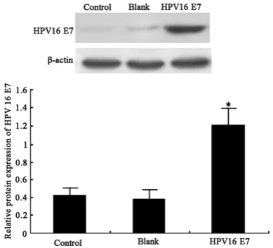
[image:2.629.100.295.82.259.2]Total RNA were extracted from Hela cells, fol-lowed by cDNA synthesis. Real-time PCR was performed using specific primers of miR-21 Figure 1. HPV6 E7 protein expression. A. Western
bation, Transwell chambers were removed and rinsed by PBS. Cells on the upper surface were removed. The whole chamber was fixed in cold ethanol and stained by crystal violet for 30 min. The number of migrated cells on the bottom surface was counted under an inverted micro-scope. 10 fields were randomly selected from each sample, with triplicates.
Caspase-3 activity assay
The activity of caspase-3 in all cells was quanti-fied using test kit (R&D, US) following manual instruction. In brief, cell were firstly digested by trypsin (Sigma, US) and centrifuged at 600 g for 5 min. After discarding the supernatant, cell lysis buffer was added for rupture the cell on ice. After centrifugation at 20 000 for 5 min, 2 24-hour time interval. Super- natants were removed after 4 hours, with the addition of 150 μL DMSO to each well. The plate was vibrated for 10 min until complete resolving of crystal violet. Absorbance value at 570 nm was mea-sured by a microplate reader (BD, US) for calculating prolif-erative rate of all cells.
Transwell chamber assay
48 hours after transfection, all cells were cultured in serum-free medium for 24 hours. Transwell chamber (Hyclone, US) was pre-coated with 50 mg/L Matrigel solu-tion on the chamber bottom and upper membrane sur-face. Supernatants in the plate were removed, followed by the addition of serum-free medium containing 1% bovine serum albumin. Transwell chamber was put into 24-well plate, in which DMEM medi-um containing 10% FBS was added outside the chamber. 0.1 mL Hela cell suspension was added into each cham-ber with serum-free medium. All experiments were per-formed in triplicates. A paral-lel group was performed using uncoated Transwell chamber. After 48hour incu-(Forward, 5’-GACTT ACATG TGACC TGCCT G-3’;
Reverse, 5’-TTCCG GTTCA ACTCT CCTTA-3’) and GAPDH as the internal reference (Forward, 5’-ATCTG GAGTT TACCG CTGG-3’; Reverse, 5’-TACCG ATGTCT GGTAG ACGAT-3’). PCR condi-tions were: 55°C for 1 min, followed by 35 cycles each containing 92°C denature for 30 sec, 58°C annealing 45 sec and 72°C 35 sec. Standard curve was plotted based on CT values of standards. Semi-quantitative analysis was performed using 2-△Ct method.
Cell proliferation assay
[image:3.629.101.383.83.254.2]Hela cells (5 × 10³/mL) at log-phase were seed-ed into 96-well plate containing DMEM plus 10% FBS. After 24-hour incubation, 20 μL MTT reagents (Gibco, US) were added to each well at
Figure 2. MiR-21 expression level. *, P<0.05 compared to control group; #, P<0.05 compared to HPV16 E7 group.
[image:3.629.101.380.308.479.2]compared to controlled Hela cells (78 ± 9, P<0.05, Figure 4). The application of miR-21 inhibitor, however, decreased number of inva-sive cells (69 ± 12). These results indicated the participation of HPV16 E7 protein in the inva-sion of Hela cells, by up-regulating miR-21 expression.
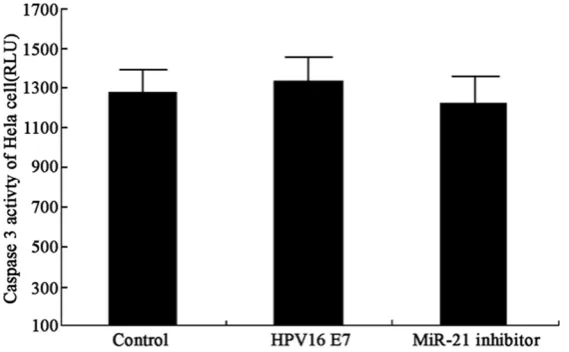
Intracellular activity of caspase-3
After overexpression of HP16 E7 protein, the up-regulation of miR-21 had no significant effects on caspase-3 activity (P>0.05, Figure 5). The transfection of miR-21 inhibitor also did not obtain statistical significance. These results collectively rejected the participation of HPV16 E7 in Hela cell apoptosis.
Discussion
The incidence of cervical cancer is increasing in recent years due to multiple factors such as viral infection, unhealthy sexual behaviors and environmental stress. In a demographic view, cervical carcinoma is most commonly occurred in young women between 30~55 years old, with primary cancer at 30~35 years and infiltra-mM Ac-DEVD-pNA was added to quantify the
OD value at 405 nm. Relative activity of cas-pase-3 was then determined.
Statistical analysis
SPSS 16.0 software was used to process all collected data, of which measurement data were presented as mean ± standard deviation (SD). Between-group-comparison was perfor- med by LSD test. A statistical significance was defined when P<0.05.
Results
HPV16 E7 protein expression in Hela cells
After transfecting HPV16 E7 expressing vector, E7 protein level was significantly facilitated in Hela cells (P<0.05, Figure 1).
MiR-21 expression in Hela cells
[image:4.629.104.378.81.323.2]Using real-time PCR to reveal the effect of HPV16 E7 protein on the expression of miR-21 in Hela cells, our results showed the over-expression of E7 significantly facilitated the expression of miR-21 in Hela cells (P<0.05,
Figure 2). The transfection of miR-21 inhibitor can remark-ably inhibited miR-21 expres- sion.
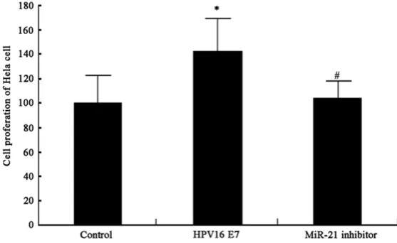
Hela cell proliferation
The overexpression of HPV16 E7 protein significantly facili-tated Hela cell proliferation (P<0.05, Figure 3). The down-regulation of miR-21 by inhibi-tor transfection depressed Hala cell proliferation. These results showed the role of HPV E7 protein in potentiating Hela cell proliferation via up-regulating miR-21 expression in tumor cells.
Cell invasion assay
We further used Transwell chamber to examine the effect on Hela cell invasion. Result showed elevated num-ber of perforated cells (128 ± 15) in E7-overexpressing cells Figure 4. Hela cell invasion after E7 over-expression. A-C showed
representa-tive staining images showing number of perforated cells in control, HPV16 E7 and miR-21 inhibitor groups, respectively. D. Quantitative data of perfo-rated cell number in each group. *, P<0.05 compared to control group; #,
caspase-3 activity. In summary, HPV16 E7 pro-tein can stimulate the expression of miR-21 in host cells, thus facilitating cervical cancer cell growth, proliferation and invasion. This study illustrated the crucial role of E7 in pathogene-sis and progression of cervical carcinoma. The exact mechanism of E7 protein in regulating miR-21, however, remained incomplete. Our results should benefit the clinical study of novel drug targets for cervical cancer.
Disclosure of conflict of interest
None.
Address correspondence to: Dr. Ping Li, Department of Obstetrics and Gynecology, Linyi People’s Hospital, Linyi City, 27 Eastern Section of Jiefang Road, Linyi 276003, Shandong, China. Tel: +86-13792967229; E-mail: [email protected]
References
[1] Munoz N and Bravo LE. Epidemiology of cervi-cal cancer in Colombia. Salud Publica Mex 2014; 56: 431-9.
[2] Jing L, Zhong X, Zhong Z, Huang W, Liu Y, Yang G, Zhang X, Zou J, Jing C, Wei X. Prevalence of human papillomavirus infection in Guangdong Province, China: a population-based survey of 78,355 women. Sex Transm Dis 2014; 41: 732-8.
[3] Shen XR, Feng R, Chai J, Cheng J, Wang DB. Modeling age-specific cancer incidences using logistic growth equations: implications for data collection. Asian Pac J Cancer Prev 2014; 15: 9731-7.
[4] Chen W, Jeronimo J, Zhao FH, Qiao YL, Valdez M, Zhang X, Kang LN, Bansil P, Paul P, Bai P, tive cancer at 45~55 years. The average age of
cervical cancer patients was becoming younger in recent years [17]. Almost 25 of all cervical cancer patients worldwide occurred in China. As most patients were already at late or termi-nal stage, the treatment strategy is somehow limited [18].
[image:5.629.101.386.83.259.2]As one important and necessary risk factor for cervical cancer, HPV infection can be divided into high-risk and low-risk groups based on the tumor site and possibility of cancer. More than 99% of cervical carcinoma, including squa-mous cell injury and gland swelling, caused serum HPV elevation [19]. HPV can enter the body via tiny injury site to infect epithelial basal cells, in which viral DNA may incorporate into host genome, causing further malignant prolif-eration of epithelial cells, and finally leading to precancerous lesion and infiltrative carcinoma. The most powerful subtypes regarding carcino-genicity included HPV16, HPV18, and HPV16 E7 protein, all of which binds onto pRB protein, leading to the release of transcriptional factor E2F family. Such E2F family number can form complexes with cyclin A, cyclin E, p21 and p27, thus making the entry into S phase of infected cells to facilitate the amplification of viral DNA [20, 21].
As one important regulatory molecule, miR par-ticipates in almost all aspects of tumor cells, including growth, proliferation, differentiation, apoptosis, angiogenesis, invasion and metas-tasis. MiR-21 can negatively regulate tumor-suppressor gene expression, and induce tumor
Peck R, Li J, Chen F, Stoler MH, Castle PE. The concordance of HPV DNA detection by Hybrid Capture 2 and careHPV on clinician- and self-collected specimens. J Clin Virol 2014; 61: 553-7.
[5] Wang SM and Qiao YL. Implementation of cer-vical cancer screening and prevention in China--challenges and reality. Jpn J Clin Oncol 2015; 45: 7-11.
[6] Pan XF, Zhao ZM, Sun J, Chen F, Wen QL, Liu K, Song GQ, Zhang JJ, Wen Y, Fu CJ, Yang CX. Acceptability and correlates of primary and secondary prevention of cervical cancer among medical students in southwest China: implications for cancer education. PLoS One 2014; 9: e110353.
[7] Arrossi S, Thouyaret L, Herrero R, Campanera A, Magdaleno A, Cuberli M, Barletta P, Laudi R, Orellana L; EMA Study team. Effect of self-col-lection of HPV DNA offered by community health workers at home visits on uptake of screening for cervical cancer (the EMA study): a population-based cluster-randomised trial. Lancet Glob Health 2015; 3: e85-94.
[8] De Vivar AD, Dawlett M, Wang JP, Jack A, Gong Y, Staerkel G, Guo M. Clinical performance of hybrid capture 2 human papillomavirus testing for recurrent high-grade cervical/vaginal in-traepithelial neoplasm in patients with an ASC-US Papanicolaou test result during long-term posttherapy follow-up monitoring. Arch Pathol Lab Med 2015; 139: 219-24.
[9] Khunamornpong S, Settakorn J, Sukpan K, Srisomboon J, Suprasert P, Siriaunkgul S. Per- formance of HPV DNA testing with hybrid cap-ture 2 in triaging women with minor cervical cytologic abnormalities (ASC-US/LSIL) in North ern Thailand. Asian Pac J Cancer Prev 2014; 15: 10961-6.
[10] Nilyanimit P. Comparison of detection sensitiv-ity for human papillomavirus between self-col-lected vaginal swabs and physician-colself-col-lected cervical swabs by electrochemical DNA chip. Asian Pac J Cancer Prev 2014; 15: 10809-12. [11] Orang AV and Barzegari A. MicroRNAs in colo-
rectal cancer: from diagnosis to targeted ther-apy. Asian Pac J Cancer Prev 2014; 15: 6989-99.
[12] Gandhi NS, Tekade RK and Chougule MB. Nanocarrier mediated delivery of siRNA/miR-NA in combination with chemotherapeutic agents for cancer therapy: current progress and advances. J Control Release 2014; 194: 238-56.
[13] Gallach S, Calabuig-Fariñas S, Jantus-Lewintre E, Camps C. MicroRNAs: promising new antian-giogenic targets in cancer. Biomed Res Int 2014; 2014: 878450.
[14] Li E, Ji P, Ouyang N, Zhang Y, Wang XY, Rubin DC, Davidson NO, Bergamaschi R, Shroyer KR, Burke S, Zhu W, Williams JL. Differential ex-pression of miRNAs in colon cancer between African and Caucasian Americans: implica-tions for cancer racial health disparities. Int J Oncol 2014; 45: 587-94.
[15] Echevarria-Vargas IM, Valiyeva F and Vivas-Mejia PE. Upregulation of miR-21 in cisplatin resistant ovarian cancer via JNK-1/c-Jun path-way. PLoS One 2014; 9: e97094.
[16] Jia W, Wu Y, Zhang Q, Gao GE, Zhang C, Xiang Y. Expression profile of circulating microRNAs as a promising fingerprint for cervical cancer diagnosis and monitoring. Mol Clin Oncol 2015; 3: 851-858.
[17] Zhang P, Xi M, Zhao L, Qiu B, Liu H, Hu YH, Liu MZ. Clinical efficacy and failure pattern in pa-tients with cervical esophageal cancer treated with definitive chemoradiotherapy. Radiother Oncol 2015; 116: 257-61.
[18] Liu Y, Zhao LJ, Li MZ, Li MX, Wang JL, Wei LH. The Number of Positive Pelvic Lymph Nodes and Multiple Groups of Pelvic Lymph Node Metastasis Influence Prognosis in Stage IA-IIB Cervical Squamous Cell Carcinoma. Chin Med J (Engl) 2015; 128: 2084-2089.
[19] Miao F, Lv T, Zhang Y, Huang Z, Wang X, Hongwei W. Induction of apoptosis in HPV16 E7 transfected human keratinocyte by ALA-mediated photodynamic therapy. Photodiagno- sis Photodyn Ther 2015; [Epub ahead of print]. [20] Zhu D, Ye M and Zhang W. E6/E7 oncoproteins
of high risk HPV-16 upregulate MT1-MMP, MMP-2 and MMP-9 and promote the migration of cervical cancer cells. Int J Clin Exp Pathol 2015; 8: 4981-9.