Original Article
A novel long noncoding RNA IRAIN regulates cell
proliferation in non small cell lung cancer
Jing Feng
1*, Yue Sun
1*, Er-Bao Zhang
2, Xi-Yi Lu
1, Shi-Dai Jin
1, Ren-Hua Guo
11Department of Oncology, First Affiliated Hospital of Nanjing Medical University, Nanjing, Jiangsu, China; 2Department of Biochemistry and Molecular Biology, Nanjing Medical University, Nanjing, Jiangsu, China. *Equal contributors.
Received August 12, 2015; Accepted September 22, 2015; Epub October 1, 2015; Published October 15, 2015
Abstract: Long non-coding RNAs (lncRNAs) are a novel class of RNA molecules defined as transcripts longer than 200 nucleotides that lack protein coding potential. LncRNA IRAIN has been verified that it is related to acute myeloid leukemia (AML) and breast cancer. However, there was no study to clarify whether it is involved in non-small cell lung cancer (NSCLC). Here, we demonstrated IRAIN as a tumor promoter in NSCLC. Its expression level was remarkably upregulated in NSCLC tissues and connected with tumor size and smoking status. Knockdown of IRAIN suppressed NSCLC cells proliferation in vitro. These data identify IRAIN as a novel promoting gene, which plays a vital role in tumorigenesis of NSCLC.
Keywords: Long non-coding RNA, IRAIN, non-small cell lung cancer, gene therapy, A549
Introduction
NSCLC is the leading cause of cancer-related
deaths worldwide [1]. The incidence of lung
cancer is still increasing remarkably in China,
in both urban and rural areas [2]. No matter
how great efforts and progressions have been
made in the study of lung cancer in recent
decades, the molecular mechanism of lung
cancer remains unclear.
Insulin-like growth factor 1 (IGF-1) signaling
mediated by IGF1R is an important growth reg
-ulatory pathway, enhanced activation of which
is thought to play a crucial role in cancer cell
proliferation, migration, and apoptosis [3-5].
IGF1R expresses in many types of cancer cells,
including NSCLC [6]. Current studies find that
IRAIN plays an important role in the regulation
of IGF-1 signaling. IRAIN is downregulated both
in leukemia cell lines and blood obtained from
high-risk AML patients. There is no large open
reading frames can be identified using soft
-ware prediction programs in IRAIN transcript
which is a 5.4 kb noncoding RNA. IRAIN is
with-in the IGF1R locus, transcribed from an with-intronic
promoter in antisense orientation as compared
to the IGF1R coding mRNA and in a
parent-of-origin specific manner. There is a single nucleo
-tide polymorphism (SNP) rs8034564: the ‘G’
genotype was favorably imprinted over the ‘A’
genotype imbalanced expressing in the two
parental alleles. In addition, it is also
downregu-lated in breast cancer as an imprinted gene [7,
8].
However, the function of IRAIN in other
malig-nancies remains unclear. A large number of
studies have demonstrated that the IGF1R
pathway is frequently dysregulated in lung can
-cer. It is necessary to clarify IRAIN whether it
is also related to lung cancer. In this study,
we characterized the differential expression of
IRAIN in a case-control study of lung cancer
samples and normal tissue specimens. More-
over, we explored if IRAIN takes part in cancer
cell proliferation, apoptosis and migration in
A549 cells.
Materials and methods
Patients
speci-mens performed no local or systemic treatment
before collecting. This research was approved
by the Research Ethics Committee of
Jiang-su province Hospital, and written informed
consent was obtained from all patients. All
tissue samples were excised and immediate-
ly stored in liquid nitrogen until detected.
Histological type and tumor-node-metastasis
(TNM) classifications were determined accord
[image:2.612.91.327.110.466.2]-ing to the National Comprehensive Cancer
Network (NCCN) criteria. These patients’ clini
-copathologic characteristics were shown in
Table 1
.
Cell culture
The human pulmonary adenocarcinoma cell
line A549 was obtained from the Chinese
Academy of Sciences Cell Bank of Type Culture
The primers were shown in
Table 2
. Ct values
were calculated using SDS 2.4 software. IRIAN
expression was normalized to that of GAPDH
with the 2^-∆Ct method.
Cell proliferation assay
To assess the effect of IRAIN expression on
A549 cells proliferation, Cell Counting Kit-8
(CCK-8, Dojindo, Kumamoto, Japan) was used
for assessing cell viability. For this assay, 5 ×
10
3cells/well in 100 UL culture medium were
seeded in 96-well plates and incubated for 24
h after transfection. WST-8 (10 UL) was added
to each well. The optical density at 450 nm was
measured after 1 hour of incubation at 37.0°C
using a Tecan Infinite M200 Multimode micro
-plate reader (Mechelen, Belgium). Each
experi-ment was performed 3 times.
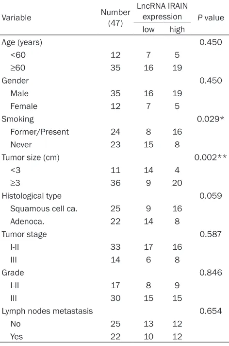
Table 1.
Correlation between IRAIN expression and
clinicopathological parameters of lung
adenocarci-noma patients
Variable Number (47)
LncRNA IRAIN
expression P value low high
Age (years) 0.450
<60 12 7 5
≥60 35 16 19
Gender 0.450
Male 35 16 19
Female 12 7 5
Smoking 0.029*
Former/Present 24 8 16
Never 23 15 8
Tumor size (cm) 0.002**
<3 11 14 4
≥3 36 9 20
Histological type 0.059
Squamous cell ca. 25 9 16
Adenoca. 22 14 8
Tumor stage 0.587
I-II 33 17 16
III 14 6 8
Grade 0.846
I-II 17 8 9
III 30 15 15
Lymph nodes metastasis 0.654
No 25 13 12
Yes 22 10 12
*P<0.05, **P<0.01.
Collection (Shanghai, China). Cells were
incubated at 37.5C with 5% CO
2in
Dulbecco’s modified Eagle’s medium
(HyClone, UT, USA), supplemented with
10% fetal bovine serum (Gibco, Auckland,
New Zealand), 100 U/mL penicillin, and
100 mg/mL streptomycin.
Transfection
IRAIN siRNA and its corresponding
nega-tive control (NC) (Invitrogen) were
trans-fected into cells for 24 h for
loss-of-func-tion experiments using Lipofectamine
2000 (Invitrogen) according to the
manu-facturer’s instructions. The NC scrambled
oligonucleotide does not encode for any
known lncRNA. Transfection efficiency
was verified by SYBR Green real-time
PCR detection of IRAIN expression. IRAIN
siRNA and its corresponding NC sequenc
-es were listed in
Table 2
.
RNA isolation,
r
everse
transcription (RT),
and
q
uantitative PCR (qPCR)
Cell migration assays
For the cell migration assay, 5 × 10
4cells
after transfection were resuspended in 100 μl
serum-free DMEM and seeded in the top
por-tion of a Transwell chamber with 8-mm pores
(Millipore). The transwell chambers were placed
in the 24-wells. The lower portion of the
cham-ber contained 600 ul 10% FBS DMEM as a
chemoattractant. After incubated at 37.0°C in
5% CO
2for 24 h, the cells on the top of the
membrane representing that those cells did
not migrate the barrier were removed with
cotton swabs. After fixed with 95% methanol,
the migrating cells at the bottom of insert
were stained with 0.2% Crystal Violet Staining
Solution (Beyotime), photographed and count
-ed under ×20 magnification. Five random fields
were analyzed for each chamber. Each experi
-ment was repeated 3 times.
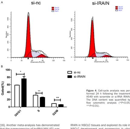
Cell cycle and apoptosis assays
For the cell cycle analysis, A549 cells transient
-ly transfected with IRAIN siRNA or NC for 24 h
were washed with PBS and fixed with ice-cold
70% ethanol at -20°C. Fixed cells were rehy
-drated in PBS and stained with PI using the
Cycle TESTTM PLUS DNA reagent kit (BD
Bio-sciences) according to the manufacturer’s pro
-tocol
.
The percentages of G0/G1, S, and G2/M
cells were counted and compared. Experiments
were performed in triplicate. APC-conjugated
Annexin V (Annexin V-APC) Kit (Cat No: 550474,
BD Pharmingen, USA) and Propidium Iodide
Staining Solution (Cat No: 556463, BD Phar-
mingen, USA) were used to detect the
percent-age of apoptotic cells according to the
manu-facturer instructions. For the apoptosis analy
-clinical pathological features, two-tailed
Chi-squared test was employed. Differences
bet-ween the groups were compared using the
Student’s t test. Results were considered sig
-nificant when
P values were <0.05.
Results
Increased expression of IRAIN in NSCLC
To explore the role of IRAIN in NSCLC, we
ana-lyzed 47 paired clinical NSCLC tissues and their
adjacent normal counterparts for IRAIN
expres-sion using qRT-PCR. IRAIN expresexpres-sion was sig
-nificantly increased in tumor samples as shown
in
Figure 1A
(P<0.01). Then we assessed its
clinical significance by evaluating the correla
-tion between its expression and
clinicopatho-logical parameters (i.e., age, gender, smoking
status, tumor size, histological type, tumor
stage, tumor grade and lymph node metasta
-sis). From the results shown in
Figure 1B
and
1C
, we found that IRAIN expression level in
NSCLC was significantly correlated with smok
-ing status (P<0.05) and tumor size (P<0.05).
However, there was no association between
IRAIN expression and other clinical characters
such as age, gender, histological type, tumor
stage, grade and lymph node metastasis in
NSCLC (
Table 1
).
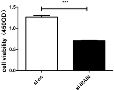
Knockdown of IRAIN suppressed proliferation
of A549 cells
[image:3.612.90.342.95.243.2]To explore the function of IRAIN in the
regula-tion of cell proliferaregula-tion, small interfering RNA
(siRNA) was transfected into A549 cells. IRAIN
expression was decreased by 41.15% in A549
cells with IRAIN siRNA transfection compared
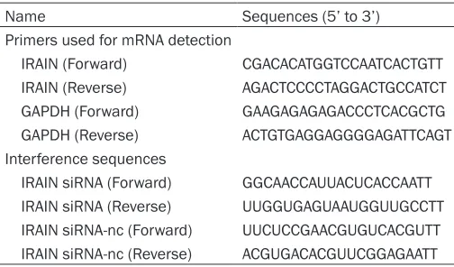
Table 2.
Sequences of primers for qRT-PCR and IRAIN
related sequence
Name Sequences (5’ to 3’)
Primers used for mRNA detection
IRAIN (Forward) CGACACATGGTCCAATCACTGTT IRAIN (Reverse) AGACTCCCCTAGGACTGCCATCT GAPDH (Forward) GAAGAGAGAGACCCTCACGCTG GAPDH (Reverse) ACTGTGAGGAGGGGAGATTCAGT Interference sequences
IRAIN siRNA (Forward) GGCAACCAUUACUCACCAATT IRAIN siRNA (Reverse) UUGGUGAGUAAUGGUUGCCTT IRAIN siRNA-nc (Forward) UUCUCCGAACGUGUCACGUTT IRAIN siRNA-nc (Reverse) ACGUGACACGUUCGGAGAATT
sis, A549 cells transfected with IRAIN
siRNA or NC for 24 h were added with
Annexin V-APC and PI. Viable, dead,
early apoptotic and late apoptotic
cells were identified with a flow
cyto-meter. Data were expressed as mean
± SEM of three independent experi-
ments.
Statistical analysis
to the scrambled siRNA at 24 h post-trans-
fection, determined by qRT-PCR (
Figure 2
,
P<0.01). The effect of IRAIN expression in cell
proliferation was evaluated by CCK-8 assay
and inhibited by 44.24% when compared with
control cells (
Figure 3
, P<0.01). In addition, the
effect of IRAIN on cell cycle and apoptosis was
analyzed by flow cytometry. There was
signi-ficant difference in G0/G1 phase (
P<0.05), S
phase (P<0.01) and G2/M phase (P<0.01)
(
Figure 4
). However, alteration of IRAIN
expres-sion did not affect apoptosis of A549 cells
(
Figure 5
,
P
>0.05). Transwell assay was per
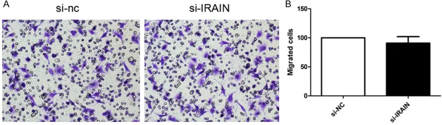
-formed to determine whether IRAIN could
pro-mote cell migration of A549 cells. As shown in
Figure 6
, knockdown of IRAIN did not decrease
cell migration compared with the controls (P
>0.05).
Discussion
The concept of functional lncRNA was first
introduced over 20 years ago, with the descrip
-tion of the X-inactive specific transcript (XIST),
which functions as a X-chromosome
suppres-sor and lacks an open reading frame [9, 10].
In recent years, emerging evidence indicates
that lncRNAs are dysregulated and fulfill impor
-tant functions in the regulation of gene
expres-sion, involved in tumorigenesis and other
bio-logical progression [11]. lncRNAs expression
profiles are altered in many types of cancers.
For example, increased expression of HOTAIR
has been noted in various primary and meta
[image:4.612.93.289.255.402.2]-static tumors, including breast, liver and the
gastrointestinal tract [12-15]. Moreover, Some
lncRNAs may play important roles in recurrent
tumor, such as ever reported H19, CRNDE, HO-
TAIRM1 or first reported AC016745.3, XLOC_
001711, RP11-128A17.1 in glioma recurrence
Figure 1. Expression pattern of IRAIN in NSCLC tumor samples compared with adjacent normal tissues, large tu-mors. (≥3 cm) compared with small tumors (<3 cm), NSCLC patients with smoking history compared with non-smokers (***P<0.0001, *P<0.05).Figure 2. Expression of IRAIN in transfected A549 cells. A549 Cells were transiently transfected with IRAIN siRNA and its corresponding NC, respectively. This effect was examined by SYBR Green real-time PCR and normalized to GAPDH expression. Data rep-resent mean ± SEM from three independent experi-ments (*P<0.05).
[image:4.612.92.287.508.662.2][16]. Another meta-analysis has demonstrated
that the overexpression of lncRNA MALAT1 was
an unfavorable prognostic factor for patients’
overall survival in NSCLC and pancreatic
can-cer [17]. Beyond MALAT1, lncRNA TUG1 [18]
and PANDAR [19] are also involved in the
pro-gression of NSCLC. They are both associated
with TNM stage, tumor size, overall survival, as
well as growth regulation. Therefore, the
identi-fication and investigation of cancer-associated
lncRNAs may become new prognostic biomark
-ers or therapeutic strategies for cancer,
includ-ing NSCLC.
IRAIN is an antisense noncoding RNA and the
full-length transcript is 5.4 kb [7]. It has been
identified that IRAIN is related to AML and
breast cancer [7, 8]. However, whether it is
correlated with NSCLC remains unclear. In this
study, we first described the characters of
IRAIN in NSCLC tissues and explored its role in
NSCLC development and progression in vitro
primarily. We discovered that IRAIN was
increased in NSCLC tissues and identified the
upregulated expression of IRAIN in bigger
tumors and smokers. However, our results are
contrary to findings from earlier studies in AML
and breast cancer. This phenomenon is
proba-bly because lncRNAs exhibit dramatically tis
-sue-specific expression patterns than
protein-coding genes [20, 21]. According these results,
we could infer that IRAIN may
present a
tissue-specific expression pattern and exhibit impor
-tant role in NSCLC development and
progres-sion. Knockdown of IRAIN inhibited cell
prolif-eration in NSCLC cells. The prolifprolif-eration of
A549 was inhibited when IRAIN was interfered
by its knockdown. The effect of IRAIN on
decreasing cells growth was exerted by block
[image:5.612.91.521.72.496.2]-ing cells in G1 phase.
IGF1R is upregulated in breast cancer [22,
23], lung cancer [24], gastrointestinal stromal
tumors [25], primary prostate cancer [26]. It is
involved in tumorigenesis [27, 28]. IGF1R plays
an important role in moderating a broad range
of cellular processes, including proliferation,
differentiation, survival, and motility [29-31].
In additional, IGF1R participates in
radioresis-tance [32] and drug resisradioresis-tance [33, 34]. It has
been identified that IRAIN plays an important
role in the formation of an intrachromosomal
enhancer/promoter loop of IGF1R. We
specu-late that the effect of IRAIN in NSCLC is
differ-ent from that in AML, namely that it actives the
pathway of IGF1R to promote the occurrence
of NSCLC while it plays a tumor suppressor in
AML. Of course it remai
ns to further confirm.
These data showed IRAIN as a possible
thera-peutic target for NSCLC, exerting its anti-tumor
effects by regulating IGF1R. The first example
of therapeutic targeting of an lncRNA in lung
cancer is decreasing the level of MALAT1 by
antisense oligonucleotides (ASOs) efficiently
reduced lung cancer metastasis in a mouse
model [35]. Therefore, IRAIN is also a potential
treatment site for gene therapy.
In conclusion, our data indicated that IRAIN
was significantly increased in NSCLC tissues
and showed significant relation to tumor size
and smoking status. Downregulated IRAIN
could suppress cell growth of NSCLC cells by
blocking cells in G1 phase. IRAIN maybe serve
as a therapeutic target for NSCLC in the future,
especially for NSCLC with bigger tumor size
and/or smoking history.
Acknowledgements
[image:6.612.95.516.71.239.2]This work was supported by the National
Natural Science Foundation of China (8107-
Figure 5. Effect of IRAIN on A549 cell apoptosis. A. Cells were harvested for cell apoptosis analysis by flow cytometry 24 h after transfection. B. Percentage of apoptotic A549 cells. Data represent mean ± SEM from three independent experiments (P>0.05). [image:6.612.91.521.305.427.2]1228), the Medical Important Talents of Jiang-
su Province (RC201157), and the Project of
Oncology Translational Medicine Central of
Jiangsu Province (BL2012008).
Disclosure of conflict of interest
None.
Address correspondence to: Dr. Ren-Hua Guo, De- partment of Oncology, First Affiliated Hospital of Nanjing Medical University, 300 Guangzhou Road, Nanjing, Jiangsu, China. Tel: 86-25-68136043; Fax: 86-25-83710040; E-mail: rhguo@njmu.edu.cn
References
[1] Siegel R, Naishadham D, Jemal A. Cancer sta-tistics, 2012. CA Cancer J Clin 2012; 62: 10-29.
[2] Yang L, Parkin DM, Li L, Chen Y. Time trends in cancer mortality in China: 1987-1999. Int J Cancer 2003; 106: 771-783.
[3] Reinmuth N, Liu W, Fan F, Jung YD, Ahmad SA, Stoeltzing O, Bucana CD, Radinsky R, Ellis LM. Blockade of insulin-like growth factor I receptor function inhibits growth and angiogenesis of colon cancer. Clin Cancer Res 2002; 8: 3259-3269.
[4] Winder T, Zhang W, Yang D, Ning Y, Bohanes P, Gerger A, Wilson PM, Pohl A, Mauro DJ, Langer C, Rowinsky EK, Lenz HJ. Germline polymor-phisms in genes involved in the IGF1 pathway predict efficacy of cetuximab in wild-type KRAS mCRC patients. Clin Cancer Res 2010; 16: 5591-5602.
[5] Furstenberger G, Senn HJ. Insulin-like growth factors and cancer. Lancet Oncol 2002; 3: 298-302.
[6] Pollak M. Insulin and insulin-like growth factor signalling in neoplasia. Nat Revi Cancer 2008; 8: 915-928.
[7] Sun J, Li W, Sun Y, Yu D, Wen X, Wang H, Cui J, Wang G, Hoffman AR, Hu JF. A novel antisense long noncoding RNA within the IGF1R gene lo-cus is imprinted in hematopoietic malignan-cies. Nucleic Acids Res 2014; 42: 9588-9601. [8] Kang L, Sun J, Wen X, Cui J, Wang G, Hoffman
AR, Hu JF, Li W. Aberrant allele-switch imprint-ing of a novel IGF1R intragenic antisense non-coding RNA in breast cancers. Eur J Cancer 2015; 51: 260-270.
[9] Brown CJ, Ballabio A, Rupert JL, Lafreniere RG, Grompe M, Tonlorenzi R, Willard HF. A gene from the region of the human X inactivation centre is expressed exclusively from the inac-tive X chromosome. Nature 1991; 349: 38-44. [10] Lee JT. Epigenetic regulation by long
noncod-ing RNAs. Science 2012; 338: 1435-1439.
[11] Ren S, Wang F, Shen J, Sun Y, Xu W, Lu J, Wei M, Xu C, Wu C, Zhang Z, Gao X, Liu Z, Hou J, Huang J, Sun Y. Long non-coding RNA metasta-sis associated in lung adenocarcinoma tran-script 1 derived miniRNA as a novel plasma-based biomarker for diagnosing prostate can-cer. Eur J Cancer 2013; 49: 2949-2959. [12] Gupta RA, Shah N, Wang KC, Kim J, Horlings
HM, Wong DJ, Tsai MC, Hung T, Argani P, Rinn JL, Wang Y, Brzoska P, Kong B, Li R, West RB, van de Vijver MJ, Sukumar S, Chang HY. Long non-coding RNA HOTAIR reprograms chromatin state to promote cancer metastasis. Nature 2010; 464: 1071-1076.
[13] Yang Z, Zhou L, Wu LM, Lai MC, Xie HY, Zhang F, Zheng SS. Overexpression of long non-cod-ing RNA HOTAIR predicts tumor recurrence in hepatocellular carcinoma patients following liver transplantation. Ann Surg Oncol 2011; 18: 1243-1250.
[14] Kogo R, Shimamura T, Mimori K, Kawahara K, Imoto S, Sudo T, Tanaka F, Shibata K, Suzuki A, Komune S, Miyano S, Mori M. Long noncoding RNA HOTAIR regulates polycomb-dependent chromatin modification and is associated with poor prognosis in colorectal cancers. Cancer Res 2011; 71: 6320-6326.
[15] Niinuma T, Suzuki H, Nojima M, Nosho K, Yamamoto H, Takamaru H, Yamamoto E, Maruyama R, Nobuoka T, Miyazaki Y, Nishida T, Bamba T, Kanda T, Ajioka Y, Taguchi T, Okahara S, Takahashi H, Nishida Y, Hosokawa M, Hasegawa T, Tokino T, Hirata K, Imai K, Toyota M, Shinomura Y. Upregulation of miR-196a and HOTAIR drive malignant character in gastrointestinal stromal tumors. Cancer Res 2012; 72: 1126-1136.
[16] Chen Y, Wu JJ, Lin XB, Bao Y, Chen ZH, Zhang CR, Cai Z, Zhou JY, Ding MH, Wu XJ, Sun W, Qian J, Zhang L, Jiang L, Hu GH. Differential lncRNA expression profiles in recurrent glio-mas compared with primary glioglio-mas identified by microarray analysis. Int J Clin Exp Med 2015; 8: 5033-5043.
[17] Zhang J, Zhang B, Wang T, Wang H. LncRNA MALAT1 overexpression is an unfavorable pro- gnostic factor in human cancer: evidence from a meta-analysis. Int J Clin Exp Med 2015; 8: 5499-5505.
[18] Zhang EB, Yin DD, Sun M, Kong R, Liu XH, You LH, Han L, Xia R, Wang KM, Yang JS, De W, Shu YQ, Wang ZX. P53-regulated long non-coding RNA TUG1 affects cell proliferation in human non-small cell lung cancer, partly through epi-genetically regulating HOXB7 expression. Cell Death Disease 2014; 5: e1243.
prognosis of non-small cell lung cancer and affects cell apoptosis by regulating Bcl-2. Cell Death Disease 2015; 6: e1665.
[20] Derrien T, Johnson R, Bussotti G, Tanzer A, Djebali S, Tilgner H, Guernec G, Martin D, Merkel A, Knowles DG, Lagarde J, Veeravalli L, Ruan X, Ruan Y, Lassmann T, Carninci P, Brown JB, Lipovich L, Gonzalez JM, Thomas M, Davis CA, Shiekhattar R, Gingeras TR, Hubbard TJ, Notredame C, Harrow J, Guigó R. The GENCODE v7 catalog of human long noncoding RNAs: analysis of their gene structure, evolution, and expression. Genome Res 2012; 22: 1775-1789.
[21] Cabili MN, Trapnell C, Goff L, Koziol M, Tazon-Vega B, Regev A, Rinn JL. Integrative annota-tion of human large intergenic noncoding RNAs reveals global properties and specific subclasses. Genes Dev 2011; 25: 1915-1927. [22] Kleinberg DL, Wood TL, Furth PA, Lee AV.
Growth hormone and insulin-like growth factor-I in the transition from normal mammary de-velopment to preneoplastic mammary lesions. Endocr Rev 2009; 30: 51-74.
[23] Renehan AG, Egger M, Minder C, O’Dwyer ST, Shalet SM, Zwahlen M. IGF-I, IGF binding pro-tein-3 and breast cancer risk: comparison of 3 meta-analyses. Int J Cancer 2005; 115: 1006-1007; author reply 1008.
[24] Tsuta K, Mimae T, Nitta H, Yoshida A, Maeshima AM, Asamura H, Grogan TM, Furuta K, Tsuda H. Insulin-like growth factor-1 receptor protein ex-pression and gene copy number alterations in non-small cell lung carcinomas. Hum Pathol 2013; 44: 975-982.
[25] Tarn C, Rink L, Merkel E, Flieder D, Pathak H, Koumbi D, Testa JR, Eisenberg B, von Mehren M, Godwin AK. Insulin-like growth factor 1 re-ceptor is a potential therapeutic target for gas-trointestinal stromal tumors. Proc Natl Acad Sci U S A 2008; 105: 8387-8392.
[26] Hellawell GO, Turner GD, Davies DR, Poulsom R, Brewster SF, Macaulay VM. Expression of the type 1 insulin-like growth factor receptor is up-regulated in primary prostate cancer and commonly persists in metastatic disease. Cancer Res 2002; 62: 2942-2950.
[27] Kang HS, Ahn SH, Mishra SK, Hong KM, Lee ES, Shin KH, Ro J, Lee KS, Kim MK. Association of polymorphisms and haplotypes in the insu-lin-like growth factor 1 receptor (IGF1R) gene with the risk of breast cancer in Korean wom-en. PLoS One 2014; 9: e84532.
[28] Gately K, Forde L, Cuffe S, Cummins R, Kay EW, Feuerhake F, O’Byrne KJ. High coexpres-sion of both EGFR and IGF1R correlates with poor patient prognosis in resected non-small-cell lung cancer. Clin Lung Cancer 2014; 15: 58-66.
[29] Weinstein D, Sarfstein R, Laron Z, Werner H. Insulin receptor compensates for IGF1R inhibi-tion and directly induces mitogenic activity in prostate cancer cells. Endocr Connect 2014; 3: 24-35.
[30] Shirakawa J, Okuyama T, Yoshida E, Shimizu M, Horigome Y, Tuno T, Hayasaka M, Abe S, Fuse M, Togashi Y, Terauchi Y. Effects of the antitumor drug OSI-906, a dual inhibitor of IGF-1 receptor and insulin receptor, on the gly-cemic control, cell functions, and beta-cell proliferation in male mice. Endocrinology 2014; 155: 2102-2111.
[31] Xu W, Hang M, Yuan CY, Wu FL, Chen SB, Xue K. MicroRNA-139-5p inhibits cell proliferation and invasion by targeting insulin-like growth factor 1 receptor in human non-small cell lung cancer. Int J Clind Exp Pathol 2015; 8: 3864-3870.
[32] Zhang H, Zhang C, Wu D. Activation of insulin-like growth factor 1 receptor regulates the radiation-induced lung cancer cell apoptosis. Immunobiology 2015; 220: 1136-1140. [33] Lee Y, Wang Y, James M, Jeong JH, You M.
Inhibition of IGF1R signaling abrogates resis-tance to afatinib (BIBW2992) in EGFR T790M mutant lung cancer cells. Mol Carcinog 2015; [Epub ahead of print].
[34] Kim GW, Song JS, Choi CM, Rho JK, Kim SY, Jang SJ, Park YS, Chun SM, Kim WS, Lee JS, Kim SW, Lee DH, Lee JC. Multiple resistant fac-tors in lung cancer with primary resistance to EGFR-TK inhibitors confer poor survival. Lung Cancer 2015; 88: 139-146.