Original Article
Co-expression of uPAR and CXCR4 promotes tumor
growth and metastasis in small cell lung cancer
Yanlei Li, Yao Shen, Yajing Miao, Yajing Luan, Baocun Sun, Xiaofei Qiu Department of Pathology, Tianjin Medical University, Tianjin, China
Received May 6, 2014; Accepted May 21, 2014; Epub June 15, 2014; Published July 1, 2014
Abstract: Urokinase-type plasminogen activator receptor (uPAR) and C-X-C-chemokine receptor-4 (CXCR4) are con-sidered as key molecules in invasion and metastasis of several cancers via extracellular matrix degeneration and
assist tumor metastasis to specific sites by chemotaxis. However, the combined effect of uPAR and CXCR4 on small
cell lung cancer (SCLC), the most aggressive type of lung cancer, is not clear. In this study, we detected the sion of uPAR and CXCR4 in SCLC tissue samples (n = 50) by immunohistochemistry. The tumors with high expres-sion of both uPAR and CXCR4 (12/50) had larger size, higher lymph node (LN) metastasis and worse prognosis of patients than those with low expression of uPAR and CXCR4 (38/50) (P < 0.05). We further identified and isolated
the both uPAR and CXCR4 positive expression subpopulation cells (uPAR+CXCR4+ cells) from the SCLC cell line H446
by flow cytometry. The uPAR+CXCR4+ cancer cells showed a higher invasive and migrating capacity in the transwell
and wound healing assays compared with other subpopulation cells (P < 0.05). uPAR+CXCR4+ cells injected
subcu-taneously in nude mice markedly increased tumor growth and induced lung metastasis, while other subpopulation cells did not. In conclusion, these data suggest that uPAR and CXCR4 co-expression predicts worse prognosis of SCLC patients. uPAR+CXCR4+ cells promote the tumor growth and play a potential role in metastasis of SCLC.
Keywords: uPAR, CXCR4, small cell lung cancer, metastasis
Introduction
Small cell lung cancer (SCLC) is a type of highly aggressive neuroendocrine tumor exhibiting a uniformly poor prognosis because of its rapid growth and early metastasis. Therefore, eluci-dating the process of tumor initiation and metastasis of SCLC is necessary for novel ther-apy development. In our previous study, we
identified a population of uPAR+ sphere-forming cells that exhibited stem cell-like properties in
H446 SCLC cells [1]. The urokinase plasmino
-gen activator receptor [uPAR or cluster of dif
-ferentiation 87 (uPAR)] is a glycoprotein 55 kDa to 60 kDa in size that belongs to the Ly-6 fami
-ly [2]. The expression and activation of uPA sys -tem plays an important role in tumorigenicity, and high endogenous levels of uPAR are
asso-ciated with advanced cancers [3]. These uPAR+ cells may play an important role in SCLC initial and development.
SCLC possesses a high propensity for early and widespread metastases, particularly in the
bone and bone marrow, liver, adrenal glands,
and brain [4, 5]. Stromal-derived factor-1
(SDF-1), the natural ligand for C-X-C-chemokine receptor-4 (CXCR4), can be found in these
tis-sues [6]. Therefore, cancer cells expressing
CXCR4 may playan important role in metasta-sis of SCLC. Evidence is growing on the CXCR4/
SDF-1 axis regulation of the migration and metastasis of a variety of cancers [7, 8]. CXCR4
is a seven-transmembrane G protein-coupled receptor expressed by various solid and liquid
tumors, such as breast cancer [6], prostate cancer [9], and acute and chronic leukemia [10, 11]. Hermann et al. [12] found that CD133+/ CXCR4+ as well as CD133+/CXCR4- CSC were both capable of inducing an orthotopic primary
tumor. However, only the co-implantation of
CD133+/CXCR4+ cells induced metastatic
spread of the primary tumor. Other investiga-tors have demonstrated CXCR4-mediated cell migration, integrin activation, and adhesion to
uPAR and CXCR4 in small cell lung cancer
and migratory ability of CXCR4-expressing cells
[15]. We hypothesized that a subset of uPAR+
cells that co-expresses CXCR4 capable of form-ing tumor metastasis may exist in SCLC.
In the present study, we investigated the signifi -cance of uPAR and CXCR4 expression in SCLC.
In addition, we identified a subpopulation of
uPAR+CXCR4+ cells that plays a potential role in tumor metastasis.
Materials and methods Patients
This study included 50 primary human SCLC from patients who underwent surgical
resec-tion in General Hospital of Tianjin Medical
University in China from 1999 to 2009. The patients included 40 men and 10 women; the median age of the patients was 56 years (range: 38 years to 76 years). A total of 41 patients were considered lymph node metastasis-posi-tive and 9 as lymph node metastasis-negametastasis-posi-tive. The pathological diagnosis was counter-checked by two senior pathologists; follow-ups were conducted by telephone, which were sent to obtain information on the patients’ out-comes. The median follow-up time was 31.5 months (range: 8 months to 69 months). Overall survival was calculated from the time of sur-gery to the time of death or the date of the last follow-up. Patients who were alive at the last follow-up were censored. The entire survey was conducted with the approval of the Ethics Committee of Tianjin Medical University.
Cell line
SCLC cell line H446 was purchased from
the Cell Resource Center (IBMS, CAMS/PUMC, Beijing China) and were cultured in RPMI-1640 medium (Neuronbc Laboratories Co., Ltd. Beijing) supplemented with 10% fetal bovine
serum (Thermo Scientific HyClone), in a humidi
-fied atmosphere with 5% CO2 at 37°C.
Immunohistochemistry
All human small cell lung cancer and xenograft
tumors paraffin-embedded tissues were cut with a thickness of 4 μm. Antigen retrieval was
accomplished by heat retrieval. Tissue sections
were placed in a 0.01 M citrate buffer at pH 6.0
and then heated at a temperature ranging from 98°C to 100°C for 15 min in a microwave oven. Endogenous peroxidase activity was blocked
using 3% hydrogen peroxide (in fresh methanol) for 15 min at room temperature. Then tissue sections were stained for primary antibodies
specific for uPAR (mouse monoclonal, 1:100, American Diagnostica, No. 3936), and CXCR4 (mouse monoclonal, 1:100, R&D, Clone 44716).
As a secondary antibody, horseradish
peroxi-dase (HRP) labeled rabbit anti-mouse IgG (Dako
Envision plus System) was used. Positive
stain-ing was visualized with DAB. Images were cap -tured by an Olympus BX41 light microscope. Tumor cells with cytoplasmatic and/or mem-brane immunohistochemical expression was considered positive cells. The percentage of positive tumor cells was counted in three
sepa-rate fields and at least 1000 adjacent cells in
the area with the highest density of positive cells for each slide. The numbers of positively labeled tumor cells were scored as follows: 0, 0%; 1, 1%-10%; 2, 11%-33%; 3, 34%-66%; and 4, 67%-100%. The intensity of staining was also evaluated and graded from 1 to 3, where 1 indi-cates weak staining; 2, moderate staining; and 3, strong staining. The two values obtained were multiplied to calculate a receptor score
(maximum value, 12). For statistical analysis,
the samples were grouped into negative (score
≤ 2) or positive (score > 2). Slides were evalu -ated by two blinded observers.
Flow cytometry analysis
For flow cytometry and cell sorting, H446 cells
were collected and washed with PBS. Incubation with the antibody uPAR (mouse monoclonal,
1:100, American Diagnostic, No. 3936) was
applied at 1:100 dilutions to the cells and the
FITC conjugated rabbit anti-mouse IgG (Dako)
was used as secondary antibody, and then CXCR4 antibody was added, which directly con-jugated with the PE (PE-concon-jugated CXCR4 anti-body from Biosynthesis Biotechnology Co., Ltd., Beijing. bs-1011R-PE). The cells were incubat-ed for 30 min at room temperature in the dark, followed by three washes of PBS, and then
resuspended in 600 μl of PBS. All samples were analyzed and sorted by a FACS Calibur flow cytometer (BD Bioscience) with Cell Quest software (BD Biosciences).
Tumor cell invasion assay
Invasion assay was performed with the
Transwell chamber with 8 μm pores (Corning). Fifty microliters diluted matrigel (2 mg/ml, BD
inner surface. Isolated cells at a concentration of 105/ml resuspended in RM1640 were placed on the top chamber. RM1640 was added to the bottom chamber. After 48 h, non-invading cells were removed from the top of the Matrigel with a cotton-tipped swab. Invading cells at the
bot-tom of the Matrigel were fixed in methanol and
stained with Crystal violet. The invasiveness was determined by counting the penetrated
cells under a microscope at × 200 magnification of 5 random fields in each well. Each experi -ment was performed in triplicate.
Wound assay to assess cell migration
Isolated four subpopulation cells (1 × 10⁵) for wound-healing assays (conventional scrape motility assays) were plated in twelve-well plates for 24 h and the cells reached 90%
con-fluence, we used sterile pipette tips to scratch
the wound uniformly. Cell motility was assessed by measuring the movement of cells into a scraped wound. The speed of wound closure was monitored after 48 h by measuring the dis-tance of the wound from 0 h. Each experiment was conducted in triplicate.
Tumorigenicity assay in nude mice
[image:3.612.93.522.73.431.2]All protocols were approved by Institutional Animal Care and Use Committee and carried out according to institutional guidelines. Animal experiments were performed on four weeks old
Figure 1. Immunohistochemistry of uPAR and CXCR4 in SCLC specimens. A: uPAR positive expression. B: uPAR
nega-tive expression. C: CXCR4 posinega-tive expression. D: CXCR4 neganega-tive expression. uPAR immunoreactivity was observed
in the membrane and CXCR4 was observed in the membrane and cytoplasm of tumor cells (× 400, Scale bar = 50
μm).
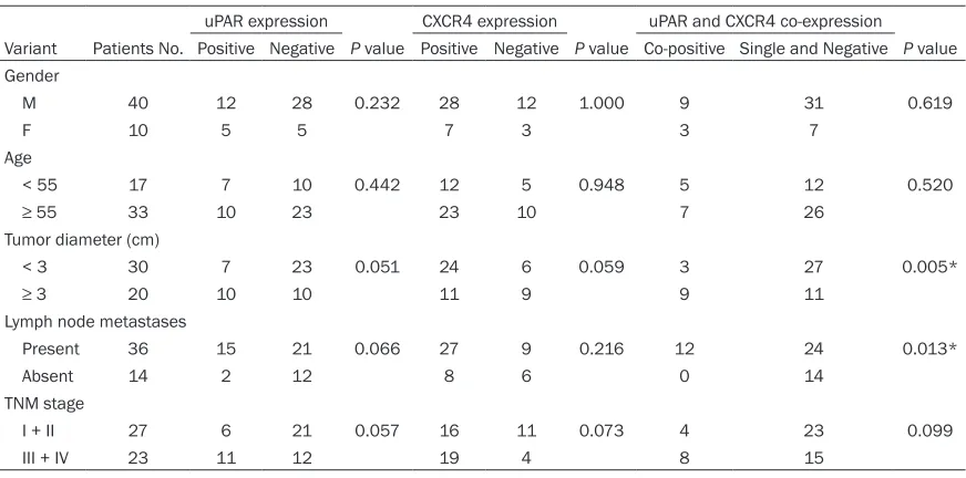
Table 1. Expression of uPAR and CXCR4 in SCLC specimens
[image:3.612.90.303.528.569.2]uPAR and CXCR4 in small cell lung cancer
Balb/c nude mice purchased from Beijing HFK Bio-Technology. Co, LTD (Beijing, China) with six
animals per group. Sorted cells were implanted
subcutaneously in the right flanks of nude mice
with 5 × 10⁵ in 100 μl RPMI1640 and 100 μl of
matrigel (BD Biosciences), making a 1:1 mix -ture. Tumors were measured once a week. After
8 weeks, mice were sacrificed and tumors were
removed and then subjected to immunohisto-chemical analysis. Tumor volume = 0.5 × length × width2.
Statistical analysis
All data in the study were evaluated with SPSS
version 17 software (SPSS Inc.). Data are pre
-sented as means ± SD. When two groups were compared, the Student t-test was used. The χ2 test was performed to determine correlations among the various parameters. Cumulative
survival rate was assessed by the
Kaplan-Meier method and analyzed by log-rank test.
Differences were considered significant at
value of P ≤ 0.05.
Results
uPAR and CXCR4 expressions in SCLC samples
From our and other researches previous study
we hypothesis uPAR and CXCR4 play important role in SCLC development. To determine the
expression and clinical significance of uPAR
and CXCR4 in SCLC, we analyzed the
[image:4.612.91.527.81.297.2]expres-sion of uPAR and CXCR4 by using immunohisto-chemistry in 50 clinical specimens of SCLC. uPAR was positive in 17 (34%) of the SCLC cases. uPAR expression was mainly observed in the membrane of tumor cells and the uPAR positive expression were generally tended to exist in foci near the invasive front of the carci-noma (Figure 1A). CXCR4 immunoreactivity was observed in the membrane and cytoplasm of tumor cells (Figure 1B), a strong CXCR4 expression was observed in 35 (70%) SCLC cases. There were 12 (24%) cases co-expressed uPAR and CXCR4 (Table 1). We examined the relationship in uPAR, CXCR4 expression and clinicopathological factors. As shown in Table 2, the tumor diameter of the uPAR and CXCR4 co-expression group was markedly larger than that of the negative uPAR and/or CXCR4 expres-sion group (P = 0.005). uPAR and CXCR4 co-expression was correlation to lymph node metastases (P = 0.013). No significant differ -ence was found between uPAR or CXCR4 single positive expression and the clinical parameters studied. Moreover, the association of uPAR, CXCR4 expression with patients’ overall surviv-al was further evsurviv-aluated. The uPAR expression observed correlation to survival, the mean sur-vival time was 22.250 ± 4.468 months in the positive uPAR expression group, but it was 34.762 ± 3.476 months in the negative uPAR expression group (P = 0.041; Figure 2A). There was no correlation between CXCR4 and surviv-al (P = 0.104; Figure 2B). The mean survival Table 2. Correlations of uPAR and CXCR4 expression with clinicopathologic parameters
uPAR expression CXCR4 expression uPAR and CXCR4 co-expression
Variant Patients No. Positive Negative P value Positive Negative P value Co-positive Single and Negative P value Gender
M 40 12 28 0.232 28 12 1.000 9 31 0.619
F 10 5 5 7 3 3 7
Age
< 55 17 7 10 0.442 12 5 0.948 5 12 0.520
≥ 55 33 10 23 23 10 7 26
Tumor diameter (cm)
< 3 30 7 23 0.051 24 6 0.059 3 27 0.005*
≥ 3 20 10 10 11 9 9 11
Lymph node metastases
Present 36 15 21 0.066 27 9 0.216 12 24 0.013*
Absent 14 2 12 8 6 0 14
TNM stage
I + II 27 6 21 0.057 16 11 0.073 4 23 0.099
III + IV 23 11 12 19 4 8 15
time of the uPAR and CXCR4 Co-expression
group was 19.625 ± 3.803 months that signifi -cant shorter than the single and co-negative expression group (29.351 ± 2.944 months) (P
= 0.033; Figure 2C).
Identify of uPAR+CXCR4+ subpopulation exist in
SCLC cell line H446
The co-expression of uPAR and CXCR4 in SCLC tissues showed correlation with tumor metas-tasis. To further demonstrate uPAR and CXCR4 involve in metastasis in SCLC, We stained both
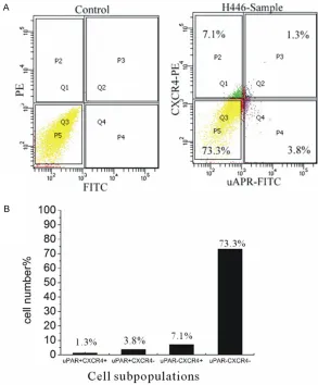
uPAR and CXCR4 fluorescent antibodies in SCLC cell line H446, analyzed by using a high-speed fluorescence-activated cell sorter.
Results showed there were contain uPAR+CX-
CR4+, uPAR+CXCR4-, uPAR-CXCR4+ and uPAR -CXCR4- four subpopulations in cell line H446 (Figure 3A). The proportion of uPAR+CXCR4+, uPAR+CXCR4-, uPAR-CXCR4+ and uPAR-CXCR4
-four subpopulations in H446 were 1.3%, 3.8%,
7.1% and 73.3% respectly (Figure 3B).
Invasion and migration capacity of uPAR+CX-
CR4+ subpopulation sorted from SCLC cell line
H446
The isolated four subpopulation cells were compared for invasion using the transwell inva-sion assay. As demonstrated via the transwell
[image:5.612.90.520.70.500.2]assay, more significant number of uPAR+CXCR4+ cells passed through the upper membrane pre-treated with matrigel compared with than the
Figure 2. Kaplan-Meier analysis of overall survival
uPAR and CXCR4 in small cell lung cancer
other groups, uPAR-CXCR4- cells showed the fewest cells migrated to the bottom chamber invasive capacity (Figure 4A). We further observed uPAR+CXCR4+ cells (65.60 ± 9.91) showed higher invasion capacity than uPAR+CXCR4- cells (31.00 ± 9.77) (P < 0.05; Figure 3B), uPAR-CXCR4+ (21.80 ± 4.81) cells
showed no significant difference in invasion
capacity than uPAR-CXCR4- cells (16.60 ± 7.09) (P < 0.05; Figure 4B). Cell migration capacity was evaluated by the wound healing assay, also known as the “scratch” assay. We observed the accordance results with invasive capacity that uPAR+CXCR4+ cells showed the highest migra-tion capacity than the other subpopulamigra-tions (migration distance were 100.50 ± 8.63, 82.80
± 8.81, 64.80 ± 6.97 and 53.4 ± 8.96 μm
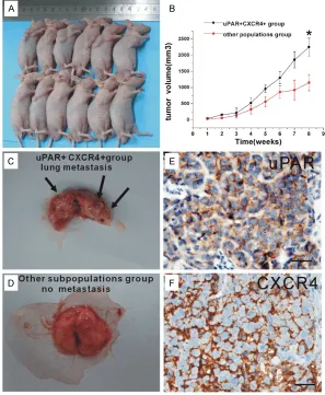
Figure 5B). Three mice in the uPAR+CXCR4+ cells group developed lung metastases (Figure 5C) whereas mice that received other subpopu-lation cells showed no trace of metastasis (Figure 5D). The lung metastatic tumor high expressed uPAR and CXCR4 (Figure 5E, 5F). These data support the hypothesis that uPAR+CXCR4+ cells may represent a distinct, migrating cell population.
Discussion
In human malignancies, uPAR overexpression is associated with an increased propensity for
cancer progression and metastasis [16, 17].
[image:6.612.92.385.72.426.2]Studies indicated intact uPAR and its cleaved forms are associated with poor prognosis in
Figure 3. FACS analysis of H446 cell line double-labeled with uPAR-FITC and CXCR4-PE, antibody. A: H446 cells staining mouse IgG as isotype control ana
-lyzed by FACS (left). H446 cell line double-labeled with uPAR-FITC and CXCR4-PE antibody analyzed by FACS (right). B: The proportion of uPAR+CXCR4+ (1.3%),
uPAR+CXCR4- (3.8%), uPAR-CXCR4+ (7.1%) and uPAR-CXCR4- (73.3%) four
sub-populations in H446.
respectively, P < 0.05, Fig- ure 4C). These results sup-ports that the strong inva-sive and migrate activity of cancer cells are mediated by uPAR and CXCR4, the function of CXCR4 maybe depend on uPAR triggered.
Growth and metastatic capacity of uPAR+CXCR4+
subpopulation from SCLC cell line H446 in vivo
To further confirm the
NSCLC and SCLC [18, 19]. However, the asso -ciation was mainly proved by detection of high levels of uPAR in patient blood but not in tumor tissue. In the present study, immunohistochem-ical staining showed 17 (17/50, 34%) cases were positive for the uPAR expression in SCLC
tissues. High expression of uPAR in SCLC patients was significantly correlated with short -er survival times than that of uPAR negative expression (P = 0.041). Thus, uPAR may be an independent prognostic indicator in SCLC. Invasion and metastasis are not random, but,
rather, highly organ-specific and pathological
processes. Chemokine receptors are deemed crucial in the homing mechanisms of hemato-poietic cells and metastasis of solid tumors
such as breast and ovarian cancers [20, 21].
Recent reports suggested that CXCR4 may be a key regulator of tumor invasiveness leading to
local progression and tumor metastasis [22]. In
SCLC, the distant organ sites most commonly affected are the lymph nodes, bone marrow,
and brain, all of which exhibit high SDF-1 con
-tents [23]. The specific receptor of SDF-1,
CXCR4, is also involved in SCLC metastasis. Previous immunohistochemical studies showed
[image:7.612.94.523.71.412.2]that SCLC cells expressed CXCR4 [24]. Here, we
Figure 4. Cell invasion and migration capability in vitro was detected by transwell and wound healing assays. A: The transmembrane cells of uPAR+CXCR4+, uPAR+CXCR4-, uPAR-CXCR4+ and uPAR-CXCR4- group (× 100, Scale bar
= 50 μm). B: The transmembrane numbers of uPAR+CXCR4+, uPAR+CXCR4-, uPAR-CXCR4+ and uPAR-CXCR4- group
were 86.50 ± 11.32, 40.25 ± 10.15, 21.34 ± 5.35 and 15.45 ± 7.82 respectively. The transmembrane number of uPAR+CXCR4+ group cells was significant more than the other subpopulations group cells (P < 0.05, respectively).
Furthermore, the transmembrane cells of uPAR+CXCR4+ group was significant more than the uPAR+CXCR4- group (P
= 0.01). However, the difference between uPAR-CXCR4+ and uPAR-CXCR4- group was not significant (P = 0.212). C:
The cell migration distances of uPAR+CXCR4+, uPAR+CXCR4-, uPAR-CXCR4+ and uPAR-CXCR4- group were 100.50 ±
8.63, 82.80 ± 8.81, 64.80 ± 6.97 and 53.4 ± 8.96 μm respectively. The cell migration distances of uPAR+CXCR4+
group cells was significant farther than the other subpopulations group cells (P < 0.05, respectively). (× 100, Scale
uPAR and CXCR4 in small cell lung cancer
uPAR+CXCR4+ cells from cell line H446. The uPAR+CXCR4+ cells, although a minor propor-tion, demonstrated stronger migratory and metastatic capacities than other subpopula-tion cells. Particularly we observed uPAR+CX- CR4+ cells showed higher invasion capacity than uPAR+CXCR4- cells (P < 0.05), and uPAR -CXCR4+ cells showed no significant difference in invasion capacity with uPAR-CXCR4- cells (P <
0.05). These results confirmed our hypothesis
that CXCR4+ cell motility maybe triggered by detected CXCR4 in SCLC specimens. There
were no significant relationship between CXCR4
expression and clinical data. But interestingly, we found evidence in SCLC specimens that links several conventional clinical factors to uPAR and CXCR4 co-expression. Tumor mean diameter of uPAR and CXCR4 co-expression group was larger than that of single or negative expression group (P = 0.005), indicating that uPAR and CXCR4 positive cells may lead to
[image:8.612.89.386.74.435.2]fast-er tumor growth. High expression of uPAR in
Figure 5. The xenograft and lung metastasis forming of uPAR+CXCR4+ and
other subpopulation cells in vivo. A: Subcutaneous implantation of 5 × 10⁵
uPAR+CXCR4+ cells or other subpopulationcells in BALB/c nude mice led to
tumor formation (6/6, 5/6, respectly). B: BALB/c nude mice shows that after 8 weeks injected, growth curve of xenograft tumors volume of uPAR+CXCR4+
group exhibit significant larger than other subpopulationgroup (2227.6 ± 211.35, 1201.0 ± 170.36 mm3, respectively, P < 0.01). C: After 8 weeks, mice
were sacrificed, lung metastasis foci were founded in uPAR+CXCR4+ cells group
(arrow indicated). D: Other subpopulation cells group showed no trace of metas -tasis. E: Immunohistochemistry showed uPAR positive expressed in lung
metas-tasis tumor tissue. F: Immunohistochemistry showed CXCR4 positive expressed in lung metastasis tumor tissue (× 400, Scale bar = 50 μm).
SCLC patients was also
sig-nificantly correlated with
ly-mph node metastases indi-cating that uPAR and CXCR4 positive cells may play an important role in tumor metastasis. uPAR and CXCR4 co-expression group also showed shorter survival times than that of single and co-negative uPAR or CXCR4 expression (P = 0.033). Therefore, uP- AR and CXCR4 co-expres-sion might be a reliable prognostic biomarker for tumor growth or metasta-sis in SCLC patients.
Studies analyzing uPAR and CXCR4 at the cellular and molecular level have revealed multiple functions of them in tumor character-istics. A study performed by Gutova and colleagues
[25] provided evidence that
the uPAR+ subpopulation of cancer cells is resistant to traditional chemotherapi- es. Using urokinase recep-tor antibody or transfected with antisense uPAR vec-tors in cell line can inhibit in vivo tumorigenicity and
metastases [26, 27]. There
was also research
[3] Dass K, Ahmad A, Azmi AS, Sarkar SH and Sarkar FH. Evolving role of uPA/uPAR system in
human cancers. Cancer Treat Rev 2008; 34: 122-136.
[4] Bezwoda WR, Lewis D and Livini N. Bone mar -row involvement in anaplastic small cell lung
cancer. Diagnosis, hematologic features, and
prognostic implications. Cancer 1986; 58: 1762-1765.
[5] Minna JD, Roth JA and Gazdar AF. Focus on
lung cancer. Cancer Cell 2002; 1: 49-52.
[6] Muller A, Homey B, Soto H, Ge N, Catron D,
Buchanan ME, McClanahan T, Murphy E, Yuan W, Wagner SN, Barrera JL, Mohar A, Verastegui E and Zlotnik A. Involvement of chemokine re-ceptors in breast cancer metastasis. Nature 2001; 410: 50-56.
[7] Manu KA, Shanmugam MK, Rajendran P, Li F, Ramachandran L, Hay HS, Kannaiyan R, Swamy SN, Vali S, Kapoor S, Ramesh B, Bist P, Koay ES, Lim LH, Ahn KS, Kumar AP and Sethi
G. Plumbagin inhibits invasion and migration of breast and gastric cancer cells by downregu-lating the expression of chemokine receptor CXCR4. Mol Cancer 2011; 10: 107.
[8] Uygur B and Wu WS. SLUG promotes prostate cancer cell migration and invasion via CXCR4/ CXCL12 axis. Mol Cancer 2011; 10: 139.
[9] Hart CA, Brown M, Bagley S, Sharrard M and
Clarke NW. Invasive characteristics of human prostatic epithelial cells: understanding the metastatic process. Br J Cancer 2005; 92: 503-12.
[10] Burger JA and Bürkle A. The CXCR4 chemokine receptor in acute and chronic leukaemia: a marrow homing receptor and potential
thera-peutic target. Br J Haematol 2007; 137:
288-296.
[11] O’Hayre M, Salanga CL, Kipps TJ, Messmer D, Dorrestein PC and Handel TM. Elucidating the
CXCL12/CXCR4 signaling network in chronic lymphocytic leukemia through phosphopro-teomics analysis. PLoS One 2010; 5: e11716.
[12] Hermann PC, Huber SL, Herrler T, Aicher A, Ellwart JW, Guba M, Bruns CJ and Heeschen C. Distinct populations of cancer stem cells de -termine tumor growth and metastatic activity in human pancreatic cancer. Cell Stem Cell 2007; 1: 313-323.
[13] Hartmann TN, Burger JA, Glodek A, Fujii N and
Burger M. CXCR4 chemokine receptor and in-tegrin signaling co-operate in mediating adhe-sion and chemoresistance in small cell lung cancer (SCLC) cells. Oncogene 2005; 24: 4462-4471.
[14] Burger M, Glodek A, Hartmann T, Schmitt-Gräff A, Silberstein LE, Fujii N, Kipps TJ and Burger JA. Functional expression of CXCR4 (CD184)
on small-cell lung cancer cells mediates
migra-uPAR. Through in vivo experiments, we found that both uPAR+CXCR4+ subpopulation and other subpopulation cells were capable of
inducing orthotropic primary tumors. However,
the uPAR+CXCR4+ cells were capable of spread-ing from the primary tumor to form metastatic lesions, while the other subpopulation cells, although containing uPAR or CXCR4 positive cells could not form metastatic lesions, sug-gesting that both uPAR and CXCR4 expression are essential for metastatic spread. In the pres-ent study, we provide evidence for a metastatic subpopulation uPAR+CXCR4+ existence in the
SCLC cell line H446. The interaction of uPAR
and CXCR4 in SCLC should be investigated in future studies.
In summary, uPAR and CXCR4 co-expression plays a critical role in tumor development and progression in SCLC. The existence of the uPAR+CXCR4+ cell subpopulation is possibly
responsible for tumor metastasis. Further
examination of such cell subpopulation in SCLC cells will provide important clues on the malig-nant progression and therapy target of SCLC cancer.
Acknowledgements
This study was supported by the Research Program of the Applied Basic and Cutting-edge Technologies of Tianjin under Contract No.
14JCZDJC35500.
Disclosure of conflict of interest
None.
Address correspondence to: Dr. Xiaofei Qiu, Depart-ment of Pathology; Tianjin Key Laboratory of Cellular
and Molecular Immunology, Tianjin Medical University, Tianjin 300070, China. Tel: 86 22 23542527; Fax: 86 22 23542527; E-mail: qiouxf@ tijmu.edu.cn
References
[1] Qiu X, Wang Z, Li Y, Miao Y, Ren Y and Luan Y. Characterization of sphere-forming cells with stem-like properties from the small cell lung
cancer cell line H446. Cancer Lett 2012; 323:
161-170.
[2] Ploug M and Ellis V. Structure-function rela-tionships in the receptor for urokinase-type plasminogen activator. Comparison to other members of the Ly-6 family and snake venom
alpha-neurotoxins. FEBS Lett 1994; 349:
uPAR and CXCR4 in small cell lung cancer
[22] Burger JA and Kipps TJ. CXCR4: a key receptor
in the crosstalk between tumor cells and their microenvironment. Blood 2006; 107: 1761-1767.
[23] Kryczek I, Wei S, Keller E, Liu R and Zou W. Stroma-derived factor (SDF-1/CXCL12) and
human tumor pathogenesis. Am J Physiol Cell Physiol 2007; 292: C987-995.
[24] Kijima T, Maulik G, Ma PC, Tibaldi EV, Turner
RE, Rollins B, Sattler M, Johnson BE and Salgia R. Regulation of cellular proliferation, cytoskel-etal function, and signal transduction through
CXCR4 and c-Kit in small cell lung cancer cells.
Cancer Res 2002; 62: 6304-6311.
[25] Gutova M, Najbauer J, Gevorgyan A, Metz MZ,
Weng Y, Shih CC and Aboody KS. Identification
of uPAR-positive chemoresistant cells in small cell lung cancer. PLoS One 2007; 2: e243.
[26] Rabbani SA and Gladu J. Urokinase receptor antibody can reduce tumor volume and detect the presence of occult tumor metastases in vivo. Cancer Res 2002; 62: 2390-2397.
[27] Go Y, Chintala SK, Mohanam S, Gokaslan Z, Venkaiah B, Bjerkvig R, Oka K, Nicolson GL,
Sawaya R and Rao JS. Inhibition of in vivo tu-morigenicity and invasiveness of a human glio-blastoma cell line transfected with antisense uPAR vectors. Clin Exp Metastasis 1997; 15: 440-446.
tion, integrin activation, and adhesion to stro-mal cells. Oncogene 2003; 22: 8093-8101.
[15] Montuori N, Bifulco K, Carriero MV, La Penna C, Visconte V, Alfano D, Pesapane A, Rossi FW,
Salzano S, Rossi G and Ragno P. The cross-talk between the urokinase receptor and fMLP re-ceptors regulates the activity of the CXCR4 chemokine receptor. Cell Mol Life Sci 2011; 68: 2453-2467.
[16] de Bock CE and Wang Y. Clinical significance of
urokinase-type plasminogen activator receptor (uPAR) expression in cancer. Med Res Rev 2004; 24: 13-39.
[17] Mazar AP. Urokinase plasminogen activator re-ceptor choreographs multiple ligand interac-tions: implications for tumor progression and therapy. Clin Cancer Res 2008; 14: 5649-5655.
[18] Almasi CE, Hoyer-Hansen G, Christensen IJ and Pappot H. Prognostic significance of urokinase
plasminogen activator receptor and its cleaved forms in blood from patients with non-small cell lung cancer. APMIS 2009; 117: 755-761.
[19] Almasi CE, Drivsholm L, Pappot H, Hoyer-Hansen G and Christensen IJ. The liberated
domain I of urokinase plasminogen activator receptor--a new tumour marker in small cell lung cancer. APMIS 2013; 121: 189-196.
[20] Sun X, Cheng G, Hao M, Zheng J, Zhou X, Zhang J, Taichman RS, Pienta KJ and Wang J.
CXCL12/CXCR4/CXCR7 chemokine axis and cancer progression. Cancer Metastasis Rev 2010; 29: 709-722.
[21] Scotton CJ, Wilson JL, Milliken D, Stamp G and Balkwill FR. Epithelial cancer cell migration: a