RESEARCH ARTICLE
Identification and characterization of the NMDA receptor and
its role in regulating reproduction in the cockroach
Diploptera punctata
Juan Huang1, Ekaterina F. Hult1, Elisabeth Marchal1,2and Stephen S. Tobe1,*
ABSTRACT
The NMDA receptor (NMDAR) plays important roles in excitatory neurotransmission and in the regulation of reproduction in mammals. NMDAR in insects comprises two subunits, NR1 and NR2. In this study, we identified two NR1 paralogs and eleven NR2 alternatively spliced variants in the cockroachDiploptera punctata. This is the first report of NR1 paralogs in insects. The tissue distributions and expression profiles ofDpNR1A,DpNR1BandDpNR2in different tissues were also investigated. Previous studies have demonstrated NMDA-stimulated biosynthesis of juvenile hormone (JH) in the corpora allata through the influx of extracellular Ca2+inDiploptera punctata. However, our data show that the transcript levels ofDpNR1A,DpNR1BandDpNR2were low in the corpora allata. MK-801, a high-affinity antagonist of NMDAR, did not show any effect on JH biosynthesisin vitro. In addition, neither partial knockdown ofDpNR2norin vivotreatment with a physiologically relevant dose of MK-801 resulted in any significant change in JH biosynthesis or basal oocyte growth. Injection of animals with a high dose of MK-801 (30 µg per animal per injection), which paralyzed the animals for 4–5 h, resulted in a significant decrease in JH biosynthesis on days 4 and 5. However, the reproductive events during the first gonadotrophic cycle in female D. punctatawere unaffected. Thus, NMDAR does not appear to play important roles in the regulation of JH biosynthesis or mediate reproduction of femaleD. punctata.
KEY WORDS: NMDA receptor, JH biosynthesis, MK-801, Reproduction,D. punctata
INTRODUCTION
L-glutamate (Glu), a major excitatory amino acid transmitter, mediates
diverse physiological functions in the vertebrate nervous system (Mahesh and Brann, 2005). The Glu receptors (GluRs) have been classified into three major subtypes: theα -amino-3-hydroxy-5-methyl-4-isoxazolepropionic acid (AMPA) receptor, N-methyl-D-aspartate
(NMDA) receptor and kainate receptor (Madden, 2002). The NMDA receptors (NMDARs) are distinguished from other ionotropic receptors by their unique properties, including selective agonists and antagonists, high Ca2+ permeability and
voltage-dependent Mg2+blockade (McBain and Mayer, 1994). The unique
properties of NMDAR allow it to play key roles in excitatory neurotransmission and important neurological processes, including learning, memory and behavior (Mussig et al., 2010; Newcomer and Krystal, 2001; Xia et al., 2005).
NMDAR in vertebrates is composed of two subunits, NR1 and NR2, and in some cases NR3 subunits (Madden, 2002). The NR1 subunit is essential for the basic channel activity of NMDAR, whereas the NR2 subunit contributes to enhance and modulate the receptor function (Sydow et al., 1996). Although much is known about the function of NMDAR in vertebrates, little information is available on NMDARs in insects. Thus far, two subunits, NR1 and NR2, have been identified in insects. The NR1 and NR2 subunits were previously reported to be distributed throughout the brain of
Drosophila melanogasterandApis mellifera(Wu et al., 2007; Xia
et al., 2005; Zannat et al., 2006).
The importance of NMDAR in the regulation of reproduction of mammals is well known. NMDAR mediates reproduction through the regulation of pulse and surge gonadotropin-releasing hormone (GnRH)/luteinizing hormone (LH) secretion (Maffucci et al., 2009; Mahesh and Brann, 2005). In insects, juvenile hormones (JHs) are key regulators of growth, development, metamorphosis, aging, caste differentiation and reproduction (Goodman and Granger, 2005; Hartfelder, 2000). As a result of the importance of JH in physiological processes, its biosynthesis is tightly regulated by many factors, including neuropeptides (allatostatins, allatotropins) (Stay and Tobe, 2007) and neurotransmitters (octopamine, dopamine and glutamate) (Granger et al., 1996; Pszczolkowski et al., 1999; Thompson et al., 1990). Chiang et al. (2002) demonstrated that JH biosynthesis by corpora allata (CA) of the cockroach, Diploptera punctata, is stimulated by an NMDA-induced influx of Ca2+ ions and this elevation was significantly
reduced by NMDAR antagonists including Mg2+, MK-801 or
conantokin T. Furthermore, a Drosophila larval mutant for
NMDAR1showed reduced mRNA levels of the gene encoding JH
acid methyltransferase (JHAMT), a key regulatory enzyme of JH biosynthesis in this species (Huang et al., 2011). These studies suggest that NMDAR plays a role in the regulation of JH biosynthesis.
As a high-affinity antagonist of NMDARs, MK-801 has been used to study the function of NMDAR in both vertebrates and invertebrates (Rawls et al., 2009; Sircar et al., 1987; Troncoso and Maldonado, 2002). In addition to the inhibitory effect of MK-801 in NMDA-stimulated JH biosynthesis inD. punctata, MK-801 was found to influence ovarian development and vitellogenesis in the flesh flyNeobellieria bullataand the locustSchistocerca gregaria
(Begum et al., 2004; Chiang et al., 2002). In S. gregaria, the inhibition of vitellogenesis was overcome by treatment with JH. A later study on the butterflyBicyclus anynanaand the cricketGryllus
bimaculatusshowed that MK-801 affects JH biosynthesisin vitro
and JH titres in both species, and subsequently regulates insect reproduction (Geister et al., 2008).
Diploptera punctata is a well-known model for studying the
physiology of JH biosynthesis and regulation; in this animal, JH Received 8 October 2014; Accepted 26 January 2015
1
Department of Cell and Systems Biology, University of Toronto, Toronto, Canada M5S 3G5.2Department of Biology, Zoological Institute, KU Leuven, Leuven B-3000, Belgium.
*Author for correspondence ([email protected])
The
Journal
of
Experimental
biosynthesis is high and stable, and the reproductive events correlate very well with rates of JH production (see review by Marchal et al., 2013a). In this study, we chose D. punctata as our model to determine the role of NMDAR in the regulation of JH biosynthesis and reproduction. We identified the genes encoding the subunits of NMDAR inD. punctata, and examined the expression of NMDAR in several tissues. In addition, we investigated the roles of NMDAR in the regulation of JH biosynthesis, vitellogenesis and oocyte growthin vivousing RNA interference (RNAi) and MK-801.
RESULTS
Identification of DpNR1 subunits
TwoDpNR1were identified with open reading frames of 2871 bp
(DpNR1A) and 2703 bp (DpNR1B). This was accomplished
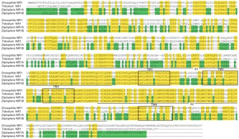
employing a degenerate PCR approach with adult brain cDNA. 5′-RACE and 3′-RACE experiments were performed to complete the sequences. The twoDpNR1sequences were deposited in NCBI GenBank (accession numbers KJ747198 and KJ747199). Unlike the alternative splicing variants of NR1 genes in other insects, the differences between the two variants inD. punctataare distributed throughout the whole gene (Fig. 1) and may be the result of gene duplication. A phylogenetic tree of NR1 was constructed by maximum-likelihood methods (Fig. 2). Generally, the sequences of NR1 were grouped based on insect orders, and the two DpNR1s (Blattodea) cluster together with confidence. However, the relationship between orders is not well resolved, as indicated by low bootstrap values at deeper nodes. The lack of power to resolve these nodes could be explained by insufficient sampling among hemimetabolous insects.
Identification of DpNR2 subunits
DpNR2 undergoes alternative splicing, generating 11 different
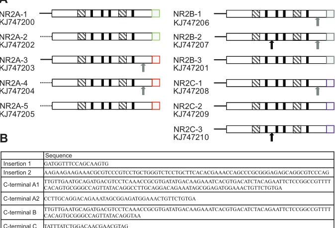
transcripts. Full-length cDNAs for all 11 variants have been isolated (GenBank accession numbers KJ747200 to KJ747210). In the 11 transcripts, there are two 5′untranslated region, two insertions, and four different 3′ ends (Fig. 3A). The sequence of insertions, deletions and different 3′ends are shown in Fig. 3B. All 11DpNR2
variants contain the domain structure, four hydrophobic regions (TM1–TM4) and two ligand binding domains. Amino acid sequence comparisons between DpNR2A-1 and NR2 subunits
fromD. melanogaster,A. melliferaandA. aegyptiwere determined
(supplementary material Table S1). DpNR2A-1 shares the highest similarity with NR2 subunits fromA. mellifera.
Expression ofDpNR1A,DpNR1BandDpNR2
Previous studies of NMDAR in insects have focused on its function in brain (Wu et al., 2007; Xia et al., 2005; Zannat et al., 2006). In the present study, we were interested in assessing the role of NMDAR in other tissues. We therefore determined the localization and relative abundance ofDpNR1A,DpNR1BandDpNR2mRNA in day 4 adult male and female cockroaches using q-RT-PCR (Fig. 4). Specific q-RT-PCR primers for each DpNR1 variant were designed to determine whether transcripts for the differentDpNR1paralogs are present in the same tissue. Q-RT-PCR primers for DpNR2 are located in the conserved region ofDpNR2common to all putative splice variants. In mated female cockroaches,DpNR1A,DpNR1B
andDpNR2were highly expressed in brain, followed by nerve cord.
A previous study has shown that NMDA stimulated JH biosynthesis by the CA of Diploptera (Chiang et al., 2002). However, the
Drosophila NR1 -MAMAEFVFCRPLFGLAIVLLVAPIDAAQRHTASDNPSTYNIGGVLSNSDSEEHFSTTIKHLNFDQQYVPRKVTYYDKTIRMDKNPIKTVFNVCDKLIENRVYAVVVSHEQTSGDLSPAAVSYTSGFY Tribolium NR1 ---MFVFVIFALNWLTIRADLWS---SNNPTVFNIGGVLSSNESEYYFKETIAHLNFDSQYVPKGVTYYDTAILMDPNPIKTALNVCKYLITSRVYAVVVSHPLTG-DLSPAAVSYTSGFY
Diploptera NR1A MNILIYLVFYSVCVLIPKLMADPKPQGASDMRRRNNPTYFNIGGVLSNMESQMIFKETISHLNFNSQYVPKGVTYDSTAISMDPNPIRTALNVCNYLINQSVYAVVVSHPLTG-DLSPAAVSYTSGFY
Diploptera NR1B ---MSRLQLNLVMLVSALQG----VVLHNPTYVNIGGLFSTMESQAFFRETVAKLNVNNDII---YDSTVTLMDVNPIRTALKVCNNIINQSVYAVVVSHSYNG-DLSPAAVSHTSGFF
Drosophila NR1 SIPVIGISSRDAAFSDKNIHVSFLRTVPPYYHQADVWLEMLSHFAYTKVIIIHSSDTDGRAILGRFQTTSQTYYDDVDVRATVELIVEFEPKLESFTEHLIDMKT-AQSRVYLMYASTEDAQVIFRDA
Tribolium NR1 HIPVIGISSRDSAFSDKNIHVSFLRTVPPYSHQADVWVEMLKHFNYKKVIFIHSSDTDGRALLGRFQTTSQSLEDDVEIKVQVESIIEFEPGLETFKEQLSDMKN-AQSRVYLMYASKTDAQVIFRDA Diploptera NR1A HIPVIGISSRDSAFSDKNIHVSFLRTVPPYSHQADVWMEFLKRFNYLKVIFIHSSDTDGRALLGRFQS-SQNLEEDVETKVQVETVIEFEPGLDSFTDQLLEVKN-LQSRVYLMYAGKTDSEVIFRDA Diploptera NR1B HIPVIGISSRDSAFSDKNIHVSFLRRVPPYSHQADVWIKILRHFRFRRFILIHSSDKDGRALMRRIQSISQNMEE--EMKVLAEAVTEFEPGLESFADQLQILRNSTQSHVFLLYAKKNDSEIIFRDA
Drosophila NR1 GEYNMTGEGHVWIVTEQALFSNNTPDGVLGLQLEHAHSDKGHIRDSVYVLASAIKEMISNETIAEAPKDCGDSAVNWESGKRLFQYLKSRNIT-GETGQVAFDDNGDRIYAGYDVINIREQQKKHVVG Tribolium NR1 AEFNMTDAGYAWIVTEQALVANNIPEGILGLRLVNATNEKAHIKDSIYVLASALRDLNQTKEITEAPKDCDDSGQIWETGRDLFDFIKKQVLMNGETGKVAFDDQGDRINAEYNIVNIQRKRKQVTVG Diploptera NR1A GLLNMTGLTYVWIVTEQALEASNVPIGTLGLKLVNATREEDHIKDSIYVLASALRVMNRTEVITKAPKDCGDSGHIWETGKTLFDYIKKQNLVNGSTGNVAFDDNGDRINAEYDIINVQSNQTQISVG
Diploptera NR1B QKLNLTGISYVWIVTEQALDADNIPVGTLGLRLVNATNEKIHIKDSIYVFKSAFLQMNKTELITSPPRDCGDSAHVWETGKSLFEIIRKQNLVKGETGNVAFDEKGDRINAEYDIININAESIQSTVG
Drosophila NR1 KFSYDSMRAKMRMRINDSEIIWPGKQRRKPEGIMIPTHLRLLTIEEKPFVYVRRMGDDEFRCEPDERPCPLFNNSDATAN--EFCCRGYCIDLLIELSKRINFTYDLALSPDGQFGHYILRNNTGAMT Tribolium NR1 KFFFNRTSNKMRLAVDENNILWPGRQHVKPEGFMIPTHLKVLTIEEKPFVYVRKLVEPQDVCTAEEIPCPHFNATQDLAG--SYCCKGYCMDLLKELSKKINFTYSLALSPDGQFGNYIIRNSS--GS
Diploptera NR1A QYHFSFEQNRMALKLDKDAIRWPGNTSQAPVGILIPTHLKVLTIEEKPFVYVRETQN-EDDCTPEEIPCPHFNNSADNNDFVKYCCKGFCMDLLKELSAKINFTYNLELSPDGQFGSYMVRNSS--MG Diploptera NR1B KYHYSNENNQMKLTLESDAIVWPGGIHNYNWGQSTPNHLKVLTIEEKPFVYVREIND-NDECSPNEISCARFNSSDFSD---SYCCKGYCIDLLKELATRINFTYHVGLSPDGQFGDYVLKENS--LG
Drosophila NR1 LRKEWTGLIGELVNERADMIVAPLTINPERAEYIEFSKPFKYQGITILEKKPSRSSTLVSFLQPFSNTLWILVMVSVHVVALVLYLLDRFSPFGRFKLSHSDSNEEKALNLSSAVWFAWGVLLNSGIG
Tribolium NR1 GKKEWTGLIGELVGERADMIVAPLTINPERAEFIEFSKPFKYQGITILEKKPSRSSTLVSFLQPFSNTLWILVMVSVHVVALVLYLLDRFSPFGRFKLANTDGTEEDALNLSSAIWFAWGVLLNSGIG Diploptera NR1A GKKEWTGLIGELVNERADMIVAPLTVNPERAEFIEFSKPFKYQGITILEKKPSRSSTLVSFLQPFSNTLWILVMVSVHVVALVLYLLDRFSPFGRFKLANTDGTEEDALNLSSAIWFAWGVLLNSGIG Diploptera NR1B PKKEWTGLIGELVNERADMIVAPLTINLERAEFIEFSKPFKYQGITILEKKPLQSTTVVSFLQPFSNTLWILVLVSVNVVALVLYLLDRFSPFSLLKRTNSEAAEENALNMQSALWFAWSVLLNSGIG
Drosophila NR1 EGTPRSFSARVLGMVWAGFAMIIVASYTANLAAFLVLERPKTKLSGINDARLRNTMENLTCATVKGSSVDMYFRRQVELSNMYRTMEANNYATAEQAIQDVKKGKLMAFIWDSSRLEYEASKDCELVT Tribolium NR1 EGTPRSFSARVLGMVWAGFAMIIVASYTANLAAFLVLERPKTKLTGINDARLRNTMENLTCATVKGSAVDMYFRRQVELSNMYRTMEANNYNTAEDAIEDVKVGKLMAFIWDSSRLEFEAAQDCELVT
Diploptera NR1A EGTPRSFSARVLGMVWAGFAMIIVASYTANLAAFLVLERPKTKLTGINDARLRNTMENLTCATVKGSAVDMYFRRQVELSNMYRTMEANNYDTAEEAIQDVKNGKLMAFIWDSSRLEYEAAQDCELVT
Diploptera NR1B EGTPKSCCTRVLGMIWAGFAMIIFASYTANLAAFLVLARPKTKLTGINDARLRNTMENLTYATVRGSAVDMYFQRQAELSNMYRTMEANNYDTAEAAIRDVKNGKLMAFIWDSARLEYEAAQDCELVT
Drosophila NR1 AGELFGRSGYGIGLQKGSPWTDAVTLAILEFHESGFMEKLDKQWIFHGHVQQNCELFEKTPNTLGLKNMAGVFILVGVGIAGGVGLIIIEVIYKKHQVKKQKRLDIARHAADKWRGTIEKRKTIRASL
Tribolium NR1 AGELFGRSGYGIGLQKGSPWADDITLAILDFHESGFMESLDNKWILQGNVQQ-CEQFEKTPNTLGLKNMAGVFILVAAGIVGGIGLIVIEMAYKKHQIKKQKRMELARHAADKWRGCVEKRKTLRASA
Diploptera NR1A AGELFGRSGYGIGLKKGSPWADAVTLAILDFHESGVMESLDNRWILQGNLQQ-CEQFEKTPNTLGLKNMAGVFILVAAGTVGGIGLIIIEMAYKKHQIRKQKRMELARHAADKWRGAIEKRKTLRATI Diploptera NR1B SGELFGHSGYGIGLTKGSRWADAVTIAILNFHESGVIDSLDHRWILQNNVQH-CDKSDKPQHTLGLKNMAGVFMLVAGGIIGGSGLIIVEVIYRRHQIRKERKLRVARYAAHKWRGIVQKRKILRATL
Drosophila NR1 AMQRQYNVGLNSTHAPGTISLAVDKRRYPRLGQRLGPERAWPGDAADVLRIRRPYELGNPGQSPKVMAANQPGMPMPMLGKTRPQQSVLPPRYSPGYTSDVSHLVV* Tribolium NR1 TTQRR--IKSNGVNDPATISLAVDKYQR---IGGPERAWPGDSD--IRQRRVEDSGGVQPVPRYLPAYTSDVSHLIV*--- Diploptera NR1A AAQRR--LKANGVNDPATVSLSVDALPRPCM-EARSPARAWPGGCD--VRQRTHHAEDTNMPYGTVLPDMIV*--- Diploptera NR1B CAQRG--LCA---VSLSLDSIPQDGT-DIRKLKRVWPGD*---
VLGMVWAGFAMIIVASYTA VLGMVWAGFAMIIVASYTA VLGMVWAGFAMIIVASYTA VLGMIWAGFAMIIFASYTA
KALNLSSAVWFAWGVLLNSGIG DALNLSSAIWFAWGVLLNSGIG DALNLSSAIWFAWGVLLNSGIG NALNMQSALWFAWSVLLNSGIG
NMAGVFILVGVGIAGGVGLIII NMAGVFILVAAGIVGGIGLIVI NMAGVFILVAAGTVGGIGLIII NMAGVFMLVAGGIIGGSGLIIV
TM1
LWILVMVSVHVVALVLYLL LWILVMVSVHVVALVLYLL LWILVMVSVHVVALVLYLL LWILVLVSVNVVALVLYLL
TM2
TM3
TM4
S1
[image:2.612.62.547.401.684.2]S2
Fig. 1. Amino acid sequence alignment of the twoDiplopteraNR1 subunits and homologous receptors fromD. melanogasterandT. castaneum. DiplopteraNR1A and NR1B aligned withD. melanogasterDNR1 (GenBank acc. no. NP_730940.1) andT. castaneumTNR1 (GenBank acc. no. XP_969654.1,
predicted sequence). Conservatively substituted residues are highlighted in yellow, and the different residues betweenDpNR1AandDpNR1Bin green. Three
putative hydrophobic transmembrane regions (TM1, TM3, and TM4) and one hydrophobic pore-forming segment (TM2) are highlighted in the boxes.
Agonist-binding domain (S1 and S2 domains) are underlined.
The
Journal
of
Experimental
transcript level ofDpNR1A,DpNR1BandDpNR2in the CA was relatively low compared with other tissues. In male cockroaches, the highest transcript levels of DpNR1A,DpNR1B and DpNR2were measured in the brain (Fig. 4). Interestingly, the transcript level of
DpNR2in testes was very low, whereas levels of mRNA encoding
both NR1 isoforms were high.
To further study the function of NMDAR in D. punctata, we determined the developmental profile of DpNR1A, DpNR1B and
DpNR2in the brain, CA and testes.DpNR1AandDpNR1Bwere stably
transcribed in the brain of day 0 to day 7 post-emergence mated female
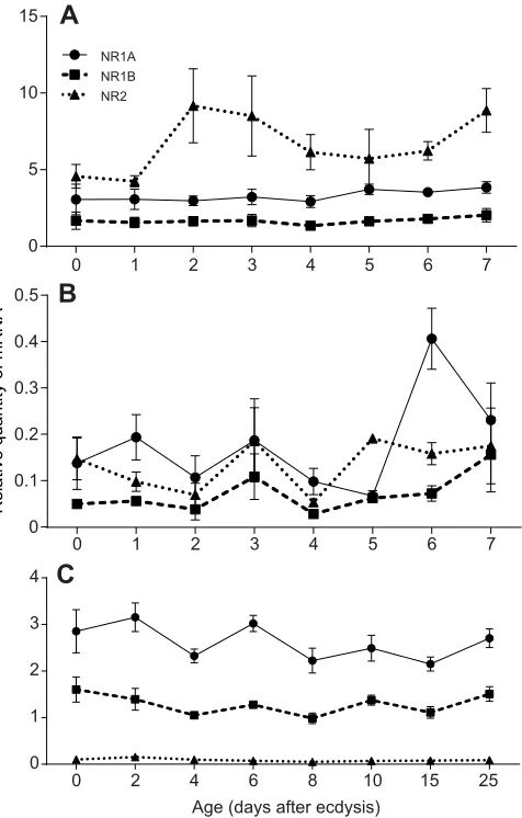
D. punctata.DpNR2mRNA in the brain showed a slight increase on
days 2, 3 and 7, but none of the changes was significant (Fig. 5A). In the CA, the transcript levels ofDpNR1BandDpNR2remained very low and did not show any significant change during the first gonadotrophic cycle (Fig. 5B).DpNR1AmRNA levels exhibited a significant increase on day 6. In male cockroaches, we studied the expression of NMDAR in testes of males of differing ages. The transcript level ofDpNR2was very low for all ages assayed (Fig. 5C).
0.2
D. punctata NR1B KJ747199
A. pisum XP_001949860.2
T. castaneum XP_969654.1
M. rotundata XP_003706090.1
M. domestica XP_005184853.1
B. terrestris XP_003394459.1
A. californica NM001204482.1
A. mellifera NP_001011573.1
D. melanogaster NP_730940.1
A. gambiae XP_321646.3
D. punctata NR1A KJ747198
C. floridanus EFN73528.1
A. aegypti XP_001653302.1
B. mori XP_004931410.1
D. plexippus EHJ78211.1
C. capitata XP_004524047.1
B. dorsalis AGS58420.1 100
38
94 82
100 100
99
96 31
100
84 51
54
[image:3.612.48.361.52.339.2]100
Fig. 2. Phylogram depicting the relationship between the NR1 subunits fromDiplopteraand orthologues of this receptor from other insects.Phylogenetic analysis was conducted in PhyML 3.0 using WAG substitution model with 100 bootstrap replicates. Poorly aligned amino acids were eliminated by eye, resulting in 854 positions. The bar
represents 0.2 substitutions per site. The sea slug,Aplysia
californicawas used as outgroup to root the tree.
NR2A-1
NR2A-2
NR2A-3
NR2A-4
NR2A-5
NR2B-1
NR2B-3
NR2C-2 NR2C-1
NR2C-3 NR2B-2 KJ747200
KJ747202
KJ747203
KJ747204
KJ747205
KJ747206
KJ747207
KJ747201
KJ747208
KJ747209
KJ747210
A
B
Sequence
Insertion 1 GATGGTTTCCAGCAAGTG
Insertion 2 AAGAAGAAGAAACGCGTCCCGTCCTGCTGGGTCTCCTGCTTCACACGAAACCAGCCCGCGGGAGAGCAGGCGTCCCAG
C-terminal A1 TTGTTGAATGCAGATGACGTCCTCAAACCGCGTGATATGACAAGAAATCACGTGACATCTACAGAATTCTCCGGCCGTTTT
CACAGTGCGGGCCAGTTATACAGGCCTTGCAGGACAGAAATAGCGGAGATGGAAACTGTTCTGTGA
C-terminal A2 CCTTGCAGGACAGAAATAGCGGAGATGGAAACTGTTCTGTGA
C-terminal B
C-terminal C
TTGTTGAATGCAGATGACGTCCTCAAACCGCGTGATATGACAAGAAATCACGTGACATCTACAGAATTCTCCGGCCGTTTT CACAGTGCGGGCCAGTTATACAGGTAA
TATTTATCTGGACAACGAACGTAG
Fig. 3. Schematic representation of the structures of the 11 variants of NR2.(A) The 5′ end untranslated region is indicated with solid or dashed lines, whereas the open reading frames (ORFs) are indicated with boxes. Alternative splicing generated different NR2 variants, including
two 5′untranslated regions, two insertions and four
different 3′ends. The position of insertion 1 is
indicated with a black arrow and insertion 2 with a gray arrow. The insertions do not change the reading frame of the translated product. The alternative C-terminal sequences are shown as colored boxes: A1 (green), A2 (red), B (gray) and C ( purple). Four putative transmembrane segments
(TMI–IV) are shown by bold black lines. The
agonist-binding domains S1 and S2 are indicated by the hatched boxes. The accession number is displayed under the gene number. (B) The sequences of insertions, deletions and the alternatively spliced C-termini.
The
Journal
of
Experimental
[image:3.612.49.386.479.708.2]Effect ofDpNR2dsRNA on JH biosynthesis and oocyte growth
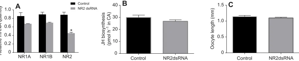
To investigate the role of NMDAR in reproduction,DpNR2was silenced using RNAi. The injection of 2 μg ofDpNR2dsRNA on days 0, 1, 2 and 3 resulted in a 48.7% knockdown ofDpNR2mRNA levels in brains of day 4 females (Fig. 6A). Knockdown ofDpNR2
also resulted in a decrease in the transcript levels ofDpNR1Aand
DpNR1B. However, JH biosynthetic activity in the CA and basal
oocyte length in dsRNA-treated animals did not show any significant difference from control animals (Fig. 6B,C). A similar result was also observed in animals treated withDpNR1BdsRNA (supplementary material Fig. S1).
In vitroeffect of the NMDAR antagonist MK-801 on JH biosynthesis
A previous study showed a dose-dependent decrease in JH biosynthesisin vitroinG. bimaculatusCA as a function of MK-801 concentration (Geister et al., 2008). Thus, we determined thein
vitroeffect of MK-801 on JH biosynthesis. Surprisingly, the rates of
JH biosynthesis remained at the same level irrespective of the
concentration of MK-801 (Fig. 7). MK-801 did not appear to have any significant effect on JH biosynthesisin vitrobyD. punctataCA.
In vivoeffect of NMDAR antagonist MK-801 on JH biosynthesis and ovarian development
The effect of NMDAR on JH biosynthesis and ovarian development was also determined by injection of the NMDAR non-competitive antagonist MK-801 into adult females. To determine the optimal dose, newly molted adult females were injected with 0 (control), 3, 12 and 30 μg MK-801 on days 0, 1 and 3. JH biosynthesis and basal oocyte length were measured on day 4. As shown in supplementary material Fig. S2, there was no significant effect on JH biosynthesis in animals treated with 3 μg or 12 μg MK-801.
To further study the effect of MK-801 on reproduction, mated female cockroaches were injected with 30 μg MK-801 on days 0, 1 and 3 following eclosion. Rates of JH biosynthesis and basal oocyte lengths in control and treated animals were determined from day 4 to day 8. As shown in Fig. 8A, application of MK-801 resulted in a 34% and 26% decrease of JH biosynthesis in day 4, and 5 animals, respectively. On day 6, however, MK-801 treated
Br NC CA Fb MG MT Ov Br Te AG
0 1 2 3
Br NC CA Fb MG MT Ov Br Te AG
0 0.5 1.0 1.5 2.0 2.5
Br NC CA Fb MG MT Ov Br Te AG
0 1 2 3 4 5
A
B
C
Female Male
Relative quantity of
NR1B
mRNA
Relative quantity of
NR2
mRNA
Relative quantity of
NR1A
[image:4.612.52.299.55.418.2]mRNA
Fig. 4. Graphic representation of the relative tissue distribution of NR1 and NR2 transcripts.(A)DpNR1Atranscript levels, (B)DpNR1Btranscript
levels and (C)DpNR2transcript levels in tissues of day 4 adult male and mated
femaleD. punctata. Relative mRNA quantity was normalized against levels of
TubulinandEF1αmRNA (Marchal et al., 2013b). The data represents an average of three pools (10 animals per pool), run in triplicate. Br, brain; NC, nerve cord; CA, corpora allata; Fb, fat body; Ov, ovary; MG, midgut; MT, Malpighian tubules; AG, accessory gland and Te, testes. Values represent mean±s.e.m.
0 1 2 3 4 5 6 7
0 5 10 15
NR1A NR1B NR2
A
0 1 2 3 4 5 6 7
0 0.1 0.2 0.3 0.4 0.5
B
Relative quantity of mRNA
0 2 4 6 8 10 15 25
0 1 2 3
4
C
Age (days after ecdysis)
Fig. 5. Relative transcript levels ofDpNR1A,DpNR1BandDpNR2. Diploptera punctatamRNA levels in (A) brains and (B) CA of mated females from day 0 to day 7 after ecdysis and (C) testes of different ages
of males normalized against levels ofTubulinandEF1amRNA (Marchal
et al., 2013b). The data represent the average of three biologically independent pools (10 animals per pool), run in triplicate. Values represent mean±s.e.m.
The
Journal
of
Experimental
[image:4.612.322.560.58.431.2]animals showed higher rates of JH biosynthesis than control animals. On days 7 and 8, there was no significant difference in JH biosynthesis between control and MK-801-treated animals. Furthermore, MK-801 did not have any effect on basal oocyte length (Fig. 8B). All animals oviposited on day 8. To study the effect of MK-801 on vitellogenin (Vg) synthesis, we determined the transcript level ofDpVgin the fat body of day 4 animals. No significant change was found in the transcription ofDpVgin MK-801-treated animals (Fig. 8C).
DISCUSSION
We have identified two distinctNR1genes and 11NR2variants in
D. punctata. The major functional domains appear to be well
conserved in bothDpNR1andDpNR2amino acid sequences (Figs 1 and 3). The protein contains three hydrophobic transmembrane regions (TM1, 3–4), a hydrophobic pore-forming segment (TM2), and two ligand binding domains (S1 and S2) (Figs 1 and 3) (Dingledine et al., 1999; Kuryatov et al., 1994; Stern-Bach et al., 1994). The asparagine residue, which was predicted to control Ca2+
permeability and voltage-dependent Mg2+blockade, is present in
[image:5.612.57.559.59.163.2]the TM2 domain of DpNR1 (N630 in DpNR1A and N608 in DpNR1B) (Burnashev et al., 1992). The overall amino acid sequence identity between DpNR1, DpNR2 and NMDAR subunits in other species is shown in supplementary material Table S1. DpNR1A has higher sequence identity to NR1 in other species than DpNR1B.Both DpNR1 and DpNR2 subunits show highest homology to NMDAR ofA. mellifera.
The two NR1 subunits are 65.6% identical at the amino acid level and the differences are distributed throughout the entire gene, which suggests that the twoNR1 paralogs result from gene duplication (Fig. 1). The otherNR1paralogs were isolated from the Zebrafish (Cox et al., 2005); they encode the same length of protein with only a few amino acid differences. No NR1 paralogs other than in Zebrafish have been identified in vertebrate or invertebrate species. Phylogenetic analysis of DpNR1 with NR1s from other insect species shows that the two DpNR1s are grouped, which suggests that these paralogs result from a gene duplication event in D.
punctatathat did not occur in other insects. However, given that our
dataset was composed primarily of higher insects, and the small number of insect species in which NR1 has been identified, it is difficult to make definitive conclusions about the origin of the paralogs. DpNR2 undergoes alternative splicing, generating 11 different transcripts that may encode nine protein isoforms (Fig. 3). Unlike NR2 in Drosophila in which alternative splicing occurs mainly at the 5′untranslated region,DpNR2undergoes alternative splicing principally at the 3′-end (Xia et al., 2005).
Tissue distribution shows that bothDpNR1andDpNR2mRNA accumulated in brain and nerve cord, which is consistent with the role of NMDAR in learning and memory (Xia et al., 2005). The NR1 subunit is essential for the basic channel activity of NMDAR, whereas the NR2 subunit is regarded as the rate-limiting molecule in controlling the optimal channel properties of NMDAR (Monyer et al., 1992; Sprengel et al., 1998; Tang et al., 1999). InD. punctata, we observed that bothDpNR1paralogs are stably expressed in brain, whereas the relative DpNR2 mRNA level underwent a slight change, probably to regulate the activity of NMDAR (Fig. 5A). In rats, D-aspartic acid (D-Asp) can induce testosterone synthesis
through the activation of NMDAR in testis (Santillo et al., 2014). A high transcript level ofDpNR1was observed in adultD. punctata
testes, indicating that NMDAR may also play a role in regulation of reproduction in our model insect (Fig. 5B). However, the transcript level of DpNR2 in testes was negligible compared with that of
DpNR1, which suggests that NR1 may form a homomeric
functional channel, as observed in an earlier study in which expression of Drosophila NR1 alone produced a weak but significant NMDA response (Ultsch et al., 1993; Xia et al., 2005). An alternative explanation for the low expression ofDpNR2in the testes could be the existence of one or more additional DpNR2
subunits inD. punctata. Four distinct NR2 subunits (A–D) were identified in mammalian species. All four NR2 subunits are expressed in brain (Cull-Candy et al., 2001). In testis, however, expression of the NR2 subunits varies (Hu et al., 2004; Santillo et al., 2014). In the rat testis, onlyNR2Aand NR2Dare strongly
NR1A NR1B NR2
0 0.2 0.4 0.6 0.8 1.0
* Control NR2 dsRNA
Control NR2dsRNA Control NR2dsRNA
0 10 20 30 40
0 0.5 1.0 1.5
A
B
C
Relative mRNA
quantity
Oocyte length (mm)
JH biosynthesis (pmol h
–
1 in CA)
Fig. 6. The effect ofDpNR2dsRNA treatment on JH biosynthesis and basal oocyte growth, and the interactions among these genes in mated femaleD. punctata.(A) Relative quantity ofDpNR1A,DpNR1BandDpNR2mRNA levels in brain between control and dsRNA-treated animals. mRNA levels were
normalized against levels ofTubulinandEF1amRNA (Marchal et al., 2013b). The data represent the average of three biologically independent pools (five animals
each pool) run in triplicate. (B) JH biosynthesis by CA from control and dsRNA-treated animals. (C) Basal oocyte length in control and dsRNA-treated animals.
Values represent means±s.e.m. *P<0.05 compared with control.
Control –8 –7 –6 –5 –4
0 10 20 30 40 50
log[MK-801]
JH biosynthesis (pmol h
–
1 in CA)
Fig. 7.In vitroeffect of MK-801 on JH biosynthesis in CA.CA were dissected from day 4 mated females. The JH biosynthesis in normal medium without MK-801 was determined as control. Each data point represents mean±
s.e.m. (N=12).
The
Journal
of
Experimental
[image:5.612.63.289.542.695.2]expressed, whereas NR2B and NR2C are undetectable (Santillo et al., 2014). In mouse testis, by contrast, only theNR2Bsubunit is highly expressed (Hu et al., 2004). Thus, it is possible that an additionalNR2gene is expressed in the testis ofD. punctata.
The function of NMDAR in reproduction is well studied in mammals (Mahesh and Brann, 2005). However, clear evidence linking NMDAR to reproduction in insects remains limited. The possible role of NMDAR in reproduction of insects was described by Chiang et al. (2002). In that study, NMDA stimulates JH biosynthesis by inducing a Ca2+-influx from the extracellular
environment into the CA ofD. punctata; this stimulation could be significantly reduced by MK-801 treatment (Chiang et al., 2002). This experiment was performed in a minimum incubation medium (150 mmol l−1 NaCl, 12 mmol l−1 KCl, 4 mmol l−1 HEPES,
100 nmol l−1dopamine, 5 μmol l−1glycine, 2% Ficoll). However,
following incubation of CA in TC199 medium, which better mimics the cockroach hemolymph environment, we found that MK-801 did not show any significant effect on JH biosynthesis (Fig. 7). Chiang et al. (2002) also reported that the stimulation by NMDA of JH biosynthesis is age dependent, which is suggested to be controlled by the expression of NMDAR. However, our study has clearly demonstrated that the transcript levels ofDpNR1andDpNR2in the CA are very low throughout the first gonadotrophic cycle and did not exhibit any significant changes. Those results suggest that although NMDA is able to stimulate JH biosynthesis in vitro, NMDAR may not function as a regulator of JH biosynthesis inD.
punctata. To answer this question, the role of NMDAR in JH
biosynthesis and reproduction was further examined by the administration of MK-801in vivoinD. punctata.
MK-801 did not have anin vivoeffect on JH biosynthesis at doses up to 12 μg per animal (about 60 μg g−1body mass) (supplementary
material Fig. S2). InD. punctata, the production of vitellogenin in fat body and the uptake of vitellogenin by the basal oocytes are JH-dependent events (Marchal et al., 2013a; Rankin and Stay, 1984; Stay and Tobe, 1978). Although a higher dose of MK-801 resulted in a significant decrease in JH biosynthesis on days 4 and 5, the change in transcript level ofDpVgwas not significant relative to controls (Fig. 8C), and the pattern of JH biosynthesis during the first gonadotrophic cycle is similar to that in control animals (Fig. 8A). Oocyte growth in treated animals is also similar to the controls (Fig. 8B). All females oviposited on day 8. Overall, administration of MK-801 did not have a significant effect on reproduction in femaleD. punctata. This is consistent with the MK-801 results. In
G. bimaculatus, in which MK-801 treatmentin vitroresulted in a
dose-dependent inhibition of JH biosynthesis by the CA, egg size and egg number were significantly affected by the application of
MK-801 (Geister et al., 2008). Thus, the regulatory role of NMDAR on JH biosynthesis appears to be species specific.
It is interesting that treatment of animals with a high dose of
MK-801in vivosignificantly reduced JH biosynthesis on days 4 and 5,
even though MK-801 did not show any significant effect on JH biosynthesisin vitro. Partial knockdown ofDpNR2did not show any effect on JH biosynthesis or on basal oocyte growth (Fig. 6). How does MK-801 change the rate of JH biosynthesis whereas NMDAR does not appear to have an in vivoeffect on JH biosynthesis or reproduction? To date, there is no clear evidence showing that the effect of MK-801 on JH biosynthesis is mediated through NMDAR. In rats, injection of 0.2 μg g−1MK-801 resulted in a failure to elevate
LH, FSH or progesterone (Luderer et al., 1993). In rats and mice, a dose of 0.1 μg g−1MK-801 was the maximum dose that could be used
without causing sensorimotor impairments and/or signs of intoxication (Van der Staay et al., 2011). Injection of 30 μg of MK-801 paralyzed the cockroaches for 4–5 h. Therefore, it is possible that the significant decrease in JH biosynthesis on days 4 and 5 inD.
punctatawas the result of physiological stress induced by MK-801
treatment, rather than the action of NMDAR.
In conclusion, two NR1 paralogs and 11 NR2 alternative splicing variants have been identified in D. punctata. The expression of NMDAR subunits suggests that NMDAR may play a role in the reproduction of male cockroaches. However, although a previous study suggested that NMDAR mediates JH biosynthesis in D.
punctata in vitro, our data in the female cockroach reveal a different
story. Neitherin vitrotreatment of MK-801 nor partial knockdown of
DpNR2 has any effect on JH biosynthesis. The decrease in JH
biosynthesis at a high dose in MK-801-treated animals appears to result from physiological stress, rather than a direct action on NMDAR. In addition, no reproductive events were affected following the blocking of NMDAR activity through RNAi or MK-801 treatment. A re-examination of the function of NMDA receptors in the reproduction of insects now appears to be appropriate and timely.
MATERIALS AND METHODS Insects
Diploptera punctataEschscholtz 1822 were reared in cages and fed with lab
chow and waterad libitumat 27–28°C in a dark room. Newly molted male and female adult cockroaches were picked from the colony and raised in separate containers. Mated status in the females was confirmed by the presence of a spermatophore.
Tissue collection
Cockroach tissues were dissected under a dissecting microscope. Basal oocyte length was measured to determine the physiological age of female cockroaches. Selected tissues were dissected and cleaned in
4 5 6 7 8 4 5 6 7
0 10 20 30 40 50 60 70
Age (days after ecdysis)
Control MK-801
*** **
*
1.2 1.4 1.6 1.8 2.0 2.2
Control MK-801 0
0.5 1.0 1.5
A
B
C
JH biosynthesis (pmol h
–
1 in CA)
Basal oocyte length (mm)
[image:6.612.72.543.57.166.2]Relative quantity of vitellogenin mRNA
Fig. 8.In vivoeffect of MK-801 on JH biosynthesis, basal oocyte growth and relative vitellogenin mRNA levels.Females were injected with MK-801
on days 0, 1 and 3 following ecdysis (30 µg/animal). Control was injected with ddH2O. JH biosynthesis (A) and oocyte length (B) were determined on day
4 to day 8. All animals oviposited on day 8. The mRNA level ofDpVgwas determined in the fat body of day 4 animals. (C) mRNA levels were normalized to levels
ofTubulinandEF1amRNA (Marchal et al., 2013b). The data represent the average of three biologically independent pools (five animals each pool), run in
triplicate. Values represent means±s.e.m. (A)N≥15; (B)N≥10. *P<0.05, **P<0.01, ***P<0.001.
The
Journal
of
Experimental
sterile cockroach ringer solution (150 mmol l−1NaCl, 12 mmol l−1KCl, 10 mmol l−1CaCl2·2H2O, 3 mmol l−1MgCl2·6H2O, 10 mmol l−1HEPES,
40 mmol l−1Glucose, pH 7.2), flash-frozen in liquid nitrogen to prevent RNA degradation and stored at−80°C until further processing.
RNA extraction and cDNA synthesis
Selected tissues were collected from adult females and males for gene sequencing, tissue distribution and developmental profiling. RNA extraction and cDNA synthesis were performed as described by Marchal et al. (2013b). For the tissue distribution and developmental profiling, three biologically independent pools of 10 animals each were collected. For RNAi and MK-801 treatment experiments, three biologically independent pools of brain and fat body were collected, each pool containing tissue from five animals.
Sequencing ofDpNR1A,DpNR1BandDpNR2
Degenerate primer sequences were designed forDpNR1A,DpNR1Band
DpNR2 based on conserved amino acid sequences of several insect
orthologs. Primers used for degenerate PCR are listed in supplementary material Table S2. Partial sequences were obtained using these primers in a standard T-gradient PCR using Taq DNA polymerase (Sigma-Aldrich) and
aD. punctatabrain cDNA sample. After purification, the resulting DNA
fragments were subcloned into a pJET 1.2/blunt cloning vector (CloneJet PCR Cloning Kit, Thermo Scientific) and sequenced following the protocol outlined in the ABI PRISM BigDye Terminator Ready Reaction Cycle Sequencing Kit (Applied Biosystems). The complete sequence ofDpNR1A,
DpNR1BandDpNR2was obtained using 5′-RACE (Rapid Amplification of
cDNA Ends) and 3′-RACE strategies, following the protocol outlined in the Roche 5′/3′RACE Kit. Primers used for RACE are listed in supplementary material Table S2.
Phylogenetic analysis
For the phylogenetic analysis ofDpNR1genes, the NR1 sequences of 15 insect species (identified or predicted) were used. These sequences were aligned using ClustalW as implemented within MEGA 6.06 (Tamura et al., 2013). Poorly aligned positions and gaps were removed, which resulted in 854 amino acid residues. The obtained alignment was used to construct a phylogenetic tree in PhyML 3.0 (Guindon and Gascuel, 2003) based on the maximum-likelihood principle, using the WAG substitution model (Whelan and Goldman, 2001). Four substitution rate categories were used to estimate the gamma parameter shape with 100 bootstrap replicates to assess branch support (Felsenstein, 1985; Yang, 1994). The resulting tree was then rooted using the sea slug,Aplysia californicaas outgroup.
Quantitative Real Time-PCR
Primers used for q-RT-PCR are shown in supplementary material Table S3. Primer sets were validated by determining relative standard curves for each gene transcript using a five-fold serial dilution of a calibrator cDNA sample. Efficiency and correlation coefficient (R²) can be found in supplementary material Table S3. Reactions were performed in triplicate on a CFX384 Touch™ Real-Time PCR Detection System (Bio-Rad) as described previously by Marchal et al. (2013b). Target specificity was confirmed by running a few representative q-RT-PCR products on an agarose gel containing GelRed (Biotium). The optimal housekeeping genes for target gene profiling and RNAi experiments were chosen according to a previous study (Marchal et al., 2013b). The quantity of mRNA for each tested gene relative to reference genes was determined as described by Vandesompele et al. (2002).
RNA interference
Primers forDpNR2and controlpJETdsRNA constructs with T7 promoters are shown in supplementary material Table S4. Double-stranded RNA (dsRNA) constructs were prepared using the MEGAscript RNAi Kit (Ambion). A PCR using forward and reverse primers with attached T7 promoters was performed to amplify the fragment, which was subcloned and sequenced to verify the presence of the T7 promoter. The amplified fragment was used in an RNA transcription reaction which was incubated
overnight to obtain a high yield of annealed dsRNA construct. A nuclease digestion was subsequently performed to remove ssRNA and DNA remaining in the product. The dsRNA was further purified according to the manufacturer’s instructions (Ambion). Concentration of the dsRNA construct was determined using a Nanodrop instrument (Thermo Fisher Scientific Inc., Canada). Five-fold diluted dsRNA was run on a 1.2% agarose gel to examine the quality and integrity of the construct.
Newly molted adult female cockroaches (day 0) were injected with 2 μg of eitherDpNR2dsRNA orpJETdsRNA diluted in 5 μl of cockroach saline. This treatment was repeated on days 1, 2 and 3, and the effect ofDpNR2
dsRNA was determined on day 4. Brains were dissected and stored in liquid nitrogen prior to RNA extraction. CA were dissected and cleaned in TC199 medium (GIBCO; 1.3 mmol l−1Ca2+, 2% Ficoll, methionine-free) for use in the radiochemical assay (RCA) (see below). Basal oocyte length was measured during dissection.
MK-801in vitroassay
To determine thein vitroeffect of MK-801 on JH biosynthesis, CA were incubated in TC199 medium (M 7653 (Sigma), supplemented with CaCl2to
a final concentration of 1.3 mmol l−1 and fortified with 2% Ficoll)
containing different concentrations of MK-801; JH production was examined using a radiochemical assay (RCA). The concentrations of MK-801 ranged from 10−4mol l−1to 10−8mol l−1. JH biosynthesis in medium
without MK-801 was used as a control. RCA was performed as described by Feyereisen and Tobe (1981) and modified by Tobe and Clarke (1985).
MK-801in vivoassay
MK-801 was dissolved in ddH2O to a concentration of 6 μg μl−1 and
injected into animals using a Hamilton syringe. Newly molted adult females were injected with 30 μg of MK-801 on days 0, 1 and 3, and the effect of MK-801 was determined from day 4 to day 8. Fat body was dissected from day 4 animals and stored in liquid nitrogen prior to RNA extraction. CA were dissected and cleaned in TC199 medium (GIBCO; 1.3 mmol l−1Ca2+, 2%
Ficoll, methionine-free) for use in the RCA (see below). Basal oocyte length was also measured.
Acknowledgements
The authors are very grateful to Dr Jennifer Mitchell for use of the q-RT-PCR equipment.
Competing interests
The authors declare no competing or financial interests.
Author contributions
J.H. and S.S.T. conceived and designed the experiments. J.H., E.F.H. and E.M. performed the experiments. J.H., E.F.H. and E.M. analyzed the data. J.H. and S.S.T. interpreted the findings being published. J.H. wrote the paper. E.F.H., E.M. and S.S.T. revised the paper. S.S.T. was the Principal Investigator, responsible for the management of the projects that provided financial support. All authors read and approved the final manuscript.
Funding
This work was supported by a Discovery grant, no. 9408-08 from the Natural Sciences and Engineering Research Council of Canada to S.S.T, and the China Scholarship Council doctoral award (J.H.).
Supplementary material
Supplementary material available online at
http://jeb.biologists.org/lookup/suppl/doi:10.1242/jeb.115154/-/DC1
References
Begum, M., Breuer, M., Kodrik, D., Rahman, M. M. and De Loof, A.(2004). The
NMDA receptor antagonist MK-801 inhibits vitellogenesis in the flesh fly
Neobellieria bullataand in the desert locustSchistocerca gregaria.J. Insect
Physiol.50, 927-934.
Burnashev, N., Schoepfer, R., Monyer, H., Ruppersberg, J., Gunther, W., Seeburg,
P. and Sakmann, B.(1992). Control by asparagine residues of calcium permeability
and magnesium blockade in the NMDA receptor.Science257, 1415-1419. Chiang, A.-S., Lin, W.-Y., Liu, H.-P., Pszczolkowski, M. A., Fu, T.-F., Chiu, S.-L.
and Holbrook, G. L.(2002). Insect NMDA receptors mediate juvenile hormone
biosynthesis.Proc. Natl. Acad. Sci. USA99, 37-42.
The
Journal
of
Experimental
Cox, J. A., Kucenas, S. and Voigt, M. M.(2005). Molecular characterization and embryonic expression of the family of N-methyl-D-aspartate receptor subunit genes in the zebrafish.Dev. Dyn.234, 756-766.
Cull-Candy, S., Brickley, S. and Farrant, M.(2001). NMDA receptor subunits:
diversity, development and disease.Curr. Opin. Neurobiol.11, 327-335.
Dingledine, R., Borges, K., Bowie, D. and Traynelis, S. F.(1999). The glutamate
receptor ion channels.Pharmacol. Rev.51, 7-61.
Felsenstein, J.(1985). Confidence limits on phylogenies: an approach using the
bootstrap.Evolution39, 783-791.
Feyereisen, R. and Tobe, S. S.(1981). A rapid partition assay for routine analysis of
juvenile hormone release by insect corpora allata.Anal. Biochem.111, 372-375.
Geister, T. L., Lorenz, M. W., Hoffmann, K. H. and Fischer, K.(2008). Effects of
the NMDA receptor antagonist MK-801 on female reproduction and juvenile hormone biosynthesis in the cricket Gryllus bimaculatus and the butterfly Bicyclus anynana.J. Exp. Biol.211, 1587-1593.
Goodman, W. G. and Granger, N. A. (2005). The juvenile hormones. In
Comprehensive Molecular Insect Science(ed. L. I. Gilbert, K. Iatrou and S. S.
Gill), pp. 319-408. Oxford: Elsevier.
Granger, N. A., Sturgis, S. L., Ebersohl, R., Geng, C. and Sparks, T. C.(1996).
Dopaminergic control of corpora allata activity in the larval tobacco hornworm,
Manduca sexta.Arch. Insect Biochem. Physiol.32, 449-466.
Guindon, S. and Gascuel, O.(2003). A simple, fast, and accurate algorithm to
estimate large phylogenies by maximum likelihood.Syst. Biol.52, 696-704.
Hartfelder, K.(2000). Insect juvenile hormone: from“status quo”to high society.
Braz. J. Med. Biol. Res.33, 157-177.
Hu, J. H., Yang, N., Ma, Y. H., Jiang, J., Zhang, J. F., Fei, J. and Guo, L. H.(2004).
Identification of glutamate transporters and receptors in mouse testis. Acta
Pharmacol. Sin.25, 366-371.
Huang, J., Tian, L., Peng, C., Abdou, M., Wen, D., Wang, Y., Li, S. and Wang, J. (2011). DPP-mediated TGFbeta signaling regulates juvenile hormone biosynthesis by activating the expression of juvenile hormone acid methyltransferase.Development138, 2283-2291.
Kuryatov, A., Laube, B., Betz, H. and Kuhse, J.(1994). Mutational analysis of the
glycine-binding site of the NMDA receptor: structural similarity with bacterial amino acid-binding proteins.Neuron12, 1291-1300.
Luderer, U., Strobl, F. J., Levine, J. E. and Schwartz, N. B.(1993). Differential
gonadotropin responses to N-methyl-D,L-aspartate in metestrous, proestrous, and ovariectomized rats.Biol. Reprod.48, 857-866.
Madden, D. R. (2002). The structure and function of glutamate receptor ion
channels.Nat. Rev. Neurosci.3, 91-101.
Maffucci, J. A., Noel, M. L., Gillette, R., Wu, D. and Gore, A. C.(2009). Age- and
hormone-regulation of N-methyl-D-aspartate receptor subunit NR2b in the anteroventral periventricular nucleus of the female rat: implications for reproductive senescence.J. Neuroendocrinol.21, 506-517.
Mahesh, V. B. and Brann, D. W.(2005). Regulatory role of excitatory amino acids in
reproduction.Endocrine28, 271-280.
Marchal, E., Hult, E. F., Huang, J., Stay, B. and Tobe, S. S.(2013a).Diploptera
punctataas a model for studying the endocrinology of arthropod reproduction and
development.Gen. Comp. Endocr.188, 85-93.
Marchal, E., Hult, E. F., Huang, J. and Tobe, S. S.(2013b). Sequencing and
validation of housekeeping genes for quantitative real-time PCR during the gonadotrophic cycle ofDiploptera punctata.BMC Res. Notes6, 237.
McBain, C. J. and Mayer, M. L.(1994). N-methyl-D-aspartic acid receptor structure
and function.Physiol. Rev.74, 723-760.
Monyer, H., Sprengel, R., Schoepfer, R., Herb, A., Higuchi, M., Lomeli, H.,
Burnashev, N., Sakmann, B. and Seeburg, P. H.(1992). Heteromeric NMDA
receptors: molecular and functional distinction of subtypes. Science 256, 1217-1221.
Mussig, L., Richlitzki, A., Rossler, R., Eisenhardt, D., Menzel, R. and Leboulle, G. (2010). Acute disruption of the NMDA receptor subunit NR1 in the honeybee brain selectively impairs memory formation.J. Neurosci.30, 7817-7825.
Newcomer, J. W. and Krystal, J. H.(2001). NMDA receptor regulation of memory
and behavior in humans.Hippocampus11, 529-542.
Pszczolkowski, M. A., Lee, W.-S., Liu, H.-P. and Chiang, A.-S.(1999).
Glutamate-induced rise in cytosolic calcium concentration stimulates in vitro rates of juvenile hormone biosynthesis in corpus allatum of Diploptera punctata. Mol. Cell.
Endocrinol.158, 163-171.
Rankin, S. M. and Stay, B.(1984). The changing effect of the ovary on rates of
juvenile hormone synthesis inDiploptera punctata.Gen. Comp. Endocrinol.54, 382-388.
Rawls, S. M., Thomas, T., Adeola, M., Patil, T., Raymondi, N., Poles, A., Loo, M.
and Raffa, R. B.(2009). Topiramate antagonizes NMDA- and AMPA-induced
seizure-like activity in planarians.Pharmacol. Biochem. Behav.93, 363-367. Santillo, A., Falvo, S., Chieffi, P., Burrone, L., Chieffi Baccari, G., Longobardi, S.
and Di Fiore, M. M.(2014). D-aspartate affects NMDA receptor-extracellular
signal-regulated kinase pathway and upregulates androgen receptor expression in the rat testis.Theriogenology81, 744-751.
Sircar, R., Rappaport, M., Nichtenhauser, R. and Zukin, S. R.(1987). The novel
anticonvulsant Mk-801: a potent and specific ligand of the brain phencyclidine sigma-receptor.Brain Res.435, 235-240.
Sprengel, R., Suchanek, B., Amico, C., Brusa, R., Burnashev, N., Rozov, A.,
Hvalby, O., Jensen, V., Paulsen, O., Andersen, P. et al.(1998). Importance of
the intracellular domain of NR2 subunits for NMDA receptor functionin vivo.Cell
92, 279-289.
Stay, B. and Tobe, S. S.(1978). Control of juvenile hormone biosynthesis during the
reproductive cycle of a viviparous cockroach. II. Effects of unilateral allatectomy, implantation of supernumerary corpora allata, and ovariectomy.Gen. Comp.
Endocrinol.34, 276-286.
Stay, B. and Tobe, S. S.(2007). The role of allatostatins in juvenile hormone
synthesis in insects and crustaceans.Annu. Rev. Entomol.52, 277-299.
Stern-Bach, Y., Bettler, B., Hartley, M., Sheppard, P. O., O’Hara, P. J. and
Heinemann, S. F.(1994). Agonist selectivity of glutamate receptors is specified
by two domains structurally related to bacterial amino acid-binding proteins.
Neuron13, 1345-1357.
Sydow, S., Köpke, A. K. E., Blank, T. and Spiess, J.(1996). Overexpression of a
functional NMDA receptor subunit (NMDAR1) in baculovirus-infected
Trichoplusia niinsect cells.Mol. Brain Res.41, 228-240.
Tamura, K., Stecher, G., Peterson, D., Filipski, A. and Kumar, S.(2013). MEGA6:
Molecular Evolutionary Genetics Analysis version 6.0. Mol. Biol. Evol. 30, 2725-2729.
Tang, Y. P., Shimizu, E., Dube, G. R., Rampon, C., Kerchner, G. A., Zhuo, M., Liu, G.
and Tsien, J. Z.(1999). Genetic enhancement of learning and memory in mice.
Nature401, 63-69.
Thompson, C. S., Yagi, K. J., Chen, Z. F. and Tobe, S. S.(1990). The effects of
octopamine on juvenile hormone biosynthesis, electrophysiology, and cAMP content of the corpora allata of the cockroachDiploptera punctata.J. Comp.
Physiol. B160, 241-249.
Tobe, S. S. and Clarke, N.(1985). The effect of L-Methionine concentration on
juvenile hormone biosynthesis by corpora allata of the cockroachDiploptera
punctata.Insect Biochem.15, 175-179.
Troncoso, J. and Maldonado, H.(2002). Two related forms of memory in the crab
Chasmagnathus are differentially affected by NMDA receptor antagonists.
Pharmacol. Biochem. Behav.72, 251-265.
Ultsch, A., Schuster, C. M., Laube, B., Betz, H. and Schmitt, B.(1993). Glutamate
receptors ofDrosophila melanogaster. Primary structure of a putative NMDA receptor protein expressed in the head of the adult fly.FEBS Lett.324, 171-177.
Van der Staay, F. J., Rutten, K., Erb, C. and Blokland, A.(2011). Effects of the
cognition impairer MK-801 on learning and memory in mice and rats.Behav. Brain Res.220, 215-229.
Vandesompele, J., De Preter, K., Pattyn, F., Poppe, B., Van Roy, N., De Paepe,
A. and Speleman, F.(2002). Accurate normalization of real-time quantitative
RT-PCR data by geometric averaging of multiple internal control genes.Genome Biol.
3, 1-12.
Whelan, S. and Goldman, N.(2001). A general empirical model of protein evolution
derived from multiple protein families using a maximum-likelihood approach.Mol.
Biol. Evol.18, 691-699.
Wu, C.-L., Xia, S., Fu, T.-F., Wang, H., Chen, Y.-H., Leong, D., Chiang, A.-S. and
Tully, T.(2007). Specific requirement of NMDA receptors for long-term memory
consolidation inDrosophilaellipsoid body.Nat. Neurosci.10, 1578-1586. Xia, S., Miyashita, T., Fu, T.-F., Lin, W.-Y., Wu, C.-L., Pyzocha, L., Lin, I.-R.,
Saitoe, M., Tully, T. and Chiang, A.-S.(2005). NMDA receptors mediate
olfactory learning and memory inDrosophila.Curr. Biol.15, 603-615.
Yang, Z.(1994). Maximum likelihood phylogenetic estimation from DNA sequences
with variable rates over sites: approximate methods.J. Mol. Evol.39, 306-314.
Zannat, M. T., Locatelli, F., Rybak, J., Menzel, R. and Leboulle, G.(2006).
Identification and localisation of the NR1 sub-unit homologue of the NMDA glutamate receptor in the honeybee brain.Neurosci. Lett.398, 274-279.