Acta Crystallographica Section E Structure Reports
Online
ISSN 1600-5368
Dinicotinium sulfate
S. Athimoolam and R. K. Rajaram*
Department of Physics, Madurai Kamaraj University, Madurai 625 021, India
Correspondence e-mail: [email protected]
Key indicators
Single-crystal X-ray study T= 293 K
Mean(C–C) = 0.008 A˚ Rfactor = 0.062 wRfactor = 0.205
Data-to-parameter ratio = 11.4
For details of how these key indicators were automatically derived from the article, see http://journals.iucr.org/e.
#2005 International Union of Crystallography
Printed in Great Britain – all rights reserved
In the title complex, 2C6H6NO2 +
SO4 2
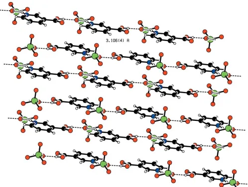
, the carboxyl groups of the two nicotinium cations (AandB) are twisted from the pyridinium ring, with dihedral angles of 9.1 (5) and 7.0 (9), respectively. Packing involves classical O—H O and N— H O hydrogen bonds. CationsAare interlinked through an O atom of the sulfate anion, forming an infinite chain running along thebaxis and leading to cationic layers separated by a distance of 3.106 (4) A˚ . Cations Bform an inversion-related closed hydrogen-bonded loop.
Comment
Hypercholesterolemia is a relevant risk factor with regard to the development of atherosclerotic diseases. Nicotinic acid, a B vitamin, also known as niacin, is a lipid-lowering agent widely used to treat hypertriglyceridemia by the inhibition of lipolysis in adipose tissue. The nicotinic acid complex 5-methylpyrazine-2-carboxylic acid 4-oxide is a commonly used drug for hypercholesterolemia (Lorenzenet al., 2001). Coor-dination complexes of nicotinic acid with metals such as Sn possess antitumour activity greater than the well known cis -platin or doxorubicin (Gielen et al., 1992). Also, the enzyme nicotinic acid mononucleotide adenylyltransferase is essential for the synthesis of nicotinamide adenine dinucleotide in all living cells and is a potential target for antibiotics (Kimet al., 2004). Because of their pharmacological importance, nicotinic acid and related compounds are the object of extensive study. The crystal structures of nicotinic acid (Wright & King, 1953; Kutoglu & Scheringer, 1983), dinicotinic acid (Takusagawaet al., 1973), isonicotinic acid (Takusagawa & Shimada, 1976), isonicotinic acid hydrazide (Bhat et al., 1974), nicotinium tetrachlorocuprate(II) (Choi et al., 2002), 2-aminonicotinic acid (Dobson & Gerkin, 1997), 6-aminonicotinic acid hydro-chloride (Giantsidis & Turnbull, 2000), nicotinic acid hydro-chloride hydrochloride (Na¨ttinen & Rissanen, 2003), 3,5-dinitro-benzoic acid nicotinic acid (Zhu & Zheng, 2004), 2-(methyl-sulfanyl)nicotinic acid (Basavoju et al., 2005), nicotinamide (Wright & King, 1954), 1-methylnicotinamide iodide, chloride and picrate (Freeman & Bugg, 1974), and dinicotinamidium squarate (Bulut et al., 2003) have been reported previously. The crystal structure of nicotinic acid complexed with the protein leghaemoglobin (Ellis et al., 1997) and the haem– nicotinate interaction in leghaemoglobin (Patel et al., 2000) were also studied. As part of our investigations of nicotinic acid complexes with inorganic acids, nicotinic acid was treated with sulfuric acid, and the crystal structure of the resulting salt is reported here.
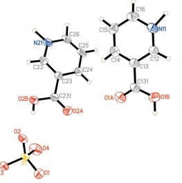
The title compound, (I), contains two nicotinium cations (A andB) and a sulfate anion in the asymmetric unit (Fig. 1). The protons from the sulfuric acid are transferred to the nicotinic acid, forming two nicotinium cations. The least-squares plane through cation A makes an angle of 50.8 (1) with that for cationB. CationsAandBare stacked close to thez= 0 andz =1
2planes, respectively. The C—N—C bond angle and the C—
N bond distances in both cations confirm protonation on the N atom of the aromatic rings. The pyridinium rings are essen-tially planar, with a maximum deviation of 0.007 (3) A˚ for N11 in cationAand 0.018 (4) A˚ for C23 in cationB. As found in other nicotinic acid–inorganic acid complexes, the carboxyl group is twisted from the pyridinium ring plane, with dihedral angles of 9.1 (5) and 7.0 (9)for cationsAandB, respectively. Protonation of the O atoms of the carboxyl groups is unam-biguously confirmed from the C—O bond distances and C— C—O bond angles. Deviation of the carbonyl atom O1A [0.191 (9) A˚ ] from the pyridinium plane is slightly greater than that of O1B[0.157 (9) A˚ ] in cationA. This is reversed in cation B, with O2B [0.235 (9) A˚ ] deviating further from the ring
plane than O2A [0.149 (9) A˚ ], as found in 2-aminonicotinic acid (Dobson & Gerkin, 1997) (Table 1). The SO4
2 anion shows nearly tetrahedral symmetry. The crystal is stabilized by extensive hydrogen bonding through three O atoms of the sulfate anions (Fig. 2). TheBcations are interlinked through N—H O and O—H O hydrogen bonds to the anions, forming an inversion-related closed hydrogen-bonded loop (Fig. 3). In residueA, the cations form infinite chains running along thebaxis by connecting to adjacent cations through the
organic papers
Acta Cryst.(2005). E61, o2764–o2767 Athimoolam and Rajaram 2C
[image:2.610.108.229.86.175.2]5H6NO2+SO42
o2765
Figure 2
The packing of the molecule, viewed down thebaxis, with hydrogen bonds drawn as dashed lines.
Figure 1
[image:2.610.317.563.198.473.2] [image:2.610.50.313.452.717.2]The asymmetric unit of the title compound, showing the atom-numbering scheme and 50% probability displacement ellipsoids.
Figure 3
[image:2.610.316.565.517.723.2]O1 atom of the sulfate anion, viz. N11—H11 O1 H1B— O1B(x, y + 1, z), leading to cationic layers separated by 3.106 (4) A˚ but not linked by any hydrogen bonding (Fig. 4 and Table 2).
Experimental
The title compound was crystallized from a solution of a mixture of nicotinic acid and sulfuric acid in a 2:1 stoichiometric ratio. Slow evaporation of the mixture at room temperature produced colourless block-like crystals, one of which was cut and used to collect the intensity data.
Crystal data
2C5H6NO2+SO42
Mr= 344.30
Triclinic,P1
a= 6.9663 (7) A˚
b= 8.3306 (14) A˚
c= 12.5098 (12) A˚
= 101.253 (11)
= 95.746 (8)
= 102.685 (11)
V= 686.85 (15) A˚3 Z= 2
Dx= 1.665 Mg m 3
Dm= 1.654 (8) Mg m
3 Dmmeasured by flotation using a
mixture of CCl4and CHBr3 MoKradiation
Cell parameters from 25 reflections
= 10.0–14.3
= 0.28 mm1 T= 293 (2) K Block, colourless 0.250.220.19 mm
Data collection
Nonius MACH3 four-circle diffractometer
!–2scans
Absorption correction: scan (Northet al., 1968)
Tmin= 0.898,Tmax= 0.947 3062 measured reflections 2414 independent reflections 1650 reflections withI> 2(I)
Rint= 0.020 max= 25.0
h=1!8
k=9!9
l=14!14 3 standard reflections
frequency: 60 min intensity decay: none
Refinement
Refinement onF2 R[F2> 2(F2)] = 0.062 wR(F2) = 0.205
S= 1.13 2409 reflections 211 parameters
H-atom parameters constrained
w= 1/[2
(Fo2) + (0.0826P)2 + 2.2093P]
whereP= (Fo2+ 2Fc2)/3 (/)max< 0.001
max= 0.53 e A˚
3 min=0.59 e A˚
3
Table 1
Selected geometric parameters (A˚ ,).
N11—C12 1.331 (7) N11—C16 1.342 (7) C131—O1A 1.204 (7) C131—O1B 1.311 (7)
N21—C22 1.338 (7) N21—C26 1.345 (7) C231—O2A 1.193 (6) C231—O2B 1.318 (6)
C12—N11—C16 122.9 (5) O1A—C131—O1B 124.9 (5)
C22—N21—C26 121.9 (5) O2A—C231—O2B 125.0 (5)
C14—C13—C131—O1A 9.6 (8) C14—C13—C131—O1B 171.9 (5)
C24—C23—C231—O2A 2.3 (8) C24—C23—C231—O2B 175.1 (5)
Table 2
Hydrogen-bond geometry (A˚ ,).
D—H A D—H H A D A D—H A
N11—H11 O1i
0.86 1.91 2.755 (6) 165 O1B—H1B O1ii
0.82 1.76 2.582 (6) 178 O2B—H2B O2 0.82 1.72 2.528 (5) 168 N21—H21 O3iii
0.86 1.80 2.641 (6) 167
Symmetry codes: (i) xþ1;yþ1;z; (ii) xþ1;y;z; (iii) xþ2;yþ1;zþ1.
Five strong reflections were omitted from the final refinement as they showedIo<<Ic. This may be due to primary extinction. All H
atoms were placed in geometrically calculated positions, with C—H = 0.93 A˚ , N—H = 0.86 A˚ and O—H = 0.82 A˚, and allowed to ride on their carrier atoms, withUiso(H) = 1.2Ueq(C) and 1.5Ueq(N,O).
Data collection: CAD-4 EXPRESS (Enraf–Nonius, 1994); cell refinement:CAD-4 EXPRESS; data reduction:XCAD4(Harms & Wocadlo, 1995); program(s) used to solve structure:SHELXTL/PC (Bruker, 2000); program(s) used to refine structure:SHELXTL/PC; molecular graphics: SHELXTL/PC and PLATON (Spek, 2003); software used to prepare material for publication:SHELXTL/PC.
The authors thank the Department of Science and Tech-nology, Government of India, for establishing a Single-Crystal Diffractometer facility at the School of Physics, Madurai Kamaraj University, Madurai, through the FIST programme.
References
Basavoju, S., Reddy, C. M. & Desiraju, G. R. (2005).Acta Cryst.E61, o822– o823.
Bhat, T. N., Singh, T. P. & Vijayan, M. (1974).Acta Cryst.B30, 2921–2922. Bruker (2000). SHELXTL/PC. Version 6.10. Bruker AXS Inc., Madison,
Wisconsin, USA.
Bulut, A., Yesilel, O. Z., Dege, N., Icbudak, H., Olmaz, H. & Bujukgungor, O. (2003).Acta Cryst.C59, o727–o729.
Choi, S. N., Lee, Y. M., Lee, H. W., Kangb, S. K. & Kim, Y. I. (2002).Acta Cryst.
E58, m583–m585.
Dobson, A. J. & Gerkin, R. E. (1997).Acta Cryst.C53, 1427–1429.
[image:3.610.315.564.70.258.2]Ellis, P. J., Appleby, C. A., Guss, J. M., Hunter, W. N., Ollis, D. L. & Freeman, H. C. (1997).Acta Cryst.D53, 302–310.
Figure 4
Enraf–Nonius (1994). CAD-4 EXPRESS. Version 5.1/1.2. Enraf–Nonius, Delft, The Netherlands.
Freeman, G. R. & Bugg, C. E. (1974).Acta Cryst.B30, 431–443. Giantsidis, J. & Turnbull, M. M. (2000).Acta Cryst.C56, 334–335.
Gielen, M., Khloufi, A. E., Biesemans, M. & Willem, R. (1992).Polyhedron,
11, 1861–1868.
Harms, K. & Wocadlo, S. (1995).XCAD4.University of Marburg. Germany. Kim, H. L., Yoon, H. J., Ha, J. Y., Lee, B. I., Lee, H. H., Mikami, B. & Suh, W. S.
(2004).Acta Cryst.D60, 948–949.
Kutoglu, A. & Scheringer, C. (1983).Acta Cryst.C39, 232–234.
Lorenzen, A., Stannek, C., Lang, H., Andrianov, V., Kalvinsh, I. & Schwabe, U. (2001).Mol. Pharmacol.59, 349–357.
Na¨ttinen, K. I. & Rissanen, K. (2003).CrystEngComm,5, 326–330. North, A. C. T., Phillips, D. C. & Mathews, F. S. (1968).Acta Cryst.A24, 351–
359.
Patel, N., Jones, D. K. & Raven, E. L. (2000).Eur. J. Biochem.267, 2581–2587. Spek, A. L. (2003).J. Appl. Cryst.36, 7–13.
Takusagawa, F., Hirotsu, K. & Shimada, A. (1973).Bull. Chem. Soc. Jpn,46, 2292–2299.
Takusagawa, F. & Shimada, A. (1976).Acta Cryst.B32, 1925–1927. Wright, W. B. & King, G. S. D. (1953).Acta Cryst.6, 305–310. Wright, W. B. & King, G. S. D. (1954).Acta Cryst.7, 283–288.
Zhu, J. & Zheng, J. M. (2004).Jiegou Huaxue(Chin. J. Struct. Chem.),23, 417– 420. (In Chinese.)
organic papers
Acta Cryst.(2005). E61, o2764–o2767 Athimoolam and Rajaram 2C
sup-1 Acta Cryst. (2005). E61, o2764–o2767
supporting information
Acta Cryst. (2005). E61, o2764–o2767 [https://doi.org/10.1107/S1600536805023871]
Dinicotinium sulfate
S. Athimoolam and R. K. Rajaram
Dinicotinium sulfate
Crystal data
2C5H6NO2+·SO42−
Mr = 344.30 Triclinic, P1 a = 6.9663 (7) Å b = 8.3306 (14) Å c = 12.5098 (12) Å α = 101.253 (11)° β = 95.746 (8)° γ = 102.685 (11)° V = 686.85 (15) Å3
Z = 2 F(000) = 356
Dx = 1.665 Mg m−3
Dm = 1.654 Mg m−3
Dm measured by flotation using a mixture of
CCl4 and CHBr3
Mo Kα radiation, λ = 0.71073 Å Cell parameters from 25 reflections θ = 10.0–14.3°
µ = 0.28 mm−1
T = 293 K Block, colorless 0.25 × 0.22 × 0.19 mm
Data collection
Nonius MACH3 four-circle diffractometer
Radiation source: fine-focus sealed tube Graphite monochromator
ω–2θ scans
Absorption correction: ψ scan (North et al., 1968)
Tmin = 0.898, Tmax = 0.947
3062 measured reflections
2414 independent reflections 1650 reflections with I > 2σ(I) Rint = 0.020
θmax = 25.0°, θmin = 2.6°
h = −1→8 k = −9→9 l = −14→14
3 standard reflections every 60 min intensity decay: none
Refinement
Refinement on F2
Least-squares matrix: full R[F2 > 2σ(F2)] = 0.062
wR(F2) = 0.205
S = 1.13 2409 reflections 211 parameters 0 restraints
Primary atom site location: structure-invariant direct methods
Secondary atom site location: difference Fourier map
Hydrogen site location: inferred from neighbouring sites
H-atom parameters constrained w = 1/[σ2(F
o2) + (0.0826P)2 + 2.2093P]
where P = (Fo2 + 2Fc2)/3
(Δ/σ)max < 0.001
Δρmax = 0.53 e Å−3
supporting information
sup-2 Acta Cryst. (2005). E61, o2764–o2767
Special details
Geometry. All e.s.d.'s (except the e.s.d. in the dihedral angle between two l.s. planes) are estimated using the full covariance matrix. The cell e.s.d.'s are taken into account individually in the estimation of e.s.d.'s in distances, angles and torsion angles; correlations between e.s.d.'s in cell parameters are only used when they are defined by crystal symmetry. An approximate (isotropic) treatment of cell e.s.d.'s is used for estimating e.s.d.'s involving l.s. planes.
Refinement. Refinement of F2 against ALL reflections. The weighted R-factor wR and goodness of fit S are based on F2,
conventional R-factors R are based on F, with F set to zero for negative F2. The threshold expression of F2 > σ(F2) is used
only for calculating R-factors(gt) etc. and is not relevant to the choice of reflections for refinement. R-factors based on F2
are statistically about twice as large as those based on F, and R- factors based on ALL data will be even larger.
Fractional atomic coordinates and isotropic or equivalent isotropic displacement parameters (Å2)
x y z Uiso*/Ueq
N11 0.2066 (7) 0.9219 (6) −0.0206 (4) 0.0355 (11)
H11 0.1538 0.9746 −0.0634 0.043*
C12 0.1797 (8) 0.7556 (7) −0.0536 (4) 0.0322 (12)
H12 0.1026 0.6984 −0.1209 0.039*
C13 0.2665 (8) 0.6683 (7) 0.0124 (4) 0.0319 (12) C14 0.3770 (8) 0.7564 (7) 0.1119 (5) 0.0375 (13)
H14 0.4368 0.6999 0.1575 0.045*
C15 0.3997 (9) 0.9278 (8) 0.1445 (5) 0.0394 (14)
H15 0.4737 0.9870 0.2123 0.047*
C16 0.3125 (9) 1.0108 (8) 0.0766 (5) 0.0394 (14)
H16 0.3266 1.1267 0.0975 0.047*
C131 0.2393 (8) 0.4808 (7) −0.0222 (5) 0.0355 (13) O1A 0.2902 (7) 0.3992 (5) 0.0400 (4) 0.0515 (12) O1B 0.1521 (7) 0.4182 (5) −0.1244 (3) 0.0488 (11)
H1B 0.1362 0.3154 −0.1387 0.073*
N21 0.6370 (7) 0.7967 (6) 0.5124 (4) 0.0344 (11)
H21 0.7086 0.8762 0.5649 0.041*
C22 0.7216 (8) 0.6820 (7) 0.4584 (4) 0.0317 (12)
H22 0.8568 0.6907 0.4765 0.038*
C23 0.6081 (7) 0.5512 (6) 0.3762 (4) 0.0282 (11) C24 0.4074 (8) 0.5456 (7) 0.3463 (5) 0.0359 (13)
H24 0.3288 0.4603 0.2894 0.043*
C25 0.3272 (8) 0.6704 (7) 0.4034 (5) 0.0390 (14)
H25 0.1948 0.6702 0.3840 0.047*
C26 0.4430 (8) 0.7910 (7) 0.4867 (5) 0.0359 (13)
H26 0.3883 0.8712 0.5270 0.043*
C231 0.6902 (8) 0.4115 (7) 0.3203 (4) 0.0307 (12) O2A 0.5922 (6) 0.2982 (5) 0.2487 (3) 0.0463 (11) O2B 0.8761 (6) 0.4250 (5) 0.3616 (3) 0.0412 (10)
H2B 0.9085 0.3378 0.3370 0.062*
sup-3 Acta Cryst. (2005). E61, o2764–o2767
Atomic displacement parameters (Å2)
U11 U22 U33 U12 U13 U23
N11 0.030 (2) 0.039 (3) 0.045 (3) 0.017 (2) 0.013 (2) 0.014 (2) C12 0.024 (3) 0.039 (3) 0.036 (3) 0.014 (2) 0.010 (2) 0.004 (2) C13 0.029 (3) 0.034 (3) 0.036 (3) 0.012 (2) 0.012 (2) 0.008 (2) C14 0.036 (3) 0.044 (3) 0.037 (3) 0.015 (3) 0.007 (2) 0.014 (3) C15 0.039 (3) 0.046 (3) 0.028 (3) 0.007 (3) 0.004 (2) 0.000 (2) C16 0.036 (3) 0.039 (3) 0.045 (3) 0.014 (3) 0.017 (3) 0.001 (3) C131 0.031 (3) 0.037 (3) 0.044 (3) 0.014 (2) 0.012 (3) 0.011 (3) O1A 0.060 (3) 0.044 (2) 0.054 (3) 0.019 (2) 0.003 (2) 0.015 (2) O1B 0.065 (3) 0.032 (2) 0.046 (3) 0.012 (2) 0.001 (2) 0.0026 (19) N21 0.040 (3) 0.033 (2) 0.031 (2) 0.017 (2) 0.004 (2) 0.0017 (19) C22 0.028 (3) 0.033 (3) 0.036 (3) 0.012 (2) 0.006 (2) 0.006 (2) C23 0.027 (3) 0.030 (3) 0.030 (3) 0.010 (2) 0.008 (2) 0.008 (2) C24 0.030 (3) 0.031 (3) 0.044 (3) 0.008 (2) 0.001 (2) 0.005 (2) C25 0.022 (3) 0.036 (3) 0.065 (4) 0.017 (2) 0.009 (3) 0.014 (3) C26 0.032 (3) 0.032 (3) 0.054 (4) 0.018 (3) 0.020 (3) 0.017 (3) C231 0.031 (3) 0.029 (3) 0.035 (3) 0.011 (2) 0.007 (2) 0.005 (2) O2A 0.035 (2) 0.043 (2) 0.052 (3) 0.0152 (19) −0.001 (2) −0.012 (2) O2B 0.031 (2) 0.035 (2) 0.055 (3) 0.0176 (17) 0.0018 (19) −0.0047 (19) S1 0.0318 (7) 0.0311 (7) 0.0310 (7) 0.0176 (6) 0.0028 (5) 0.0025 (5) O1 0.042 (2) 0.041 (2) 0.038 (2) 0.0115 (18) 0.0006 (18) 0.0010 (18) O2 0.031 (2) 0.035 (2) 0.053 (2) 0.0184 (17) 0.0045 (18) 0.0025 (18) O3 0.059 (3) 0.043 (2) 0.057 (3) 0.033 (2) −0.021 (2) −0.003 (2) O4 0.053 (3) 0.063 (3) 0.061 (3) 0.022 (2) 0.034 (2) 0.023 (2)
Geometric parameters (Å, º)
N11—C12 1.331 (7) N21—H21 0.8600
N11—C16 1.342 (7) C22—C23 1.375 (7)
N11—H11 0.8600 C22—H22 0.9300
C12—C13 1.380 (8) C23—C24 1.399 (7)
C12—H12 0.9300 C23—C231 1.492 (7)
C13—C14 1.372 (8) C24—C25 1.395 (8)
C13—C131 1.501 (8) C24—H24 0.9300
C14—C15 1.375 (8) C25—C26 1.347 (8)
C14—H14 0.9300 C25—H25 0.9300
C15—C16 1.371 (8) C26—H26 0.9300
C15—H15 0.9300 C231—O2A 1.193 (6)
C16—H16 0.9300 C231—O2B 1.318 (6)
C131—O1A 1.204 (7) O2B—H2B 0.8200
C131—O1B 1.311 (7) S1—O4 1.442 (4)
O1B—H1B 0.8200 S1—O2 1.469 (4)
N21—C22 1.338 (7) S1—O3 1.471 (4)
N21—C26 1.345 (7) S1—O1 1.478 (4)
supporting information
sup-4 Acta Cryst. (2005). E61, o2764–o2767
C12—N11—H11 118.5 N21—C22—H22 120.1
C16—N11—H11 118.5 C23—C22—H22 120.1
N11—C12—C13 119.8 (5) C22—C23—C24 119.1 (5)
N11—C12—H12 120.1 C22—C23—C231 122.1 (5)
C13—C12—H12 120.1 C24—C23—C231 118.7 (5)
C14—C13—C12 118.5 (5) C25—C24—C23 118.8 (5)
C14—C13—C131 120.3 (5) C25—C24—H24 120.6
C12—C13—C131 121.2 (5) C23—C24—H24 120.6
C13—C14—C15 120.4 (5) C26—C25—C24 119.5 (5)
C13—C14—H14 119.8 C26—C25—H25 120.2
C15—C14—H14 119.8 C24—C25—H25 120.2
C16—C15—C14 119.6 (5) N21—C26—C25 120.7 (5)
C16—C15—H15 120.2 N21—C26—H26 119.6
C14—C15—H15 120.2 C25—C26—H26 119.6
N11—C16—C15 118.8 (5) O2A—C231—O2B 125.0 (5)
N11—C16—H16 120.6 O2A—C231—C23 122.2 (5)
C15—C16—H16 120.6 O2B—C231—C23 112.7 (4)
O1A—C131—O1B 124.9 (5) C231—O2B—H2B 109.5
O1A—C131—C13 122.1 (5) O4—S1—O2 110.7 (3)
O1B—C131—C13 113.0 (5) O4—S1—O3 112.0 (3)
C131—O1B—H1B 109.5 O2—S1—O3 108.8 (2)
C22—N21—C26 121.9 (5) O4—S1—O1 109.7 (3)
C22—N21—H21 119.1 O2—S1—O1 106.9 (2)
C26—N21—H21 119.1 O3—S1—O1 108.6 (2)
C16—N11—C12—C13 −1.5 (8) C26—N21—C22—C23 −1.8 (8) N11—C12—C13—C14 0.9 (8) N21—C22—C23—C24 3.4 (8) N11—C12—C13—C131 −180.0 (5) N21—C22—C23—C231 −174.4 (5) C12—C13—C14—C15 0.1 (8) C22—C23—C24—C25 −2.0 (8) C131—C13—C14—C15 −179.1 (5) C231—C23—C24—C25 176.0 (5) C13—C14—C15—C16 −0.5 (9) C23—C24—C25—C26 −1.1 (9) C12—N11—C16—C15 1.0 (8) C22—N21—C26—C25 −1.5 (8) C14—C15—C16—N11 0.0 (8) C24—C25—C26—N21 2.9 (9) C14—C13—C131—O1A 9.6 (8) C22—C23—C231—O2A −179.8 (6) C12—C13—C131—O1A −169.5 (5) C24—C23—C231—O2A 2.3 (8) C14—C13—C131—O1B −171.9 (5) C22—C23—C231—O2B 2.8 (8) C12—C13—C131—O1B 9.0 (7) C24—C23—C231—O2B −175.1 (5)
Hydrogen-bond geometry (Å, º)
D—H···A D—H H···A D···A D—H···A
N11—H11···O1i 0.86 1.91 2.755 (6) 165
O1B—H1B···O1ii 0.82 1.76 2.582 (6) 178
O2B—H2B···O2 0.82 1.72 2.528 (5) 168
N21—H21···O3iii 0.86 1.80 2.641 (6) 167