Case Reports in Clinical Medicine, 2018, 7, 90-125 http://www.scirp.org/journal/crcm ISSN Online: 2325-7083 ISSN Print: 2325-7075
DOI: 10.4236/crcm.2018.72009 Feb. 7, 2018 90 Case Reports in Clinical Medicine
Ebstein’s Anomaly—An Overview
Ramachandran Muthiah
Thoothukudi Medical College Hospital, Thoothukudi, India
Abstract
Aim: To present the various echocardiographic spectrum of Ebstein’s mal-formation in adolescence and adults. Introduction: Ebstein’s anomaly has different anatomic and hemodynamic variables with clinical manifestations of cyanosis, right-sided heart failure and arrhythmias. The leaflet tethering and dysplasia, together with dilatation of the tricuspid valve ring, constitute the anatomic cause of tricuspid regurgitation observed in this condition. Case Reports: The spectrum of leaflet tethering from mild to extreme with varying degrees of tricuspid regurgitation were documented by echocardiography in a 16-year-old cyanotic male with Ebstein’s anomaly associated with an atrial septal defect and mild low tricuspid regurgitation (TR), 22-year-old acyanotic male with right-sided heart failure due to severe tricuspid regurgitation and an intact atrial septum, 55-year-old asymptomatic female with moderate high tricuspid regurgitation. The extreme form was described as an “atretic” mem-brane in a 28-year-old cyanotic male and as a “blanket” of leaflet tissue in a 30-year-old cyanotic male. Conclusion: Ebstein’s anomaly has to be suspected clinically in presence of cyanosis with a WPW (Wolf-Parkinson-White) or atrioventricular (AV) block pattern of ECG and its management is complex and must be individualized. RV (right ventricular) exclusion procedures are preferred in advanced cases.
Keywords
“Sail-Like” Deformity, “Atretic” Ebstein’s Anomaly, Inverted Ebstein’s Anomaly, Ebstein’s Mitral Valve, Kay Annuloplasty
1. Introduction
Ebstein’s anomaly is a disease of the entire right ventricle. It is a spectrum of abnormalities, characterized by apical displacement of the valve, anomalous dis-tal attachment of the leaflets, size of the functional right ventricle and degree of tricuspid regurgitation, with alteration in the left ventricle as well. It occurs with How to cite this paper: Muthiah, R. (2018)
Ebstein’s Anomaly—An Overview. Case Reports in Clinical Medicine, 7, 90-125. https://doi.org/10.4236/crcm.2018.72009
Received: January 1, 2018 Accepted: February 4, 2018 Published: February 7, 2018
Copyright © 2018 by author and Scientific Research Publishing Inc. This work is licensed under the Creative Commons Attribution International License (CC BY 4.0).
http://creativecommons.org/licenses/by/4.0/
R. Muthiah
DOI: 10.4236/crcm.2018.72009 91 Case Reports in Clinical Medicine
a prevalence of about 0.3% to 0.7% among patients with congenital cardiac dis-ease [1] and most cases occur sporadically with an equal distribution between males and females. The anatomical hallmark of this entity is the apical displace-ment of the attachdisplace-ments of septal and posterior leaflets of the tricuspid valve. The displaced tricuspid valve divides the right ventricle into two parts. The inlet portion is usually integrated functionally with the right atrium (“functional” atrialization) and the apico-trabecular and outlet portions constitute the func-tional right ventricle. The proximal atrialized ventricle has a thinner wall due to partial absence of myocardium and described as “anatomical” atrialization.
In 1988, Carpentier, et al. proposed a classification of Ebstein’s anomaly based on the morphology of right ventricle and tricuspid valve as shown in Table 1 [2].
The essence of the Ebstein’s malformation is the fact that the tricuspid valve leaflets do not attach normally at the tricuspid annulus [3]. The degree of dis-placement is variable and in one-third of all cases, the leaflets are adherent to the ventricular wall (“plastered down”) rather than truly displaced and so these cases had been reported.
2. Case Reports
1) Case 1 (16-year old cyanotic male with Ebstein’s anomaly)
A 16-year old male presented with cyanosis and he had features of an atrial septal defect such as wide, fixed splitting of second heart sound at left second in-tercostal space and a grade 2/6 systolic murmur at the lower left sternal border. 2D echocardiography revealed the features of Ebstein’s anomaly such as tether-ing of septal tricuspid leaflet (STL) to the ventricular wall associated with an os-tium secundum type atrial septal defect (ASD) and low mild tricuspid regurgita-tion jet as shown in Figures 1-3.
The patient was advised definite repair with closure of the atrial septal defect. 2) Case 2 (22-year old acyanotic male with Ebstein’s anomaly)
A 22-year old acyanotic male was presented with features of right heart failure and a grade 3/6 systolic murmur at lower left sternal border. 2D echocardiography
Table 1. Carpentier’s classification of Ebstein’s anomaly.
Type Right ventricle Tricuspid valve
A ventricle. Adequate-size right ventricle Small contractile atrialized right Moderate displacement of septal and posterior leaflets. Normal anterior leaflet.
B Large noncontractile atrialized right ventricle. Small right ventricle Marked displacement of septal and posterior leaflets. Hypoplastic adherent septal leaflet. Normal anterior leaflet.
C Large noncontractile atrialized right ventricle. Very small right ventricle leaflets. HypolasticMarked displacement of septal and posterior adherent septal and posterior leaflets. Restricted anterior leaflet motion.
D atrialized right ventricle (except for Almost completely noncontractile infundibulum)
Marked displacement of septal, posterior and anterior leaflets. Hypoplastic adherent septal and
R. Muthiah
[image:3.595.236.513.358.569.2]DOI: 10.4236/crcm.2018.72009 92 Case Reports in Clinical Medicine
Figure 1. Tilted apical view showing the well defined septal tricuspid leaf-let (STL) (lower arrow) inserting directly into the ventricular septum (IVS) and an ostium secundum type atrial septal defect (ASD) in a 16-year old cyanotic male. ATL (anterior tricuspid leaflet) is “sail-like” (upper ar-row) and anomalously attached to IVS. RA—right atrium.
Figure 2. Tilted apical view showing the tethering of septal tricuspid leaf-let (STL) along with ventricular septum (IVS)-Becker’s grade III (>50%) and an ostium secundum type atrial septal defect (ASD) in a 16-year old cyanotic male. ATL (anterior tricuspid leaflet) is “sail-like” (arrow), ano-malously attached to IVS and mimicking as a thickened moderator band. RA—right atrium.
R. Muthiah
DOI: 10.4236/crcm.2018.72009 93 Case Reports in Clinical Medicine
Figure 3. Tilted Apical view showing the mild low tricuspid regurgitation jet in a 16-year old cyanotic male. ATL (anterior tricuspid leaflet) is “sail-like” (arrow) and anomalously attached to IVS (interventricular septum). STL—septal tricuspid leaflet. RA—right atrium.
R. Muthiah
DOI: 10.4236/crcm.2018.72009 94 Case Reports in Clinical Medicine
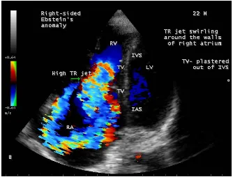
Figure 5. Apical four-chamber view showing the severe high tricuspid regurgitation jet (arrow) swirling around the walls of the right atrium in a 22-year old acyanotic male. IAS—interatrial septum. RA—right atrium.
Figure 6. Continuous Wave (CW) Doppler (green line) showing the low pressure TR (tricuspid regurgitation) jet in a 22 year old acyanotic male.
[image:5.595.206.540.71.325.2]R. Muthiah
DOI: 10.4236/crcm.2018.72009 95 Case Reports in Clinical Medicine
[image:6.595.207.540.71.322.2]Figure 7. M-mode LV (left ventricle) (green line) showing the sail-like tricuspid valve (TV) (arrows) and a normal LV function (EF 61%) in a 22-year old acyanotic male.
Figure 8. Subcostal view showing the dilated atrialized RV (right ventricle) in a 22-year old acyanotic male. Septal tricuspid leaflet (STL) is displaced 57 mm from the annulus (lower arrow).
3) Case 3 (55-year old female with Ebstein’s anomaly)
[image:6.595.207.541.371.635.2]R. Muthiah
DOI: 10.4236/crcm.2018.72009 96 Case Reports in Clinical Medicine
presented with grade 2/6 systolic murmur at the lower left sternal border. 2D echocardiography revealed a septal tricuspid leaflet tethering and a high mod-erate tricuspid regurgitation as shown in Figure 9 and Figure 10 suggesting an Ebstein’s anomaly.
The patient was advised periodic follow up since she was remaining asymp-tomatic.
4) Case 4 (28-year old male with Ebstein’s anomaly)
A 28-year old male presented with cyanosis and auscultation revealed a “sail sound” (loud tricuspid component of first heart sound due to increased tension developed by the large anterior leaflet as it reaches the limits of its systolic excur-sion—an important sign of anterior leaflet mobility), a “cadence” quality of quadruple rhythm due to wide splitting of first and second sounds (due to com-plete right bundle branch block), atrial and ventricular filling sounds (summa-tion of these sounds due to prolonged PR interval). ECG revealed the features of Ebstein’s anomaly as shown in Figure 11 and Figure 12. X-ray chest revealed the Ebstein’s configuration as shown in Figure 13. 2D echocardiography re-vealed a “sail-like” anterior tricuspid leaflet forming a “muscular curtain” in be-tween the inflow and trabecular parts of the right ventricle as an “imperforate membrane” with a “pinhole” communication, associated with a muscular VSD (ventricular septal defect) in the proximal, atrialized compartment of right ven-tricle suggesting an “atretic” (“imperforate”) Ebstein’s anomaly as shown in
Figures 14-27.
R. Muthiah
[image:8.595.209.539.69.311.2]DOI: 10.4236/crcm.2018.72009 97 Case Reports in Clinical Medicine
Figure 10. Apical view showing the high moderate tricuspid regurgitation (TR) jet (ar-row) in a 55-year old acyanotic female. RA—right atrium.
Figure 11. ECG revealed complete right bundle branch block in V1, QR complexes in
V1-V3, precordial Q wave in V1 and a same QRS pattern in V1 and lead aVR (since it
[image:8.595.210.537.352.660.2]R. Muthiah
[image:9.595.210.540.71.312.2]DOI: 10.4236/crcm.2018.72009 98 Case Reports in Clinical Medicine
Figure 12. ECG showing recurrent Mobitz I block in a 28-year old cyanotic male with Ebstein’s anomaly.
[image:9.595.210.540.358.631.2]R. Muthiah
[image:10.595.207.539.72.361.2]DOI: 10.4236/crcm.2018.72009 99 Case Reports in Clinical Medicine
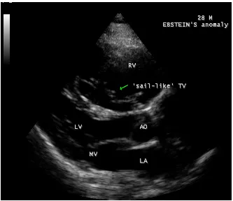
Figure 14. Tilted Parasternal long axis view showing the “sail-like” muscular skirt of tri-cuspid valve (arrow) in a 28-year old cyanotic male. AO—aorta.
[image:10.595.207.542.406.691.2]R. Muthiah
[image:11.595.208.540.72.338.2]DOI: 10.4236/crcm.2018.72009 100 Case Reports in Clinical Medicine
Figure 16. Apical view showing the redundant, “sail-like” anterior tricuspid leaflet (ATL) (right arrow) fused with the displaced (76 mm from annulus) septal tricuspid leaflet (STL) (left arrow) as an imperforate membrane in the RV cavity in a 28-year old cyanotic male. RA—right atrium.
[image:11.595.208.542.412.671.2]R. Muthiah
[image:12.595.207.540.71.326.2]DOI: 10.4236/crcm.2018.72009 101 Case Reports in Clinical Medicine
Figure 18. Apical view showing the displacement ratio with mild TR (tricuspid regurgita-tion) in a 28-year old cyanotic male. STL—septal tricuspid leaflet. RA—right atrium.
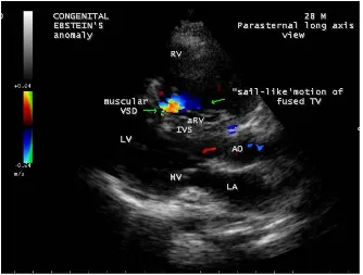
Figure 19. Parasternal long axis view showing the muscular VSD (ventricular septal de-fect) (right arrow) in the proximal compartment of RV (right ventricle) and the “sail-like” motion of fused tricuspid valve (TV) (left arrow) in a 28-year old cyanotic male. AO— aorta.
[image:12.595.207.541.372.625.2]R. Muthiah
[image:13.595.208.541.71.348.2]DOI: 10.4236/crcm.2018.72009 102 Case Reports in Clinical Medicine
Figure 20. Apical view showing the left ventricular to right atrial communication (dilated atrialized right ventricle) (lower arrow) and the displaced septal tricuspid leaflet (STL) (upper arrow) in 28-year old cyanotic male. RA—right atrium.
[image:13.595.207.540.406.693.2]R. Muthiah
[image:14.595.208.540.73.346.2]DOI: 10.4236/crcm.2018.72009 103 Case Reports in Clinical Medicine
Figure 22. Tilted Parasternal long axis view showing the position of anterior leaflet dur-ing the “sail-like” motion (arrow) in a 28-year old male [5]. AO—aorta.
[image:14.595.206.540.391.682.2]R. Muthiah
[image:15.595.209.542.71.343.2]DOI: 10.4236/crcm.2018.72009 104 Case Reports in Clinical Medicine
Figure 24. Right ventricular apical long axis view showing the displaced septal tricuspid leaflet (STL) (arrow) and a dilated atrialized RV (right ventricle) in a 28-year old cyanotic male. IAS—interatrial septum. RA—right atrium.
[image:15.595.208.540.398.694.2]R. Muthiah
[image:16.595.207.539.76.324.2]DOI: 10.4236/crcm.2018.72009 105 Case Reports in Clinical Medicine
Figure 26. Short axis view showing the “whipping motion” (arrow) of tricuspid valve across the RVOT (right ventricular outflow tract) in a 28-year old cyanotic male [6]. AO—aorta.
[image:16.595.208.541.387.693.2]R. Muthiah
DOI: 10.4236/crcm.2018.72009 106 Case Reports in Clinical Medicine
5) Case 5. (30-year old cyanotic male with Ebstein’s anomaly)
A 30-year old male was presented with marked cyanosis and no murmur and abnormal heart sounds on auscultation. 2D echocardiography revealed the fea-tures of Ebstein’s anomaly such as insertion of anterior leaflet into the trabecu-lated RV, forming a “blanket” of leaflet tissue across the inflow and trabecular parts and bulging of ventricular septum towards leftward due to marked dilata-tion of RV, and LV became “banana” shaped as shown in Figures 28-30.
The patient was palliated with bidirectional Glenn shunt and advised periodic follow up for an RV exclusion procedure.
6) Case 6. (12-year old male with inverted Ebstein’s anomaly)
A 12-year old, asymptomatic boy was presented with features of an inverted Ebstein’s anomaly on routine echocardiographic screening as shown in Figure 31 and the boy was advised periodic follow up.
7) Case 7. (Ebstein’s mitral valve in a 10-year old boy).
A 10-year old boy was presented with a grade 1/6 systolic murmur at the apex and blood chemistry revealed a positive ASO (antistreptolysin O) titer, suggest-ing a rheumatic involvement. 2D echocardiography revealed a displaced anterior mitral leaflet and it is thick, calcified and mildly regurgitant as shown in Figure 32.
[image:17.595.209.541.398.682.2]The boy was advised penicillin prophylaxis (oral penicillin V 250 mg twice daily) and periodic follow up.
R. Muthiah
[image:18.595.206.540.72.341.2]DOI: 10.4236/crcm.2018.72009 107 Case Reports in Clinical Medicine
Figure 29. Tilted apical view showing the plastering of the tricuspid valve leaflets into the ventricular wall as a “blanket” of leaflet tissue (arrows) in a 30-year old cyanotic male. Right ventricle (RV) is circular and LV (left ventricle) is banana shaped. RA—right atrium.
[image:18.595.207.540.409.679.2]R. Muthiah
[image:19.595.208.540.72.351.2]DOI: 10.4236/crcm.2018.72009 108 Case Reports in Clinical Medicine
Figure 31. Apical view showing the ventricular inversion and Ebstein’s malformation of the inverted tricuspid valve on the left-side in a 12-year old asymptomatic male. “Arrow” showing the moderator band as a feature of morphologic right ventricle (mRV). STL— septal tricuspid leaflet.
[image:19.595.207.541.424.690.2]R. Muthiah
DOI: 10.4236/crcm.2018.72009 109 Case Reports in Clinical Medicine
3. Discussion
3.1. Review of Literature
Ebstein’s anomaly was initially described by Wilhelm Ebstein at All-Saints Hos-pital in Breslau (now Wroclaw), Poland in the autopsy findings of a 19-year old cyanosed laborer, Joseph Prescher who died with a severe malformation of tri-cuspid valve in July 1864 [7][8]. The eponym “Ebstein’s disease” was coined by Arnstein in 1927 [9]. Yater and Shapiro described the radiologic and echocardi-ographic features of Ebstein’s anomaly in 1937 [10]. Tourniaire, et al diagnosed this anomaly in a living in 1949 and Engle asserted the clinical recognition of Ebstein’s anomaly in 1950 [11]. Edwards emphasized the continuity of muscles between the atrium and the “atrialized” right ventricle across the atrioventricular ring in 1979 [12].
3.2. Etiopathogenesis
The exact embryology of Ebstein’s anomaly is unknown since the normal tricus-pid valve is believed to be formed by a process of either “delamination” of the inner layer of the inlet zone of right ventricle [13][14] or “undermining” [15]. During cardiac embryogenesis, the process of valve formation is completed by the 12th week of gestation and the development of septal tricuspid leaflet is not
complete until the 16th week and the valves are better delineated in the second
and third trimester.
In Ebstein’s anomaly, “delamination” fails to occur, resulting in downward displacement of the origin of septal and inferior leaflets and the antero-superior leaflet is mostly “sail-like”, but still attached to its usual atrioventricular junction line and the mechanism for this is not entirely understood [16].
The leaflets of the tricuspid valve develop equally from the endocardial cu-shion tissue and myocardium. Endocardial cucu-shion tissue lines the right atrio-ventricular junction and acts as an adhesive to maintain the integrity of that por-tion of the subjacent myocardium which forms the tricuspid valve apparatus. Expansion in the size of right ventricular inflow tract occurs by a process called “undermining”. The subendocardial portion of the ventricular free wall becomes fenestrated and spongy since the resorption of the myocardium progressively dissolves the wall and thereby enlarges the chamber size. Undermining and re-sorption produce a flap-like muscular skirt that is attached at the annulus and is anchored to the underlying ventricular wall by numerous branching pillars of myocardium. Progressive thinning and fibroblastic ingrowth result in the for-mation of tricuspid valve leaflets as anterior leaflet forms much earlier and it is not adherent to the right ventricular wall. The late forming septal and posterior leaflets remain adherent to the underlying myocardial wall and thus, Ebstein’s malformation is thought to occur as a result of incomplete undermining of the right ventricular myocardium [17].
in-R. Muthiah
DOI: 10.4236/crcm.2018.72009 110 Case Reports in Clinical Medicine
volved in the process of connection between the future right atrium to the de-veloping right ventricle and monogenic as well as oligogenic factors play a role in its driving pathogenesis in animal models [19]. Rare cases of cardiac tran-scription factor NKX 2 - 5 mutation [20], 10 p13-p14 deletion [21] and Ip34.3- 36.11 deletion [22] have been described in the anomaly. Maternal exposure to benzodiazepins [23] and lithium carbonate therapy [24][25][26] can rarely lead to Ebstein’s anomaly in the offspring.
3.3. Morphological Features
The malformation documented by Ebstein at autopsy consists of an abnormal insertion of the tricuspid valve, the septal and posterior leaflets were adherent to the ventricular wall and the mobile free parts were displaced towards the apex of the right ventricle. The two anatomic features, i.e., valve displacement and leaflet morphology vary independently and its pathology is not uniform.
Apical displacement was determined as the distance between the anticipated normal basal attachments of the leaflets and the right ventricular apex. The de-gree of displacement was classified by Becker in 1971 as shown in Table 2[27].
The intrinsic abnormality of the tricuspid valve leaflets is generally catego-rized as “dysplasia” and it usually affects all leaflets with varying degrees as shown in Table 3 [28], mostly the anterior leaflet, often with displaced leaflets and sometimes, occasional [29].
Apical displacement always affects the septal leaflet and also involves the posterior leaflet. The leaflets are not displaced beyond the junction between the inlet and trabecular parts of the right ventricle and the point of maximum dis-placement is usually at the commissure between the two leaflets. The spectrum of leaflet tethering varies from mild to extreme. The degree of dysplasia is also categorized as I to III as shown in Table 4.
[image:21.595.207.540.648.734.2]When chordae are absent, the free leaflets insert directly into the ventricular wall. In some cases, the greater part of the affected leaflets is firmly adherent to the right ventricular wall, drape the apical trabeculations and completely blended with ventricular wall. The communication between the atrialized and functional right ventricle is confined to slits or perforations in the anterior leaflet as Ebstein originally described. When the anteromedial commissure is fused and the anterior leaflet is intact, the tricuspid orifice is “imperforate” [30] and it oc-curs in 10% of hearts with Ebstein’s anomaly. In “imperforate Ebstein’s anoma-ly”, the septal, inferior and antero-superior leaflets may fuse either completely or
Table 2. Becker’s classification of degree of valve displacement.
Grade Distance between atrioventricular junction and the apex
Grade 1 Apical displacement of <10% (or <25%) (the normal septal attachment of the tricuspid valve dipped below the Expected annular attachments)
Grade 2 displaced between 10% and 50% (or 25% to 50%)
R. Muthiah
[image:22.595.209.537.90.160.2]DOI: 10.4236/crcm.2018.72009 111 Case Reports in Clinical Medicine
Table 3. Lev and Becker’s grading of dysplasia.
Grade a focal or diffuse thickening of the leaflets Grade b deficient development of chordae and papillary muscle Grade c improper separation of the valve components from the ventricular wall
Grade d focal agenesis of valve tissue
Table 4. showing the categories of dysplasia.
Dysplasia I Fibromyxomatous thickening
II Shortening and thickening of the chordate tendineae III leaflet tethering, rudimentary or absent papillary muscles, Fibromuscular histology of anterior leaflet
in part, so that the inner surface of the inflow part of the right ventricle is formed by a “blanket” of dysplastic valve tissue towards the apex. The leaflets are said to be “plastered” out of the right ventricular myocardium, so that the fibr-ous transformation of the leaflets from the muscular precursors remain incom-plete. In severe cases, the leaflets are thickened, focally muscularized and at-tached to the underlying myocardium by numerous muscular stumps. In ex-treme cases, the fusion of leaflet tissue is so complete as a membrane-like conti-nuum and the only identified remnants of leaflet tissue are nodular fibrous ridges at the level of the displaced functional annulus. In this setting, the entire inflow tract is atrialized and the functional right ventricle consists only of trabe-cular and outflow (infundibulum) components. The designation of “atretic“ Ebs-tein’s malformation was documented by Kumar [31] to the imperforate fused tricuspid valve and its incidence becomes even higher to 29% with an inclusion of “pinhole” communication [32].
3.4. Echocardiographic Features
R. Muthiah
DOI: 10.4236/crcm.2018.72009 112 Case Reports in Clinical Medicine
[35]. The ratio between the mitral-to-apex distance and the tricuspid-to-apex distance varies from 1 to 1.2 in normal subjects and 1.8 to 3.2 in patients and it is 3.6 as in Figure 18 with Ebstein’s anomaly. The true distance in the level of in-sertion of atrioventricular valves is obtained by substracting the tricuspid-to- apex distance from the mitral-to-apex distance with a mean value of 27.25 ± 12 mm in patients with proven Ebstein’s anomaly and it is 60 mm as shown in Figure 18 compared to reference group (5.7 ± 2 mm). Kambe and coworkers calculated the distance between both atrioventricular valves directly as a mean value of 21 mm with a range of 14 to 32 mm [36]. A maximum difference in the level of valve insertion of >15 mm in children and >20 mm in adults is discriminated between normal and Ebstein’s anomaly [37][38]. Despite this fact, a patient with an “une-quivocal” Ebstein’s malformation can be encountered in whom the diagnosis can-not be made with certainity solely on the basis of apical displacement of the septal tricuspid valve leaflet. Occasionally, the leaflet attaches to the trabecular part rather than the inlet part of the septum, the conventional four-chamber view will not re-veal any septal insertion as shown in Figure 28 and Figure 29.
The anterior tricuspid leaflet is not involved in the process of downward dis-placement, it may be abnormally inserted occasionally as in Figures 1-4 and Shiina, et al documented the apical displacement of anterior tricuspid leaflet in 14% of cases echocardiographically [39]. The anterior leaflet forms a large, sail-like intracavitary curtain as in Figure 14, Figure 25 and contains muscular strands instead of consisting entirely of a fibrous membrane as in the normal tricuspid valve [40]. It is potentially mobile with a brisk sail-like movement as shown in Figures 21-24[41], free bloating with a “whipping motion” across the right ventricular outflow tract (RVOT) as shown in Figure 26 and in some cases, the movement is restricted due to its adherence to the ventricular wall as in
Fig-ure 1 and Figure 2, Figure 4 and Figure 9. It is often fenestrated, may in part be
musculaized , inserting into the trabeculations of the right ventricle (RV) as in
Figure 28 and rarely, the anterior leaflet forms an “atretic” membrane that spans
the midportion of the right ventricular cavity as in Figure 16.
In severe cases, the inferior wall of the right ventricle may consists soley of thin fibrous tissue, devoid of myocytes and thereby represent an area of aneu-rysmal dilatation as in Cases 2 (Figure 8) and 4 (Figure 20 and Figure 24). It is apparently due either to slippage of right ventricular inflow tract away from the right atrioventricular junction or to focal excessive “undermining” to myocar-dium, transmurally to the level of epicardium. A large atrialized area causes a severe reduction in the volume of the right ventricular pumping chamber and usually produces an abnormal configuration of muscular interventricular sep-tum, which bulges leftward and thereby compresses the left ventricular chamber, resulting in reversal of ventricular shapes with a “circular” right ventricle and a “banana” [42] or “crescentic” [43] left ventricle as shown in Figure 29 and
Fig-ure 30 Thus, the proximal component of the right ventricle, the “inlet portion”
R. Muthiah
DOI: 10.4236/crcm.2018.72009 113 Case Reports in Clinical Medicine
not involved and may be of normal size, but usually markedly diminished in di-mensions and in some cases, it is dilated and thin walled.
The tricuspid orifice is typically incompetent (primary tricuspid regurgitation as in Figure 3), Figure 5 and Figure 10, occasionally stenotic, and rarely im-perforate as in Figure 16 [44][45]. The true anatomic tricuspid annulus occu-pies its normal position at the right atrioventricular junction and it is less well defined than in a normal heart. The annulus tends to be appreciably dilated and contribute to the development of valvular incompetence. In extreme downward displacement of posterior and septal leaflets, the closure of the tricuspid annulus depends on the size and potential excursion of anterior leaflet. When the chordal attachments are short and the leaflets contain multiple or large fenestrations, adequate valve closure is impossible to achieve and varying degrees of regurgita-tion results. Color flow imaging and Doppler interrogaregurgita-tion can establish the rel-atively low velocity regurgitant flow as in Figure 6, which begins at the level of the displaced septal and posterior leaflets as in Figure 5 and Figure 10 and courses through the atrialized right ventricle into the right atrium proper as shown in Figure 5. Tricuspid regurgitation increases by annular dilatation [46]. During contraction of the atrium, the atrialized portion of the right ventricle balloons out and acts as a passive reservoir. Functional improvement of right ventricle depends on the severity of tricuspid regurgitation and on the ratio of the combined areas of right atrium and atrialized right ventricle relative to the areas of functional right ventricle and left ventricle [47]. Celermajer, et al. de-scribed an echocardiographic grading score for neonates with Ebstein’s anomaly as shown in the Table 5[48].
The functional impairement of right ventricle and regurgitation of tricuspid valve retard the forward flow and the overall effect is right atrial dilatation as shown in Figure 4. In many cases, the right atrial enlargement is extensive, but the mural thrombus is not a feature even in right ventricular dilatation. The en-larging right atrium becomes sufficiently compliant to accomodate a large vo-lume of regurgitant flow with little or no increase in pressure as shown in Figure 6. In patients with marked Ebstein’s malformation and severe tricuspid regurgi-tation, liver and portal circulation are extensively affected, congestive hepatos-plenomegaly and microscopic hepatic fibrosis eventually occurs in chronic cases with hypersplenism [49].
R. Muthiah
DOI: 10.4236/crcm.2018.72009 114 Case Reports in Clinical Medicine
Table 5. “Celermajer” echocardiographic grading score—GOSE (Great Ormond Street Echocardiography Score—Grade 3 and 4 have a very poor prognosis).
GOSE score Index (RA + RV): (RV + LA + LV) Risk of mortality (%)
Grade 1 Ratio < 0.5 0
Grade 2 Ratio of 0.5 to 0.99 10
Grade 3 Ratio of 1 to 1.49 44 - 100
Grade 4 Ratio ≥ 1.5 100
incidence of 4% in clinical series and 12% in autopsy studies. The hearts in which the opening is proximal to the displaced tricuspid valve, a left ventricular to right atrial shunt may occur as shown in Figure 20.
3.5. Left-Sided Ebstein’s Anomaly
Ebstein’s anomaly of inverted tricuspid valve has been described in 15% - 50% of cases of congenitally corrected transposition of great arteries [51] as in Figure 31. The anterior leaflet of inverted Ebstein’s anomaly is usually small, mal-formed and the atrialized inverted right ventricle is poorly developed, not thinned and rarely dilated [52]. The posterior wall of the inflow tract of the right ventricle is normal in inverted Ebstein (discordant atrioventricular connection) [53] and it is always abnormal in Ebstein’s anomaly of concordant connection in which the myocardium is replaced by fibrous tissue to variable degree. Rarely, a morphologic mitral valve (mMV)-right sided inverted mitral valve has anatomic features of Ebstein’s anomaly [54]. The leaflets tend to be thickened and even dysplastic in appearance [55]. Ebstein’s-like anomaly can also affect a normally positioned mitral valve as shown in Figure 32, although this is exceedingly rare, first reported by Rusahhaupt, et al in 1976 [56] and its embryologic origin is ob-scure [57]. The left atrioventricular valve incompetence is not necessarily caused by an Ebstein-like malformation, may be congenital and it is due to rheumatic involvement in this case. The anterior mitral leaflet (the aortic leaflet of mitral valve) is not particularly redundant, and its annulus inserts along the septum normally without downward displacement (offsetting). The normal “offsetting” of the mitral and tricuspid valve is maintained in the four-chamber view [58]. Only the posterior leaflet, attached to the left ventricular free wall, is involved in downward displacement. Cases have also been reported in which both the mitral and tricuspid valves were involved by Ebstein’s malformation [59][60].
3.6. Management
The treatment plan for Ebstein’s anomaly is individualized since the hemody-namic abnormality vary considerably with the extent of tricuspid valve dis-placement into the right ventricle.
3.7. Medical Therapy
[image:25.595.206.540.103.188.2]deteriorat-R. Muthiah
DOI: 10.4236/crcm.2018.72009 115 Case Reports in Clinical Medicine
ing course with severe heart failure, cyanosis and acidosis. If congestive cardiac failure is prominent, infants may require initial support with inotropic agents and long-term anticongestive measures with digoxin and diuretics. The efficacy of angiotensin-converting enzyme inhibitors in patients with Ebstein’s anomaly having right-sided heart failure is unproved and they are not recommended, in-cluding angiotensin-receptor blockers (ARBs) and β-blockers, because these drugs may increase pulmonary artery pressure, triggering further heart failure and pulmonary edema [61]. The standard heart failure therapy may be reserved for those patients who are not candidates for surgery.
In newborn infants whose primary problem is that of cyanosis, treatment may be limited to careful observation awaiting its resolution when pulmonary vascu-lar resistance decreases. In early neonatal period, maintanance of the patency of ductus arteriosus is necessary with prostaglandin E1 to ensure adequate pulmo-nary blood flow.
Occasionally, arrhythmias with or without associated Wolf-Parkinson-White syndrome will be dominant in infants. Treatment is directed primarily towards delaying conduction through the AV node with class I antiarrhythmic agents (quinidine, procainamide, flecainide).
3.8. Interventional Therapy
Downward displacement of the septal tricuspid leaflet is associated with discon-tinuity of the central fibrous body and the septal atrioventricular ring, creating a potential substrate for accessory pathways and preexcitation. The atrialized right ventricle contains right ventricular muscle fibers, which can provoke polymor-phic ventricular tachycardia [62][63] that rapidly degenerate into ventricular fi-brillation since the isolated islands of myocytes cannot anchor reentrant spir-al/scroll waves which break up immediately.
“Electrical instability” due to ventricular preexcitation (5% to 25% [64] or 20% to 30%) of cases with supraventricular reentrant tachyarrhythmias (the most frequent rhythm disorder—AVNRT (1% to 2%)) or to atrial flutter or fibrillation due to progressive right atrial dilatation is the main clinical problem. The risk of sudden death is high due to adverse association of atrial fibrillation and preexci-tation (uniformly via a right bypass tract-Type B Wolf-Parkinson White) in asymptomatic individuals.
R. Muthiah
DOI: 10.4236/crcm.2018.72009 116 Case Reports in Clinical Medicine
first-degree atrioventricular block as shown in Figure 11 occurs in 42% of pa-tients due to right atrial enlargement and structural abnormalities of the atrio-ventricular conduction system. Permanent pacing is required for 3.7% of pa-tients with Ebstein’s anomaly [71].
Selective right-sided cryo-ablation-Maze procedure may be done for patients with documented paroxysmal flutter or inducible atrial arrhythmias.
3.9. Surgical Therapy
Surgical options in Ebstein’s anomaly depend on specific circumstances and the goal should be to palliate for optimum survival, accomplished with valvuloplas-ty, Valve repair or replacement and right ventricular (RV) exclusion procedures.
3.10. Valvuloplasty
It is favoured if the anterior leaflet is suitable for use as a functional monocuspid valve. The leaflet must exhibit adequate excursion and be free of large fenestra-tions. If the leaflet is “sail-like” and free and when the tricuspid annulus is mar-kedly dilated, an aggressive “Kay annuloplasty” is preferred as for case 4.
3.11. Tricuspid Valve Repair
R. Muthiah
DOI: 10.4236/crcm.2018.72009 117 Case Reports in Clinical Medicine
of inferoseptal wall of left ventricle and inferior wall of right ventricle.
3.12. Biventricular Repair (Knott-Craig Approach)
If there is moderate to severe TR and an adequate sized functional RV, then a complete biventricular repair (Knott-Craig approach) may be considered as for case 2. In this method, the tricuspid valve is repaired and the atrial septal defect is partially closed. A RV to PA valve conduit is used along with tricuspid valve repair and currently, valve repair is preferred over valve replacement whenever feasible in Ebstein’s anomaly
3.13. Definite Repair
For older patients with persistent cyanosis or congestive cardiac failure, definite repair can be considered. Tricuspid valve replacement along with plication of some part of the atrialized right ventricle and closure of the atrial septal defect is recommended mostly as for case 1. Valve replacement may be done either a bi-oprosthetic (preferred) as for case 2 or mechanical valve. A regurgitant tricuspid valve was successfully replaced in Ebstein’s anomaly as a first time in 1962 [76].
3.14. Ventricularization
It is the reintegration of the atrialized chamber into the right ventricular cavity, which can be obtained by orthopic transposition of the detached septal and post-erior leaflets of the tricuspid valve. The reimplanted septal leaflet serves as an op-posing structure for the coaptation of the reconstructed atrioventricular valve [77].
3.15. Cone Reconstruction
When there is greater than 50% delamination of the anterior leaflet and a usable posterior leaflet, cone reconstruction is a preferred technique and it is closest to an “anatomic repair” [78]. In this procedure, all attachments to body of leaflet may be cut, but the attachments to the free edge must be left intact. When the leaflet edge is attached to the myocardium, fenestrations are performed to help free the leaflet and the height of the fenestration is typically one-fourth to one-third the distance from the leaflet edge. Care must be taken to avoid injury to the thinned underlying right ventricle (RV). The section of RV that usually needs to be remodeled includes the acute margin near the diaphragmatic surface and the goal of plication is to reduce the volume of the ventricle as well as re-moving akinetic and dyskinetic regions to improve efficacy. It also reduces the distortion of the papillary muscles and primary chordal attachments, which contribute to recurrent TR [79].
3.16. Right Ventricular Exclusion Procedures (Univentricular
Approach)
R. Muthiah
DOI: 10.4236/crcm.2018.72009 118 Case Reports in Clinical Medicine
presence of a dilated right-sided pumping chamber (the functional RV) is also considered as an ominous sign, usually impairing the surgical results.
RV exclusion approach was first reported by Starnes and associates in 1991. The procedure included patching of the tricuspid valve and a modified Bla-lock-Taussig shunt. RV exclusion procedure should be indicated for the more severe end spectrum where there is diminutive true RV, highly laminated leaflet tissue and a marked dilatation of right ventricle impinging on the left ventricular function as in case 5. The RV exclusion technique decompresses the right side, allow the left ventricle to function more effectively and decrease the incidence of atrial arrhythmias by eliminating the right atrial enlargement. RV exclusion with a fenestrated tricuspid valve patch and a systemic-to-pulmonary shunt provide an effective palliation for neonates presenting with critical Ebstein’s anomaly who had a poor outcome, but with an adequate LV size. In patients with small RV and mild TR, A modified Blalock-Taussig shunt (mBTS) is preferred. If there is moderate to severe TR and the functional RV is small, then a fenestrated RV exclusion (Starnes’ procedure) should be performed. The Starnes’ procedure consists of a patch closure of the tricuspid valve orifice, enlarging the interatrial communication and placing a systemic-to-pulmonary artery shunt. This ap-proach is particularly helpful in patients who also have an anatomic right ven-tricular outflow tract obstruction. The total RV exclusion is a modification to Starnes’ procedure, proposed by Sano and associates, in which the free wall of the right ventricle is resected and closed primarily with a polytetrafluro ethylene patch and it acts like a large RV plication.
Few patients may undergo a successful valve repair and also a potential for one and half ventricular repair after palliation by RV exclusion. The downside of RV exclusion procedure is a commitment to the single-ventricle Fontan path-way. In very severe cases, with massive right ventricular dilatation and dysfunc-tion, the Fontan procedure may be applicable. Conversion to a single-ventricle approach for symptomatic neonates also had been advocated [80]. In patients with almost imperforate Ebstein’s malformation of the tricuspid valve, a mod-ified Fontan procedure is advised, but it is very rarely required after infancy.
Chauvaud suggest that a paper thin right ventricular wall is an indication that a Glenn shunt might be needed [81]. When the right ventricle is markedly di-lated and functioning poorly, a bidirectional cavopulmonary shunt may be used after tricuspid valve repair or replacement to reduce the RV volume overload in selected patients with nonneonatal Ebstein’s anomaly.
For severe cases of Ebstein’s anomaly associated with left-sided obstructive le-sions, transplantation should be considered. However, heart transplantation is rarely necessary in the current era because of improved results with Knott-Craig and Starnes’ approaches in the most severe forms of Ebstein’s anomaly [82].
3.17. Outcome
ano-R. Muthiah
DOI: 10.4236/crcm.2018.72009 119 Case Reports in Clinical Medicine
maly has been aptly characterized as “appalling” [83]. Neonates confront a high risk of mortality and later death if they survive infancy. Transient neonatal cya-nosis that recurs a decade or more later is an occasional and distinctive feature of Ebstein’s anomaly. The prognosis for infants born with Ebstein’s anomaly is ever changing. Early reports described a 10% to 20% mortality during the first year of life without surgical interventions and poor outcomes were those asso-ciated with severe tricuspid valve displacement and dysfunction and increased cardiothoracic ratio (>0.65). For those who had surgical treatment, 54% did not survive after operation. The actual survival rate for all live-born with Ebstein’s anomaly is 67% at one year and 59% at 10 years. With application of newer sur-gical techniques, improved results have been documented in recent series [84].
Besides the fact that patients with milder form of the disease can live well into adulthood, woman become pregnant and deliver normal term infants in most cases [85]. There is an astonishing longevity noticed in patients with Ebstein’s anomaly with a survival into eighth and nineth decades [86]. The oldest record-ed patients with Ebstein’s anomaly livrecord-ed to 85 years, without cardiac symptoms until the age of 79 years [87].
3.18. Case Analysis
Ebstein’s original case was an example of obstruction at the tricuspid orifice by a membrane dividing the right ventricle into two halves as shown in Figure 16 of a 28-year old cyanotic male with ECG and X-ray characteristics as in Figures
11-13. suggesting an advanced spectrum of Ebstein’s malformation ,
necessitat-ing RV exclusion techniques such as Starnes’ procedure. The florid case of Ebs-tein’s anomaly with the insertion of leaflet tissue along with ventricular walls as a “blanket” as in Figure 28 and Figure 29 in a 30-year old cyanotic male may go for an initial palliation with bidirectional Glenn shunt (cavopulmonary anasto-mosis). The other variants of moderate degree of leaflet tethering with varying degrees of regurgitation, but an intact basal leaflet attachments with atrioventri-cular junction as in Cases 1 and 2 may need a definite repair. In Ebstein’s mitral valve as in Figure 32 in a 10-year old boy, the downward displacement of func-tional annulus > 0.8 cm/m2 is not particularly striking and tends to affect the
septal leaflet (anterior mitral leaflet) alone. The valve is thickened and mildly regurgitant due to rheumatic involvement rather than an anatomic cause.
On follow-up, cases 1 and 2 showed no further deterioration and cases 3, 6, 7 remain asymptomatic. Cases 4 and 5 are referred to surgical interventions.
4. Conclusion
R. Muthiah
DOI: 10.4236/crcm.2018.72009 120 Case Reports in Clinical Medicine
were documented by echocardiography and various treatment options were de-scribed in this spectrum.
References
[1] Simcha, A. and Bonham-Carter, R.E. (1971) Ebstein’s Anomaly. Clinical Study of 32 Patients in Childhood. British Heart Journal, 33, 46.
https://doi.org/10.1136/hrt.33.1.46
[2] Morgan, L.B. and Dearani, J.A. (2016) Ebstein’s Anomaly. Chapter 130, Thoracic Key.
[3] Ligtvoet-Gussenhoven, W.J. (1984) Echocardiography in Ebstein’s Anomaly. The-sis. University of Rotterdam.
[4] Bialostozky, D., Medrano, G.A., Munoz, L. and Contrera, S. (1972) Vectorcardio-graphic Study and Anatomic Observations in 21 Cases of Ebstein’s Malformation of the Tricuspid Valve. American Journal of Cardiology,30, 354.
https://doi.org/10.1016/0002-9149(72)90565-6
[5] Rama Chandran (2018) Ebstein’s Anomaly—“Sail-Like” Motion. https://youtu.be/-3LFiY2XNA0
[6] Rama Chandran (2018) Ebstein’s Anomaly—“Whipping Motion”. https://youtu.be/apw5g3iqI5c
[7] Mann, R.J. and Lie, J.T. (1979) The Life Story of Wilhem Ebstein (1836-1912) and His Almost Overlooked Description of a Congenital Heart Disease. Mayo Clinic Proceedings, 54, 197-204.
[8] Ebstein, W.C. (1866) On a Very Rare Case of Insufficiency of the Tricuspid Valve Caused by a Severe Congenital Malformation of the Same. Archiv fur Anatomie Physiologie, und Wissenschaffiche Medicin Leipzig, 33, 238-254.
[9] Arnsten, A. (1927) Ebstein’s Disease. Virchow’s Archiv. A. Pathological Anatomy and Histology,266, 247-254.
[10] Yater, W.M. and Shapiro, M.J. (1937) Congenital Displacement of Tricuspid Valve (Ebstein’s Disease), Review and Report of Case. Annals of Internal Medicine, 11, 1043. https://doi.org/10.7326/0003-4819-11-6-1043
[11] Engle, M.A., Payne, T.P.B., Bruins, C. and Taussig, H.B. (1950) Ebstein’s Anomaly of the Tricuspid Valve, Report of Three Cases and Analysis of Clinical Syndrome. Circulation, 1, 1246. https://doi.org/10.1161/01.CIR.1.6.1246
[12] Burchell, H.B. (1979) Editorial. Ebstein’s Disease. Mayo Clinic Proceedings, 54, 205-207.
[13] Frescura, C., Angelini, A., Daliento, L. and Thiene, G. (2000) Morphologic Aspects of Ebstein’s Anomaly in Adults. Thoracic and Cardiovascular Surgery, 48, 203-208. https://doi.org/10.1055/s-2000-6893
[14] Lamers, W.H., Viragh, S., Wessels, A., Moorman, A.F.M. and Anderson, R.H. (1995) Formation of the Tricuspid Valve in the Human Heart. Circulation, 91, 111- 121. https://doi.org/10.1161/01.CIR.91.1.111
[15] Van Mierop, L.H.S., Kutsche, L.M. and Victoria, B.E. (1989) Ebstein Anomaly. Moss’ Heart Disease in Infants, Children and Adolescents, 4th Edition, Williams And Wilkins, Baltimore, 361-371.
R. Muthiah
DOI: 10.4236/crcm.2018.72009 121 Case Reports in Clinical Medicine
[17] Edwards, W.D. (1993) Embryology and Pathologic Features of Ebstein’s Anomaly. Progress in Pediatric Cardiology, 2, 5-15.
https://doi.org/10.1016/1058-9813(93)90042-X
[18] Donegan, et al. (1968) Familial Ebstein’s Anomaly of the Tricuspid Valve. American Heart Journal,75, 375-379. https://doi.org/10.1016/0002-8703(68)90095-1
[19] Andelfinger, G.U. (2017) Molecular Pathways and Animal Models of Ebstein’s Anomaly. Springer International Publishing.
[20] Benson, D.W., Silberbach, G.M., Kavanaugh McHugh, A., Cottrill, C., Zhang, Y., Riggs, S., Smalls, O., Johnson, M.C., Watson, M.S., Seidman, J.G., Seidman, C.E., Plowden, J. and Kugler, J.D. (1999) Mutations in the Cardiac Transcription Factor NKX2.5 Affect Diverse Cardiac Developmental Pathways. Journal of Clinical Inves-tigation,104, 1567-1573.
[21] Yatsenko, S.A., Yastsenko, A.N., Szigeti, K., Craigen, W.J., Stankiewicz, P., Cheung, S.W. and Lupski, J.R. (2004) Interstitial Deletion of 10p and Atrial Septal Defect in DiGeorge 2 Syndrome. Clinical Genetics,66, 128-136.
https://doi.org/10.1111/j.1399-0004.2004.00290.x
[22] Yang, H., Lee, C.L., Young, D.C., Shortliffe, M., Yu, W. and Wright, J.R. (2004) A Rare Case of Interstitial Del (1)(p34.3p36.11) Diagnosed Prenatally. Fetal and Pe-diatric Pathology, 23, 251-255. https://doi.org/10.1080/15227950490923624
[23] Correa-Villasenor, A., Ferencz, C., Neill, C.A., Wilson, P.D. and Boughman, J.A. (1994) Ebstein’s Malformation of the Tricuspid Valve: Genetic and Environmental Factors. Teratology, 50, 137-147. https://doi.org/10.1002/tera.1420500208
[24] Cohen, L.S., Friedman, J.M., Jefferson, J.W., Johnson, E.M. and Weiner, M.L. (1994) A Reevaluation of Risk of in Utero Exposure to Lithium. JAMA (Journal of Ameri-can Medical Association),271, 146-150.
https://doi.org/10.1001/jama.1994.03510260078033
[25] Sipek, A. (1989) Lithium and Ebstein’s Anomaly. Cor et Vasa, 31, 149-156.
[26] Long, W.A. and Willis, P.W. (1984) Maternal Lithium and Neonatal Ebstein’s Anomaly: Evaluation with Cross-Sectional Echocardiography. American Journal of Perinatology, 1, 182-184. https://doi.org/10.1055/s-2007-999999
[27] Becker, A.E., Becker, M.J. and Edwards, J.E. (1971) Pathologic Spectrum and Dys-plasia of the Tricuspid Valve: Features in Common with Ebstein’s Malformation. Archives of Patholoy, 91, 167-178.
[28] Roberson, D.A. and Silverman, N.H. (1989) Ebstein’s Anomaly: Echocardiographic and Clinical Features in the Fetus and Neonates. Journal of American College of Cardiology, 14, 1300. https://doi.org/10.1016/0735-1097(89)90432-4
[29] Lev, M., Liberthson, R.R., Joseph, R.H., Seten, C.E., Kunske, R.D., Eckner, F.A.O. and Miller, R.A. (1970) The Pathologic Anatomy of Ebstein’s Disease. Archives of Patholoy,90, 334-343.
[30] Zuberbuhler, J.R., Becker, A.E., Anderson, R.H. and Lenox, C.C. (1984) Ebstein’s Malformation and the Embryological Development of the Tricuspid Valve. Pedia-tric Cardiology, 5, 289. https://doi.org/10.1007/BF02424974
[31] Kumar, A.E., Fyler, D.C., Miettinen, O.S. and Nadas, A.S. (1971) Ebstein’s Anoma-ly-Clinical Profile and Natural History. American Journal of Cardiology, 28, 84-95. https://doi.org/10.1016/0002-9149(71)90038-5
R. Muthiah
DOI: 10.4236/crcm.2018.72009 122 Case Reports in Clinical Medicine
[33] Hagan, A., Sahn, D.J. and Friedman, W.F. (1974) Cross-Sectional Echocardio-graphic Features of Ebstein’s Malformation. Circulation,49/50, 111.
[34] Silver, M.D., Lam, J.H.C., Ranganathan, N. and Wigle, E.D. (1971) Morphology of the Human Tricuspid Valve. Circulation, 43, 333.
[35] Feigenbaum, H., Armstrong, W.F. and Ryan, T. (2005) Congenital Heart Disease. Feigenbaum’s Echocardiography, 6th Edition, 564.
[36] Kambe, T., Ichimiya, S., Toguchi, M., Hibi, N., Fukui, Y., Nishimura, K., Sakamoto, N. and Hojo, Y. (1980) Apex and Subxiphoid Approaches to Ebstein’s Anomaly Using Cross-Sectional Echocardiography. American Heart Journal, 100, 53-57. https://doi.org/10.1016/0002-8703(80)90278-1
[37] Gussenhoven, F.J., Stewart , P.A., Becker, A.E., et al. (1984) “Offsetting” of the Sep-tal Tricuspid Leaflet in Normal Hearts and in Hearts with Ebstein’s Anomaly. American Journal of Cardiology,54, 172.
https://doi.org/10.1016/0002-9149(84)90324-2
[38] Ammash, N.M., Warnes, C.A., Connolly, H.M., et al. (1997) Mimics of Ebstein’s Anomaly. American Heart Journal, 134, 508.
https://doi.org/10.1016/S0002-8703(97)70088-7
[39] Shiina, A., Seward, J.B., Hagler, D.J., Tajik, A.J. and Danielson, G.K. (1983) Two-Dimensional Echocardiographic-Surgical Correlation in Ebstein’s Anomaly: Preoperative Deteminantion of Patients Requiring Tricuspid Valve Plication vs Re-placement. Circulation,68, 534-544. https://doi.org/10.1161/01.CIR.68.3.534 [40] Anderson, K.R. and Lie, J.T. (1979) Right Ventricular Myocardium in Ebstein’s
Anomaly. Mayo Clinic Proceedings,54, 181.
[41] Rusconi, P.G., Zuberbuhler, J.R., Anderson, R.H. and Rigby, M.L. (1991) Morpho-logic Echocardiographic Correlates of Ebstein’s Malformation. European Heart Journal, 12, 784.
[42] Daliento, L., Angelini, A., Ho, S.Y., Frescura, C., Turrini, P., Baratella, M.C., Thiene, G. and Anderson, R.H. (1997) Angiographic and Morphologic Features of the Left Ventricle in Ebstein’s Malformation. American Journal of Cardiology, 80, 1051- 1059. https://doi.org/10.1016/S0002-9149(97)00602-4
[43] Shiini, A., Seward, J.B., Edwards, W.D., Hagler, D.J. and Tajik, A.J. (1984) Two- Dimensional Echocardiographic Spectrum of Ebstein’s Anomaly: Detailed Ana-tomic Assessment. Journal of American College of Cardiology,3, 356-370. https://doi.org/10.1016/S0735-1097(84)80020-0
[44] Giuliani, E.R., Fuster, V. and Brandenberg, R.O. (1979) Ebstein’s Anomaly. Mayo Clinic Proceedings,54, 163.
[45] Leung, M.E., Barker, E.J., Anderson, R.H. and Zuberbuhler, J.R. (1988) Cineangio-graphic Spectrum of Ebstein’s Malformation: Its Relevance to Clinical Presentation and Outcome. Journal of American College of Cardiology,11, 154.
https://doi.org/10.1016/0735-1097(88)90182-9
[46] Dearani, J.A. and Danielson, G.K. (2003) Ebstein’s Anomaly of the Tricuspid Valve. Pediatric Cardiac Surgery,3rd Edition, 524-536.
[47] Celermajer, D.S., Cullen, S., Sullivan, I.D., et al. (1992) Outcome in Neonates with Ebstein’s Anomaly. Journal of American College of Cardiology, 19, 1041.
https://doi.org/10.1016/0735-1097(92)90291-T
R. Muthiah
DOI: 10.4236/crcm.2018.72009 123 Case Reports in Clinical Medicine
College of Cardiology, 23, 170-176. https://doi.org/10.1016/0735-1097(94)90516-9 [49] Bennett, M.R. and Shiu, M.F. (1991) Ebstein’s Anomaly Associated with
Splenome-galy and Reversible Hypersplenism. British Heart Journal, 65, 223-224. https://doi.org/10.1136/hrt.65.4.223
[50] Brickner, M.E., Hillis, L.D. and Lange, R.A. (2000) Congenital Heart Disease in Adults: Second of Two Parts. New England Journal of Medicine, 342, 334-342. https://doi.org/10.1056/NEJM200002033420507
[51] Dekker, A., Mehrizi, A. and Vengsarkar, A.S. (1965) Corrected Transposition of the Great Vessels with Ebstein Malformation of the Left Atrioventricular Valve: An Embryologic Analysis and Two Case Reports. Circulation,31, 119-126.
https://doi.org/10.1161/01.CIR.31.1.119
[52] Silverman, N.H., Gerlis, L.M., Horowitz, E.S., Ho, S.Y., Neches, W.H. and Ander-son, R.H. (1995) Pathologic Elucidation of the Echocardiographic Features of Ebs-tein’s Malformation of the Morphologically Tricuspid Valve in Discordant Atrio-ventricular Connections. American Journal of Cardiology, 76, 1277-1283. https://doi.org/10.1016/S0002-9149(99)80356-7
[53] Freedom, R.M. (2002) Discordant Atrioventricular Connection and Congenitally Corrected Transposition. Pediatric Cardiology, 2nd Edition, 1321-1335.
[54] Gerlis, L.M., Wilson, N. and Dickinson, D.F. (1986) Abnormalities of the Mitral Valve in Congenitally Corrected Transposition (Discordant Atrioventricular and Ventriculoarterial Connection). British Heart Journal, 55, 475.
https://doi.org/10.1136/hrt.55.5.475
[55] Caruso, G., Gifarelli, A., Balducci, G. and Facilone, F. (1987) Ebstein’s Malforma-tion of the Mitral Valve in Atrioventricular and Ventriculo Arterial Concordance. Pediatric Cardiology, 8, 209-210. https://doi.org/10.1007/BF02263456
[56] Rauschhaupt, D.G., Bharati, S. and Lev, M. (1976) Mitral Valve Malformation of Ebstein Type in Absence of Corrected Transposition. American Journal of Cardiol-ogy, 38, 109-112. https://doi.org/10.1016/0002-9149(76)90071-0
[57] Jacob, J.L.B., Da Silveira, L.C. and Braile, D.M. (1991) Echocardiographic and An-giographic Diagnosis of Ebstein’s Anomaly of the Mitral Valve. British Heart Jour-nal, 66, 379-380. https://doi.org/10.1136/hrt.66.5.379
[58] Leung, M., Rigby, M.L., Anderson, R.H., Wyse, R.K.H. and Macartney, F.J. (1987) Reversed Offsetting of the Septal Attachments of the Atrioventricular Valves and Ebstein’s Malformation of the Morphologically Mitral Valve. British Heart Journal, 57, 184-187. https://doi.org/10.1136/hrt.57.2.184
[59] Ferreira, S.M., Ebaid, M. and Aiello, V.D. (1991) Ebstein’s Malformation of the Tricuspid and Mitral Valves Associated with Hypoplasia of the Ascending Aorta. International Journal of Cardiology, 33, 170-172.
[60] Dusmet, M., Oberhaenski, I. and Cox, J.N. (1987) Ebstein’s Anomaly of the Tricus-pid and Mitral Valves in an Otherwise Normal Heart. British Heart Journal, 58, 400-404. https://doi.org/10.1136/hrt.58.4.400
[61] Xu, F. and Zhou, J.Z. (2015) Ebstein’s Anomaly with Refractory Right-Side Heart Failure and Leg Ulcers. Journal of Vascular Medicine And Surgery, 3, 177.
[62] Obio-Ngwa, O., Milliez, P., Richardson, A., et al. (2001) Ventricular Tachycardia in Ebstein’s Anomaly. Circulation,104, 92. https://doi.org/10.1161/hc4301.098011 [63] Lo, H., Lin, F., Jong, Y., et al. (1989) Ebstein’s Anomaly with Ventricular
R. Muthiah
DOI: 10.4236/crcm.2018.72009 124 Case Reports in Clinical Medicine
[64] Bialostozky, D., Horwitz, S. and Espino-Vela, J. (1972) Ebstein’s Malformation of the Tricuspid Valve: A Review of 65 Cases. American Journal of Cardiology, 29, 826-836. https://doi.org/10.1016/0002-9149(72)90503-6
[65] Ho, S.Y., Goltz, D., McCarthy, K., Cook, A.C., Connell, M.G., Smith, A., et al. (2000) The Atrioventricular Junction in Ebstein Malformation. Heart,444-449. https://doi.org/10.1136/heart.83.4.444
[66] Khositseth, A., Danielson, G.K., Dearani, J.A., Munger, T.M. and Porter, C.J. (2004) Supraventricular Tachyarrhythmias in Ebstein Anomaly: Management and Out-come. Journal of Thoracic and Cardiovascular Surgery, 128, 826-833.
https://doi.org/10.1016/j.jtcvs.2004.02.012
[67] Greason, K.L., Dearani, J.A., Theodoro, D.A., Porter, C.B., Warnes, C.A. and Da-nielson, G.K. (2003) Surgical Management of Atrial Tachyarrhythmias Associated with Congenital Cardiac Anomalies. Mayo Clinic Experience. Seminars in Thoracic and Cardiovascular Surgery: Pediatric Cardiac Surgery Annual,6, 59-71.
https://doi.org/10.1016/S1092-9126(03)70009-X
[68] Danielson, G.K., Driscoll, D.J., Mair, D.D., et al. (1992) Operative Treatment of Ebstein’s Anomaly. Journal of Thoracic and Cardiovascular Surgery, 104, 1195- 1202.
[69] Kugler, J.D., Gillette, P.C., Duff, D.F., et al. (1978) Elective Mapping and Surgical Division of the Bundle of Kent in a Patient with Ebstein’s Anomaly Who Required Tricuspid Valve Replacement. American Journal of Cardiology, 41, 602-605. https://doi.org/10.1016/0002-9149(78)90022-X
[70] Cappato, R., Hebe, J., Weib, C., et al. (1993) Radiofrequency Current Ablation of Accessory Pathways in Ebstein’s Anomaly. Journal of American College of Cardi-ology, 21, 172A.
[71] Allen, M.R., Hayes, D.L., Warnes, C.A. and Danielson, G.K. (1997) Permanent Pac-ing in Ebstein’s Anomaly. Pacing and Clinical Electophysiology, 20, 1243-1246. https://doi.org/10.1111/j.1540-8159.1997.tb06776.x
[72] Schiebler, G.L., Adam Jr., P., Anderson, R.C., Amplatz, K. and Lester, R.G. (1959) Clinical Study of Twenty-Three Cases of Ebstein’s Anomaly of the Tricuspid Valve. Circulation, 19, 165-187. https://doi.org/10.1161/01.CIR.19.2.165
[73] Dearani, J.A. and Danielson, G.K. (2003) Tricuspid Valve Repair for Ebstein’s Anomaly. Operative Techniques in Thoracic and Cardiovascular Surgery, 8, 188- 192. https://doi.org/10.1016/S1522-2942(03)80006-5
[74] Danielson, G.K., Maloney, J.D. and Devloo, R.A. (1979) Surgical Repair of Ebstein’s Anomaly. Mayo Clinic Proceedings, 54, 185-192.
[75] Edwards, W.D. (1989) Congenital Heart Disease. In: Schoen, F.J., Ed., Intervention-al and SurgicIntervention-al Cardiovascular Pathology: Clinical Correlations and Basic Prin-ciples, WB (Walter Burns) Saunders, Philadelphia, 281-367.
[76] Barnard, C.N. and Schrire, V. (1963) Surgical Correction of Ebstein’s Malformation with Prosthetic Tricuspid Valve. Surgery,54, 302-308.
[77] Ullman, M.V., Born, S., Sebening, C., Gorenflo, M., Ulmer, H.E. and Hagl, S. (2004) Ventricularization of the Atrialized Chamber: A Concept of Ebstein’s Anomaly Re-pair. Annals of Thoracic Surgery, 78, 918-924.
https://doi.org/10.1016/j.athoracsur.2004.02.134
R. Muthiah
DOI: 10.4236/crcm.2018.72009 125 Case Reports in Clinical Medicine
https://doi.org/10.1053/j.optechstcvs.2008.03.003
[79] Emani, S.M. and Del Nido, P. (2015) Ebstein’s Malformation of the Tricuspid Valve in Children. Mastery of Cardiothoracic Surgery, 3rd Edition, 1098-1104.
[80] Starnes, V.A., Pitlick, P.T., Bernstein, D., Griffin, M.L., Choy, M. and Shumway, N.E. (1991) Ebstein’s Anomaly Appearing in the Neonate: A New Surgical Ap-proach. Journal of Thoracic and Cardiovascular Surgery,101, 1082-1087.
[81] Chauvaud, S. (2004) Repair of Ebstein’s Anomaly. Operative Techniques in Tho-racic and Cardiovascular Surgery, 9, 194-207.
[82] Carpentier, A., Chauvaud, S., Mace, L., Relland, J., Mihaileanu, S., Marino, J.P., Abry, B. and Guibourt, P. (1988) A New Reconstructive Operation for Ebstein’s Anomaly of the Tricuspid Valve. Journal of Thoracic and Cardiovascular Surgery, 96, 92-101.
[83] Perloff, J.K. and Marelli, A.J. (2012) Ebstein’s Anomaly of the Tricuspid Valve. Per-loff’s Clinical Recognition of Congenital Heart Disease, 6th Edition, 179.
[84] Quaegebeur, J.M., Sreeram, N., Fraser, A.G., et al. (1991) Surgery for Ebstein’s Anomaly: The Clinical and Echocardiographic Evaluation of a New Technique. Journal of American College of Cardiology,17, 722-728.
https://doi.org/10.1016/S0735-1097(10)80190-1
[85] Donnelly, J.E., Brown, J.M. and Radford, D.J. (1991) Pregnancy Outcome and Ebs-tein’s Anomaly. British Heart Journal, 66, 368-371.
https://doi.org/10.1136/hrt.66.5.368
[86] Cabin, H.S., Wood, T.P., Smith, J.O. and Robertts, W.C. (1981) Ebstein’s Anomaly in the Elderly. Chest,80, 212. https://doi.org/10.1378/chest.80.2.212
[87] Seward, J.B., Tajik, A.J., Feist, D.J. and Smith, H.C. (1979) Ebstein’s Anomaly in an 85-Year Old Man. Mayo Clinic Proceedings, 54, 193.
[88] Van Son, J.A., Konstantinov, I.E. and Zimmerman, V. (2001) Wilhem Ebstein and Ebstein’s Malformation. European Journal of Cardiothoracic Surgery, 20, 1082. https://doi.org/10.1016/S1010-7940(01)00913-7


![Figure 22. Tilted Parasternal long axis view showing the position of anterior leaflet dur-ing the “sail-like” motion (arrow) in a 28-year old male [5]](https://thumb-us.123doks.com/thumbv2/123dok_us/9988875.499187/14.595.206.540.391.682/figure-tilted-parasternal-showing-position-anterior-leaflet-motion.webp)
