ORIGINAL RESEARCH
BRAIN
Can 3T MR Angiography Replace DSA for the Identification of
Arteries Feeding Intracranial Meningiomas?
H. Uetani, M. Akter, T. Hirai, Y. Shigematsu, M. Kitajima, Y. Kai, S. Yano, H. Nakamura, K. Makino, M. Azuma, R. Murakami, and Y. Yamashita
ABSTRACT
BACKGROUND AND PURPOSE: For identifying the arterial feeders of meningiomas, the usefulness of 3D TOF MRA at 3T has not been systematically investigated. This study was intended to assess whether unenhanced 3D TOF MRA at 3T can replace DSA for the identifi-cation of arteries feeding intracranial meningiomas and whether it is useful for assessing their dural attachment.
MATERIALS AND METHODS: Twenty-one consecutive patients with intracranial meningiomas (18 women, 3 men; aged 42–77 years, mean 57 years) underwent DSA, conventional MR imaging, and 3D TOF MRA. Two neuroradiologists independently evaluated the primary and secondary feeders of each tumor on maximum-intensity-projection and source MRA images. They also identified the location of dural attachments based on information from MR imaging/MRA images. Interobserver and intermodality agreement was determined by calculating thecoefficient.
RESULTS: For the identification of primary and secondary feeders on MRA images, interobserver agreement was very good (⫽0.83; 95% CI, 0.66 –1.00) and moderate (⫽0.58; 95% CI, 0.34 – 0.82) and intermodality agreement (consensus reading of MRA versus DSA findings) was excellent (⫽0.94; 95% CI, 0.84 –1.00) and good (⫽0.72; 95% CI, 0.51– 0.93), respectively. With respect to the dural attachment of meningiomas, interobserver agreement was very good (⫽0.95; 95% CI, 0.84 –1.00). The agreement in the diagnosis between MR imaging/MRA and surgery was excellent (⫽1.00).
CONCLUSIONS: Unenhanced 3D TOF MRA at 3T cannot at present supplant DSA for the identification of the feeding arteries of intracranial meningiomas. This information may be useful for evaluating their dural attachment.
ABBREVIATIONS:CI⫽confidence interval; MIP⫽maximum intensity projection; MOTSA⫽multiple overlapping thin slab acquisitions; MPR⫽multiplanar reconstruction; NSA⫽number of signal-intensity acquisitions; TOF⫽time-of-flight.
M
eningiomas are the most frequently diagnosed primary brain tumors, and account for approximately one-third of these tumors.1They tend to be hypervascular and are fed byar-teries attached to the dura. In patients with meningioma, tumor embolization is considered a useful preoperative adjuvant therapy to mitigate blood loss during subsequent surgical resection.2To
determine the need for preoperative embolization, one must un-derstand the tumor vasculature. For the selection of the optimal surgical approach to meningiomas, the precise anatomic location of the dural attachment must be understood, and the critical feed-ing arteries must be localized early.3,4
Intra-arterial DSA, the reference standard for assessing the vasculature of meningiomas, plays a role in planning the emboli-zation of large tumors and tumors whose blood supply is inacces-sible (eg, those of the base of the skull).5The inherently high
spatial and temporal resolution of DSA facilitates the identifica-tion of the feeding arteries and tumor vascularity. Because DSA is invasive, exposes the patient to radiation, requires the injection of iodinated contrast material, and does not show anatomic rela-tionship with brain structures, however, a noninvasive method is needed for the accurate diagnosis of these tumors and their pre-interventional and presurgical evaluation.
Unenhanced 3D TOF MRA is a safe, less expensive, widely used, and noninvasive imaging technique. Source images from 3D TOF MRA yield both flow and anatomic information with high spa-tial resolution.6,7As the quality and spatial resolution of 3D TOF
MRA is improved on 3T MR units,8-103D TOF MRA at 3T can be
expected to provide precise information on the vessels feeding me-ningiomas. To our knowledge, there are no comparative studies on 3D TOF MRA at 3T and DSA to assess their utility for identifying
Received April 12, 2012; accepted after revision June 19.
From the Departments of Diagnostic Radiology (H.U., M. Akter, T.H., Y.S., M.K., M. Azuma, Y.Y.), Neurosurgery (Y.K., S.Y., H.N., K.M.), and Medical Imaging (R.M.), Graduate School of Medical Sciences, Kumamoto University, Kumamoto, Japan. Please send correspondence to Toshinori Hirai, Department of Diagnostic Radiol-ogy, Graduate School of Medical Sciences, Kumamoto University, 1-1-1 Honjo, Ku-mamoto 860-8556 Japan; e-mail: t-hirai@kuKu-mamoto-u.ac.jp
the arterial feeders of meningiomas. Here we tested our hy-pothesis that unenhanced 3D TOF MRA at 3T can replace DSA for the identification of the feeders of intracranial meningio-mas and that it is useful for assessing their dural attachment.
MATERIALS AND METHODS
PatientsThis study was approved by our institutional review board. In-formed consent for imaging studies was obtained from all patients or their legal representatives. Our prospective study included 21 consecutive patients whose initial diagnosis was intracranial me-ningioma (18 women, 3 men; age range 42–77 years, mean age 57 years⫾11) and who underwent preoperative DSA and 3D TOF MRA at 3T. The interval between DSA and 3D TOF MRA ranged from 2 to 30 days (mean 11 days).
Of the 21 meningiomas, 15 were supratentorial, 5 were in-fratentorial, and 1 was supra- and infratentorial. The presenting symptoms were headache in 7 patients, visual disturbance in 5, hemiparesis in 3, gait disturbance in 3, speech disturbance in 2, and trigeminal neuralgia in 1 (Table 1).
MR Imaging and MRA
All MR imaging studies were done on a 3T scanner (Achieva 3T; Philips Medical Systems, Best, the Netherlands) using 8-channel head coils. The imaging sequences included 3-plane scout local-izers, axial spin-echo T1-weighted (TR/TE/NSA 450 ms/10 ms/1, matrix 320⫻320), turbo spin-echo T2-weighted (TR/TE/NSA 4060 ms/80 ms/1, turbo factor 9, matrix 512⫻512), FLAIR (TR/TE/NSA/TI 9000 ms/120 ms/1/2500 ms, turbo factor 15, matrix 352⫻352), 3D TOF MRA, and postcontrast T1-weighted and 3D turbo field echo (TR/TE/NSA 450 ms/10 ms/1, matrix
320 ⫻320) images. FOV was 23 cm on all conventional MR images.
The volume for 3D TOF MRA by using MOTSA was localized on a sagittal scout image. The parameters were TR/TE/NSA 20 ms/3.5 ms/1, flip angle 20°, FOV 20⫻20 cm, matrix 512⫻512, 5 slabs, voxel size 0.39⫻0.39⫻1.0 mm (reconstructed voxel size 0.25⫻0.25⫻0.5 mm), parallel imaging factor 2, and acquisition time 4 minutes 48 seconds. Cephalad saturation pulses were ap-plied to eliminate venous blood signals. When a meningioma was located at the top of the head, another 3D TOF MRA slab was rostrally added by using the same parameters. MIP and partial MIP techniques were used for the 3D display of MRA images. Coronal and sagittal images acquired with the MPR method were also reconstructed for the observers’ interpretation.
DSA Technique
After catheterization of the internal and external carotid and ver-tebral arteries via a femoral artery approach, diagnostic biplanar intra-arterial DSA (Allura Xper FD; Philips Medical Systems) was performed by a trained neuroradiologist and/or a neurosurgeon. Images were obtained with a 2048⫻2048 matrix and a 17-cm FOV. The temporal resolution of the images was 3 frames/s. For each projection we manually injected a 6- to 10-mL bolus of an undiluted iodinated contrast material with an iodine concentra-tion of 300 mg/mL (Iopamidol, Iopamiron 300; Bayer-Schering, Berlin, Germany).
Image Analysis
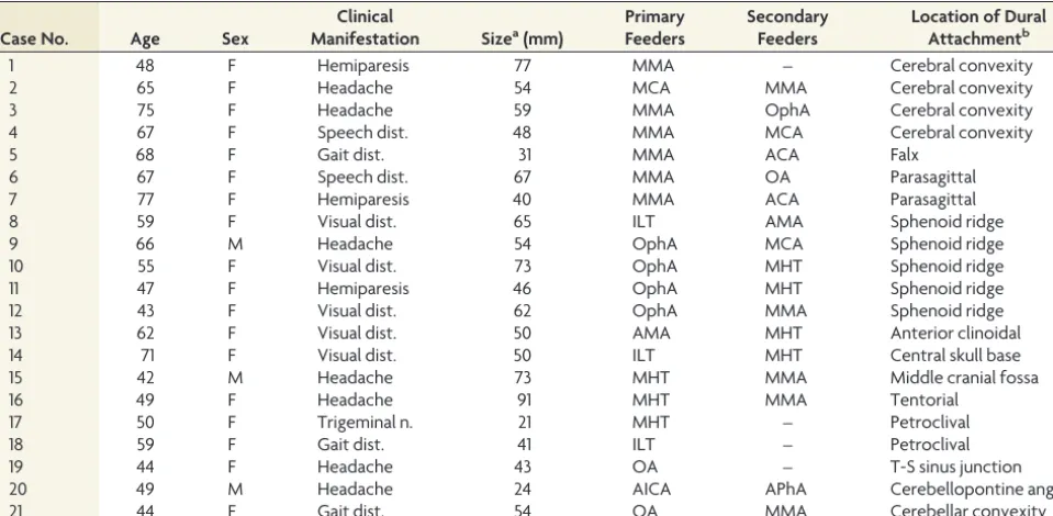
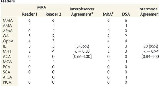
[image:2.594.54.534.56.291.2]Two independent readers (Y.K. and T.H., with 23 and 21 years of experience in neuroangiography, respectively) qualitatively eval-uated the entire series of DSA images on a PACS workstation.
Table 1: Summary of patients and intracranial meningiomas
Case No. Age Sex
Clinical
Manifestation Sizea(mm)
Primary Feeders
Secondary Feeders
Location of Dural Attachmentb
1 48 F Hemiparesis 77 MMA – Cerebral convexity
2 65 F Headache 54 MCA MMA Cerebral convexity
3 75 F Headache 59 MMA OphA Cerebral convexity
4 67 F Speech dist. 48 MMA MCA Cerebral convexity
5 68 F Gait dist. 31 MMA ACA Falx
6 67 F Speech dist. 67 MMA OA Parasagittal
7 77 F Hemiparesis 40 MMA ACA Parasagittal
8 59 F Visual dist. 65 ILT AMA Sphenoid ridge
9 66 M Headache 54 OphA MCA Sphenoid ridge
10 55 F Visual dist. 73 OphA MHT Sphenoid ridge
11 47 F Hemiparesis 46 OphA MHT Sphenoid ridge
12 43 F Visual dist. 62 OphA MMA Sphenoid ridge
13 62 F Visual dist. 50 AMA MHT Anterior clinoidal
14 71 F Visual dist. 50 ILT MHT Central skull base
15 42 M Headache 73 MHT MMA Middle cranial fossa
16 49 F Headache 91 MHT MMA Tentorial
17 50 F Trigeminal n. 21 MHT – Petroclival
18 59 F Gait dist. 41 ILT – Petroclival
19 44 F Headache 43 OA – T-S sinus junction
20 49 M Headache 24 AICA APhA Cerebellopontine angle
21 44 F Gait dist. 54 OA MMA Cerebellar convexity
Note:—F indicates female; M, male; Speech dist., speech disturbance; Visual dist., visual disturbance; Trigeminal n., trigeminal neuralgia; Gait dist., gait disturbance; MMA, middle meningeal artery; AMA, accessory meningeal artery; APhA, ascending pharyngeal artery; OA, occipital artery; OphA, ophthalmic artery; ILT, inferolateral trunk; MHT, meningo-hypophyseal trunk; MCA, middle cerebral artery; ACA, anterior cerebral artery; AICA, anterior inferior cerebellar artery; T-S sinus junction, transverse-sigmoid sinus junction; –, None.
a
Maximum diameter of the tumor. b
Disagreements were resolved by consensus. Two other readers (M.K. and Y.S., with 19 and 18 years of experience in diagnostic neuro-MR imaging, respectively) blinded to the clinical and DSA results, independently evaluated the 3D TOF MRA data on a PACS workstation. In each case, the 3D, MPR, and MRA source images and conventional MR imaging data were displayed with all regions visible. The software allowed the enlargement of regions of special interest in any given spatial orientation.
DSA and 3D TOF MRA data were assessed to identify primary and secondary feeders. Tumor feeders were defined as arterial branches continuing to or entering the tumor that originated at the external carotid artery (eg, the middle meningeal, accessory meningeal, ascending pharyngeal, or occipital artery); the internal carotid artery (eg, the ophthalmic artery, inferolateral trunk, me-ningohypophyseal trunk); the middle cerebral, anterior cerebral, posterior cerebral, or cerebellar artery (eg, the superior, anterior inferior, or posterior inferior cerebellar artery); or other arteries. When no feeders were identified, the readers recorded “none.” The volume of blood supplied to the tumor was estimated based on the feeder caliber and/or the stain volume on DSA and MRA images. The primary feeder was considered to be the artery that supplied most of the blood to the tumor; the following supplying artery was designated the secondary feeder.
Experienced neurosurgeons who performed or assisted at sur-gery determined the anatomic location of the dural attachment of each meningioma based on surgical findings. When total tumor resection was not obtained, DSA findings were also used for de-termining the dural attachment of meningiomas.11The 2 readers
(M.K. and Y.S.) who were blinded to surgical and DSA results, also independently recorded the location of the dural attachment of each tumor based on MRA and conventional MR imaging stud-ies. The location of the dural attachment was recorded as cerebral convexity, falx, parasagittal, sphenoid ridge, anterior clinoidal, central skull base, anterior cranial fossa, middle cranial fossa, ten-torial cerebelli, petroclival,
transverse-sig-moid sinus junction, cerebellopontine an-gle, cerebellar convexity, or other. Final agreements were obtained by consensus. After the blinded study the 2 readers retro-spectively and consensually reviewed addi-tional information of 3D TOF MRA com-pared with conventional MR images.
Statistical Analysis
The levels of interobserver agreement (be-tween readers 1 and 2 for DSA and MRA images) and intermodality agreement (be-tween consensus readings of MRA and DSA images) with respect to primary and sec-ondary arterial feeders were determined by calculating thecoefficient (⬍0.20⫽ poor;⫽0.21– 0.40, fair;⫽0.41– 0.60, moderate;⫽0.61– 0.80, good;⫽0.81– 0.90, very good; and⬎ 0.90, excellent agreement).
With regard to the dural attachment of the meningiomas, interobserver agreement
between the 2 readers of MRA/MR imaging studies, and the agree-ment between the diagnosis from consensus readings of MRA/MR imaging and the final diagnosis based on surgery/DSA were determined by calculating thecoefficient.
We also recorded the exact number and percentage of times when the results from the 2 readers, the 2 modalities, and the MRA/MR imaging and final diagnosis were in exact agreement, including the 95% CI. A statistical package, MedCalc for Win-dows (MedCalc Software, Mariakerke, Belgium), was used for all analyses.
RESULTS
The primary and secondary meningioma feeders identified on DSA images are shown in Table 1. At qualitative evaluation of DSA, interobserver agreement was excellent for primary and secondary feeders (⫽1.0; 95% CI, 1.0⫺1.0). Table 2 is a summary of the DSA and 3D TOF MRA findings on the 21 meningiomas.
On DSA images, 6 meningiomas were supplied primarily by the middle meningeal artery, 4 by the ophthalmic artery, 3 each by the inferolateral trunk and meningohypophyseal trunk, 2 by the ophthalmic artery, and 1 each by the middle cerebral artery, ac-cessory meningeal artery, and anterior inferior cerebellar artery (Table 1). In the analysis of the primary arterial feeders, the 2 readers reviewing MRA images agreed in 18 of 21 studies (86%); interobserver agreement was recorded as very good (⫽0.83; 95% CI, 0.66 –1.00; Figs 1 and 2). In 20 of 21 studies (95%), the MRA (consensus reading) and DSA findings of the 2 readers co-incided with respect to the primary arterial feeders. Intermodality agreement (consensus reading of MRA versus DSA findings) was excellent (⫽0.94; 95% CI, 0.84 –1.00; Table 2).
[image:3.594.221.532.482.651.2]The secondary feeders on DSA images were the middle men-ingeal artery in 5 meningiomas, the meningohypophyseal trunk in 4, the anterior cerebral artery in 2, the middle cerebral artery in
Table 2: Interobserver and intermodality agreement for the identification of primary feeders
MRA Interobserver
Agreementa MRAb DSA
Intermodality Agreementc Reader 1 Reader 2
MMA 6 6 6 6
AMA 1 1 1 1
APhA 0 1 1 0
OA 3 2 2 2
OphA 4 3 4 4
ILT 3 3 18 (86%) 3 3 20 (95%)
MHT 2 4 ⫽0.83 3 3 ⫽0.94
ACA 0 0 关0.66–1.00兴 0 0 关0.84–1.00兴
MCA 1 1 1 1
PCA 0 0 0 0
SCA 0 0 0 0
AICA 1 0 0 1
PICA 0 0 0 0
Other 0 0 0 0
Note:—Data are number of meningiomas. Data in parentheses are the percentage of times that results that were concordant, and data in brackets are 95% confidence intervals. MMA indicates middle meningeal artery; AMA, accessory meningeal artery; APhA, ascending pharyngeal artery; OA, occipital artery; OphA, ophthalmic artery; ILT, inferolateral trunk; MHT, meningohypophyseal trunk; MCA, middle cerebral artery; ACA, anterior cerebral artery; PCA, posterior cerebral artery; SCA, superior cerebellar artery; AICA, anterior inferior cerebellar artery; PICA, posterior inferior cerebellar artery.
aAgreement of MRA between Reader 1 and Reader 2. b
Consensus reading at MRA of Reader 1 and Reader 2.
2, and the ophthalmic artery, accessory meningeal artery, occipi-tal artery, and ascending pharyngeal artery in 1 each; 4 tumors exhibited no secondary feeders (Table 1). In the analysis of the secondary arterial feeders the 2 readers reviewing MRA images agreed in 15 of 21 studies (71%); interobserver agreement was recorded as moderate (⫽0.58; 95% CI, 0.34 – 0.82; Figs 1 and 2). In 16 of 21 studies (76%), the MRA (consensus reading) and DSA findings of the 2 readers coincided with respect to the secondary arterial feeders. Intermodality agreement (consensus reading of MRA versus DSA findings) was good (⫽0.72; 95% CI, 0.51– 0.93; Table 3).
Based on surgical findings with or without DSA information, dural attachment was to the sphenoid ridge in 5 meningiomas, the cerebral convexity in 4, the parasagittal and petroclival in 2 each, and the falx, anterior clinoidal, central skull base, middle cranial fossa, tentorial, transverse-sigmoid sinus junction, cerebellopon-tine angle, and cerebellar convexity in 1 each (Table 1). In the diagnosis from reading of 3D TOF MRA and conventional MR images, the 2 readers agreed in 19 of 21 studies (90%), and the interobserver agreement was recorded as very good (⫽0.95; 95% CI, 0.84 –1.00). The agreement between the diagnosis from consensus readings of MRA/MR imaging and the final diagnosis based on surgery/DSA was excellent (⫽1.00; 95% CI, 1.00 – 1.00; Table 4).
In the retrospective part of this study, dural attachments were
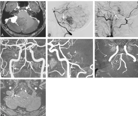
identified correctly on conventional MR imaging alone in 13 (62%) of 21 meningiomas (Fig 3). All dural attachments were identified when 3D TOF MRA was added to conventional MR images. In the 8 meningiomas where 3D TOF MRA was useful for the identification of dural attachments, their location included the sphenoid ridge (n⫽4), and parasagittal, central skull base, tentorial, and transverse-sigmoid sinus junction (n⫽1 each; Figs 1 and 2).
DISCUSSION
Compared with DSA, unenhanced 3D TOF MRA at 1.5T or less has been reported to be unsatisfactory for the depiction of arterial meningioma feeders.12,13We found that unenhanced 3D TOF
MRA at 3T is a reliable diagnostic tool for the identification of primary feeders; the agreement with DSA was excellent (⫽0.94; 95% CI, 0.84 –1.00). We attribute the excellent identification of primary feeders to the combined effect of several factors.
3T TOF MRA benefits from 2 key conditions. The theoretic signal-to-noise ratio at 3T is twice that at 1.5T, allowing for in-creased spatial resolution. The longer T1 values of tissues at 3T produce better background suppression, additional inflow en-hancement, and improved contrast-to-noise ratios.8-10In
addi-tion, the MRA sequence we used is based on a combination of MOTSA technique and short TE. In general, the sensitivity of MRA is limited because of phase dispersions from turbulence or
[image:4.594.54.532.45.343.2]flow saturation from slow flow. The MOTSA technique mini-mizes losses in signal intensity caused by spin saturation, main-tains small voxels and short TEs to minimize intravoxel phase dispersion,14,15and allows larger imaging volumes. Using these
techniques we were able to obtain high spatial resolution (recon-structed voxel size 0.25⫻0.25⫻0.5 mm), which allowed the depiction of small arterial feeders.
To evaluate the tumor feeders, our readers used axial source, MIP, and partial MIP MRA, and conventional MR images. Al-though the MIP algorithm provides angiogram-like images, the lower signal intensity feature of vessels may be lost and small or slow-flowing vessels may not be visualized.16MRA source images
contain both flow and anatomic information. Small intracranial structures can be seen because the acquisition of submillimeter sections results in high resolution and flow-related enhancement. On partial MIP images, vessels and the surrounding brain in a targeted region of interest can be seen without vessel overlap. To our observers evaluating the feeding arteries, conventional MR images were available, and some feeders could be predicted based on anatomic information provided by conventional MR images. We posit that our display method contributed to the identifica-tion of small feeders on 3D TOF MRA images.
Interobserver and intermodality agreements for secondary
feeders were slightly lower than for primary feeders. In our study, the spatial resolution of MRA was much lower than that of DSA. Compared with the voxel size of MRA images, the diameter of arterial feeders can be small, and the spatial resolution on these images is not sufficiently high enough to depict them. On unen-hanced 3D TOF MRA, the visualization of the feeders (or at least their intensity relative to stationary tissue) will depend on their orientation relative to the scan plane. If the scan plane is parallel to the flowing blood, enhancement may be lost as spins will be ex-posed to the saturating radio-frequency pulses. In addition, the details of the acquisition parameters (eg, TE, spatial resolution) as well as the vendor-specific details of the pulse sequence may have had an influence on the outcome of our study. Technical advances (eg, 32-channel coils, 7T MR imaging systems) have led to in-creases in the signal-to-noise ratio17,18and may improve the
vi-sualization of secondary feeders on MRA images. In 2 of our cases, parasitic or dural branches from the cerebral or cerebellar arteries were not secondary feeders but primary feeders. This unexpected blood supply to the tumor might have affected the readers’ inter-pretation of the MRA images.
The accurate assessment of dural attachments in meningi-omas is important for satisfactory surgical outcomes.3When
the tumors are small or located at a specific area, the dural
[image:5.594.56.534.48.351.2]attachment site may be easily identified by conventional MR imaging alone. This may not be true for large meningiomas, however. In some earlier reports, the dural attachment of me-ningiomas was identified, and hyperostosis at the dural attach-ment site is a phenomenon recognized on CT scans.19,20A
contrast-enhanced 3D MR technique (eg, fast imaging with steady-state acquisition) may provide information regarding the adhesiveness of dural attachments.21Because these are
in-direct radiologic findings, however, the accurate site at which
tumor-feeding arteries enter the tumor via the attached dura mater is not fully understood.
In our study, the addition of 3D TOF MRA to conventional MR imaging was use-ful for identifying the dural attachment in nearly one-third of the meningiomas, espe-cially large tumors in contact with several areas. We used source and partial MIP im-ages from MRA studies; they contain both anatomic and vessel information at high spatial resolution. We think that this pre-cise information contributed to the identi-fication of the dural attachment with feed-ing arteries supplyfeed-ing blood to the tumor.
Information on the vessels feeding in-tracranial hypervascular mass lesions is necessary for a differential diagnosis (eg, in-tra- versus exin-tra-axial lesions) and for plan-ning interventional procedures.22It is also
important for surgeons to know the vascu-lature ahead of time, even without preoper-ative embolization, to facilitate operpreoper-ative planning. From our study, it is not clear whether MRA appropriately triages pa-tients for beneficial preoperative DSA with or without embolization. The indication of preoperative DSA might be determined by various factors: tumor location, size and vascularity, and feeder location. This infor-mation may be obtained from MR imaging and MRA. Further investigations are re-quired to clarify the indication of preoper-ative DSA. Our results suggest that 3D TOF MRA is useful to identify the feeders of me-ningiomas. It cannot, however, replace DSA at the planning of interventional pro-cedures because its identification of other feeders is relatively limited. Compared with DSA, 3D TOF MRA also has several disad-vantages: lower spatial resolution, the lack of temporal resolution of the vascular flow dynamics, and no information about ve-nous anatomy.
Our study has some limitations. First, it is a preliminary look into a 3D TOF MRA technique at 3T in a specific subset of pa-tients. The temporal and spatial resolution of DSA could be further improved by using smaller FOVs and frame rates of up to 30/s. DSA can also be performed more selec-tively. Further comparative studies with DSA are needed to clarify the value of this MRA technique. Second, we did not compare unenhanced 3D TOF MRA with contrast-enhanced 3D MRA data. Because contrast-enhanced 3D MRA combines the T1 shortening effect of a gadolinium-based contrast agent, it might yield additional information on the feeding artery.23Third, we
did not address the actual number of vessels that supplied
indi-Table 3: Interobserver and intermodality agreement for the identification of secondary feeders
MRA Interobserver
Agreementa MRAb DSA
Intermodality Agreementc Reader 1 Reader 2
MMA 3 2 3 5
AMA 0 0 0 1
APhA 1 0 0 1
OA 0 0 0 1
OphA 1 2 1 1
ILT 0 0 15 (71%) 0 0 16 (76%)
MHT 5 3 ⫽0.58 5 4 ⫽0.72
ACA 2 2 关0.34–0.82兴 2 2 关0.51–0.93兴
MCA 2 4 2 2
PCA 0 0 0 0
SCA 0 0 0 0
AICA 0 1 1 0
PICA 0 0 0 0
None 7 7 7 4
Other 0 0 0 0
Note:—Data are number of meningiomas. Data in parentheses are the percentage of times that results that were concordant, and data in brackets are 95% confidence intervals. MMA indicates middle meningeal artery; AMA, accessory meningeal artery; APhA, ascending pharyngeal artery; OA, occipital artery; OphA, ophthalmic artery; ILT, inferolateral trunk; MHT, meningohypophyseal trunk; MCA, middle cerebral artery; ACA, anterior cerebral artery; PCA, posterior cerebral artery; SCA, superior cerebellar artery; AICA, anterior inferior cerebellar artery; PICA, posterior inferior cerebellar artery.
aAgreement of MRA between Reader 1 and Reader 2. b
Consensus reading of MRA of Reader 1 and Reader 2.
[image:6.594.53.365.65.242.2]cAgreement between the consensus reading of MRA of Reader 1 and Reader 2 and DSA.
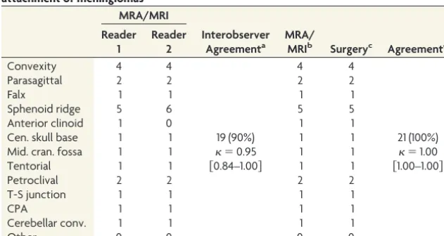
Table 4: Interobserver and intermodality agreement for the location of dural attachment of meningiomas
MRA/MRI
Interobserver Agreementa
MRA/
MRIb Surgeryc Agreementd Reader
1
Reader 2
Convexity 4 4 4 4
Parasagittal 2 2 2 2
Falx 1 1 1 1
Sphenoid ridge 5 6 5 5
Anterior clinoid 1 0 1 1
Cen. skull base 1 1 19 (90%) 1 1 21 (100%)
Mid. cran. fossa 1 1 ⫽0.95 1 1 ⫽1.00
Tentorial 1 1 关0.84–1.00兴 1 1 关1.00–1.00兴
Petroclival 2 2 2 2
T-S junction 1 1 1 1
CPA 1 1 1 1
Cerebellar conv. 1 1 1 1
Other 0 0 0 0
Note:—Data are number of meningiomas. Data in parentheses are the percentage of times that results that were concordant, and data in brackets are 95% confidence intervals. Cen. skull base indicates central skull base; Mid. cran. fossa, middle cranial fossa; T-S junction, transverse-sigmoid sinus junction; CPA, cerebellopontine angle; Cerebellar conv., cerebellar convexity
aAgreement of MRA/MRI between Reader 1 and Reader 2. b
Consensus reading of MRA/MRI of Reader 1 and Reader 2.
cFinal diagnosis of dural attachment was determined by surgical findings. When total tumor resection was not obtained, DSA findings were also used for determining the dural attachment of meningiomas.
[image:6.594.53.369.357.525.2]vidual tumors. Fourth, our data were obtained from a single-center study in a small number of patients. Additional studies with large populations are needed.
CONCLUSIONS
The excellent agreement between MRA and DSA findings sug-gests that unenhanced MRA at 3T is a reliable diagnostic tool for the identification of the primary meningioma feeders. MRA at 3T, however, cannot at present replace DSA for the identification of the feeding arteries of intracranial meningio-mas. The combined information from MRA and conventional MR imaging may be useful for evaluating the dural attachment of meningiomas.
REFERENCES
1. Wiemels J, Wrensch M, Claus EB.Epidemiology and etiology of me-ningioma.J Neurooncol2010;99:307–14
2. Gemmete JJ, Ansari SA, McHugh J, et al.Embolization of vascular tumors of the head and neck.Neuroimaging Clin N Am2009;19: 181–98
3. Adachi K, Kawase T, Yoshida K, et al.ABC surgical risk scale for skull base meningioma: a new scoring system for predicting the extent of tumor removal and neurological outcome.J Neurosurg2009;111: 1053– 61
4. Bassiouni H, Asgari S, Sandalcioglu IE, et al. Anterior clinoidal meningiomas: functional outcome after microsurgical resection in a consecutive series of 106 patients.J Neurosurg2009;111:1078 –90 5. Hu WY, TerBrugge KG.The role of angiography in the evaluation of
vascular and neoplastic disease in the external carotid artery circu-lation.Neuroimaging Clin N Am1996;6:625– 44
[image:7.594.53.534.45.447.2]6. Du C, Korogi Y, Nagahiro S, et al.Hemifacial spasm: three-dimen-sional MR images in the evaluation of neurovascular compression.
Radiology1995;197:227–31
7. Hirai T, Korogi Y, Hamatake S, et al.Three-dimensional FISP imag-ing in the evaluation of carotid cavernous fistula: comparison with contrast-enhanced CT and spin-echo MR.AJNR Am J Neuroradiol
1998;19:253–59
8. Willinek WA, Born M, Simon B, et al. Time-of-flight MR angiography: comparison of 3.0-T imaging and 1.5-T imaging– initial experience.Radiology2003;229:913–20
9. Schmitz BL, Aschoff AJ, Hoffmann MH, et al.Advantages and pit-falls in 3T MR brain imaging: a pictorial review.AJNR Am J Neuro-radiol2005;26:2229 –37
10. DeLano MC, DeMarco JK.3.0 T versus 1.5 T MR angiography of the head and neck.Neuroimaging Clin N Am2006;16:321– 41 11. Kunii N, Ota T, Kin T, et al.Angiographic classification of tumor
attachment of meningiomas at the cerebellopontine angle.World Neurosurg2011;75:114 –21
12. Kadota T, Kuriyama K, Inoue E, et al.MR angiography of meningi-oma.Magn Reson Imaging1993;11:473– 83
13. Engelhard HH.Progress in the diagnosis and treatment of patients with meningiomas. Part I: diagnostic imaging, preoperative embo-lization.Surg Neurol2001;55:89 –101
14. Blatter DD, Parker DL, Robison RO.Cerebral MR angiography with multiple overlapping thin slab acquisition. Part I. Quantitative analysis of vessel visibility.Radiology1991;179:805–11
15. Davis WL, Blatter DD, Harnsberger HR, et al.Intracranial MR
angiography: comparison of single-volume three-dimensional time-of-flight and multiple overlapping thin slab acquisition tech-niques.AJR Am J Roentgenol1994;163:915–20
16. Tsuruda J, Saloner D, Norman D.Artifacts associated with MR neu-roangiography.AJNR Am J Neuroradiol1992;13:1411–22
17. Parikh PT, Sandhu GS, Blackham KA, et al.Evaluation of image quality of a 32-channel versus a 12-channel head coil at 1.5T for MR imaging of the brain.AJNR Am J Neuroradiol2011;32:365–73 18. von Morze C, Xu D, Purcell DD, et al.Intracranial time-of-flight MR
angiography at 7T with comparison to 3T.J Magn Reson Imaging
2007;26:900 – 04
19. Akutsu H, Sugita K, Sonobe M, et al.Parasagittal meningioma en plaque with extracranial extension presenting diffuse massive hy-perostosis of the skull.Surg Neurol2004;61:165– 69
20. Bikmaz K, Mrak R, Al-Mefty O.Management of bone-invasive, hy-perostotic sphenoid wing meningiomas. J Neurosurg 2007;107: 905–12
21. Yamamoto J, Kakeda SH, Takahashi M, et al.Dural attachment of intracranial meningiomas: evaluation with contrast-enhanced three-dimensional fast imaging with steady-state acquisition (FIESTA) at 3T.Neuroradiology2010;53:413–23
22. Latchaw RE.Preoperative intracranial meningioma embolization: technical considerations affecting the risk-to-benefit ratio.AJNR Am J Neuroradiol1993;14:583– 86