ORIGINAL RESEARCH
SPINE
SAPHO Syndrome: Imaging Findings of Vertebral Involvement
XA.M. McGauvran,X A.L. Kotsenas,XF.E. Diehn,XJ.T. Wald,XC.M. Carr, andX J.M. Morris
ABSTRACT
BACKGROUND AND PURPOSE: Imaging findings in patients with a combination of synovitis, acne, pustulosis, hyperostosis, and osteitis (SAPHO) are often misinterpreted as discitis/osteomyelitis or metastases, resulting in multiple biopsies and delayed diagnosis. We have incidentally noted a semicircular morphology in vertebral body imaging in several cases of SAPHO syndrome with vertebral involvement. Our goal was to evaluate the prevalence of this distinctive morphology in these patients.
MATERIALS AND METHODS: A retrospective review of patients with SAPHO syndrome diagnosed between July 1998 and August 2013 was conducted. A descriptive analysis of MR imaging, CT, radiography, bone scanning, and PET imaging was performed for the presence and distribution of vertebral body signal intensity or attenuation changes and/or enhancement; contiguous vertebral body involvement; vertebral body collapse; endplate irregularity; disc space, facet, and spinous process involvement; subligamentous thickening; and para-spinal soft-tissue involvement.
RESULTS:Eighteen patients (16 women [89%]; mean age, 52.9 years) with SAPHO and spine involvement were included. Contiguous involvement ofⱖ2 vertebral bodies was found in 16 patients (89%), with a curvilinear or “semicircular” pattern involving portions of adjacent vertebral bodies in 10 (63%,P⫽.14). Most intervertebral discs demonstrated absence of abnormal T2 hyperintensity (73%) and enhancement (89%). Subligamentous thickening was present in 12 (67%). Paraspinal soft-tissue involvement was present in 6 (33%).
CONCLUSIONS: SAPHO syndrome should be included in the differential diagnosis in a patient with a curvilinear or semicircular pattern of vertebral involvement, contiguous vertebral body involvement, and absence of intervertebral disc edema and enhancement.
ABBREVIATION:SAPHO⫽combination of synovitis, acne, pustulosis, hyperostosis, and osteitis
T
he association of bone disease and chronic cutaneous pustular lesions has been observed since the 1960s, but it was not until 1987 that Chamot et al1first used the acronym SAPHO tode-scribe this rare group of chronic, relapsing, inflammatory osteo-articular disorders commonly associated with skin manifesta-tions. SAPHO was proposed to refer to a combination of synovitis, acne, pustulosis, hyperostosis, and osteitis as a heading for these syndromes. Cutaneous lesions are characterized by pal-moplantar pustulosis, acne conglobata, and/or hidradenitis sup-purativa. A wide variety of bone and joint manifestations has been described.
SAPHO syndrome shares some overlapping features with other spondyloarthropathies, including reactive arthritis, psori-atic arthritis, spondyloarthropathy associated with inflammatory bowel disease, and idiopathic ankylosing spondylitis. In children, the disease most commonly presents as a recurrent multifocal osteomyelitis, favoring the long bone metaphysis. This presenta-tion is in contradistincpresenta-tion to that in adults in whom the anterior chest wall, including the sternoclavicular and manubriosternal junctions, is most commonly affected.1-4The spine is also
fre-quently involved in adults. Findings of spinal involvement on conventional radiographic imaging have been reported to include vertebral body osteosclerosis, paravertebral ligament ossification, hyperostosis, and discovertebral junction lesions. Although plain radiographic findings of vertebral involvement have been well-described, the radiology literature has only a limited number of case reports and small case series describing imaging findings in detail with other modalities, specifically MR imaging.5-10These
advanced imaging findings are often misinterpreted as discitis/ osteomyelitis or metastases.11,12In our experience, this
misinter-Received October 9, 2015; accepted after revision January 14, 2016. From the Department of Radiology, Mayo Clinic, Rochester, Minnesota.
Paper previously presented at: Annual Meeting of the American Society of Neuro-radiology and the Foundation of the ASNR Symposium, May 17–22, 2014; Montreal, Quebec, Canada.
Please address correspondence to Amy L. Kotsenas, MD, Department of Radiol-ogy, Mayo Clinic, 200 First St SW, Rochester, MN 55905; e-mail: Kotsenas.amy@ mayo.edu; @AmyKotsenas
pretation can lead to unnecessary biopsies, other invasive proce-dures, and, ultimately, delayed diagnosis.
Anecdotally, we have commonly observed a “semicircular” pattern of contiguous vertebral body involvement localized to either the anterior or posterior vertebral bodies of the middle segments and adjacent anterior or posterior endplates of the sur-rounding vertebral bodies. This was first reported by Peffers et al.13
The goal of the study was to evaluate the imaging findings in a series of patients with vertebral involvement as part of SAPHO syndrome, with specific attention paid to the prevalence of this unique semicircular morphology in the vertebral bodies.
MATERIALS AND METHODS
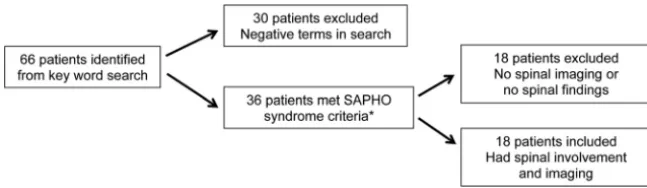
PatientsInstitutional review board approval was obtained, and informed consent was waived for this retrospective review. This study com-plied with all Health Insurance Portability and Accountability Act requirements. A search of clinical notes and the radiology infor-mation management system for patients with the possible diag-nosis of SAPHO or any combination of the terms “synovitis,” “acne,” “pustulosis,” “hyperostosis,” and “osteitis” between July 1998 and August 2013 identified 66 patients (Fig 1). Of these, 36 patients had a final clinical diagnosis of SAPHO meeting at least 1 of the 4 criteria proposed by Benhamou et al14: 1) osteoarticular
manifestations of acne conglobata, acne fulminans, or hidradeni-tis suppurativa; 2) osteoarticular manifestations of palmar plantar pustulosis; 3) hyperostosis involving either the anterior chest wall, spine, or limbs with or without dermatosis; and 4) chronic recur-rent multifocal osteomyelitis with or without dermatosis. Eigh-teen patients were further excluded because they did not have imaging of the spine.
Imaging and Analysis
We retrospectively reviewed the radiographic imaging findings, including MR imaging, CT, bone scintigraphy, and PET/CT. The images were evaluated by 2 experienced neuroradiologists in con-sensus (A.L.K. with 15 years, J.M.M. with 7 years). Biopsies, in-cluding location, number, and histopathologic and microbiologic culture results, were also recorded.
MR imaging was evaluated for vertebral body signal-intensity changes and/or enhancement; contiguous vertebral body involve-ment; the presence of vertebral body collapse; endplate irregular-ity; disc space involvement including enhancement; facet and spi-nous process involvement; subligamentous thickening; and paraspinal soft-tissue involvement. MR imaging sequences,
imag-ing parameters, and magnet strength varied because many of the scans came from outside facilities. Vertebral body signal intensity was evaluated on T1-and T2-weighted MR images T1-and on gadolinium-enhanced T1-weighted im-ages when available. The semicircular pattern of enhancement was recorded if present. The intervertebral discs were evaluated for enhancement, irregularity, associated fluid, and narrowing or wid-ening. The number and laterality of the facet joints with signal abnormalities and/or enhancement and the presence of spinous process signal changes were recorded. We looked for ligamentous thickening of the anterior or posterior longitudinal ligaments and ligamenta flava, including thickening that extended to uninvolved vertebral bodies. When there was masslike soft-tissue involve-ment, the location, maximal diameter (in millimeters), and lon-gitudinal extent of the abnormal tissues were recorded.
CT was evaluated for vertebral body sclerosis or lysis. Radio-tracer uptake, if present, was recorded for bone scintigraphy and PET/CT examinations.
Statistical Analysis
Statistical analysis was performed by using the JMP software package (Version 9.0; SAS Institute, Cary, North Carolina). The Fisher exact test was used to determine the association between the presence of vertebral involvement and sex, the semicircular pattern of involvement, disc space narrowing, and endplate irreg-ularities. Only 2-tailed tests were used. APvalue of⬍.05 was significant.
RESULTS
DemographicsOf the 36 patients with SAPHO who were identified (Fig 1), 26 (72%) were female with an age range of 11–76 years (mean, 44 years; median, 47.5 years). Eighteen patients had spinal imaging available for review, and all demonstrated spinal involvement (50%; 16 women [89%;P⫽.06]; age range, 23– 69 years).
Vertebral Lesions
A total of 104 vertebral bodies were involved in 18 patients (Table). One patient had a single level of involvement (6%), while the remaining 17 patients had a median of 5 vertebral levels involved (range, 2–13 levels; mean, 5.8⫾3.4 levels). The thoracic spine was most commonly involved (n⫽14, 78%), withⱖ2 separate thoracic segments involved in 57% of pa-tients (n⫽ 8/14). The lumbosacral spine was the next most commonly involved (n ⫽ 7, 39%), followed by the cervical spine (n⫽3, 17%). Multiple regions of the spine were involved in 7 patients (39%). Contiguous involvement ofⱖ2 vertebral bodies was found in 16 patients (89%), with a curvilinear or semicircular pattern involving the anterior or posterior por-tions of adjacent vertebral bodies in 10 of these patients (63%, P⫽.14) (Fig 2A, -B).
[image:2.594.54.378.46.140.2]MR imaging was available for 16 patients (89%). Many pa-tients came to our institution with imaging from other facilities. Therefore, there was variation in the MR imaging field strength, sequences, and imaging parameters. T1-weighted images were available in all cases; 1 patient (6%) did not have T2-weighted imaging. Vertebral body low-signal intensity on T1-weighted im-ages was seen in 15/16 patients (93%) with corresponding high-signal intensity on T2-weighted images in 12 patients (75%), compatible with bone marrow edema (Fig 2A,-B). One patient (6%) had mixed hypo-/hyperintensity on T1-weighted images, while 3 patients (18%) had mixed hypo-/hyperintensity on T2-weighted images, suggestive of concomitant bone marrow edema and cancellous bone sclerosis. Gadolinium-based con-trast was administered in 9 patients in whom MR imaging was available (56%), with 100% of patients demonstrating en-hancement in the same distribution as the signal changes de-scribed above. Vertebral body corner erosions were not ob-served in any patient.
Corresponding noncontrast CT imaging was available in 14 of the 16 patients in whom MR imaging was available. CT demon-strated sclerosis in the areas of MR signal abnormality in 100% of patients, regardless of the pattern of the MR imaging signal changes (Fig 2C). No lytic lesions were identified.
Nine patients had technetium Tc99m methylene diphosphonate 3-phase whole-body bone scans to correlate with the spi-nal MR imaging. All 9 patients demon-strated increased radiotracer on MR imaging in the vertebral bodies affected (Fig 2D). There were no discordant areas of vertebral body uptake between the 2 modalities. Extravertebral findings on bone scans are described in further detail below. In 2 of the 18 (11%) patients with spinal involvement who did not have an MR imaging, the number, level, and contiguity of vertebral lesions were de-termined solely by nuclear medicine bone scanning in 1 patient and with lumbar spine radiographs in the second. In the first patient, the bone scan dem-onstrated radiotracer uptake in the non-contiguous T5, T9, T11, and L1 vertebral bodies and right sacroiliac joint. In the second, lumbar spine radiographs dem-onstrated attenuated vertebral body sclerosis from L3 to the sacrum and par-tial fusion of the left sacroiliac joint.
Thirteen of 18 patients (72%) had plain radiographs. One patient had nor-mal findings on plain radiographic im-aging (8%). Twelve patients had verte-bral body sclerotic changes, including shiny corners (92%). Despite abnormal findings on MR imaging, no abnormal FDG uptake was seen in the 2 patients with whole-body FDG-PET/CT.
Facet Joints/Spinous Processes
There were 34 facet joints involved with T2 hyperintensity and/or enhancement in 7 patients (39%), with unilateral involvement at 6 levels (3 patients, 17%) and bilateral involvement at 14 levels (4 patients, 22%). Spinous process T2 hyperintensity or enhance-ment or both was observed at 15 levels in 5 patients (28%), all with concurrent uni- or bilateral facet joint involvement at the same levels (Fig 3).
Disc Spaces
MR imaging demonstrated at least 1 intervening or adjacent disc space that was narrowed in 8 of 16 (50%) patients, with irregular endplates observed in 7 patients (43%). The combination of disc space narrowing with endplate irregularity was noted in 5 patients who had MR imaging (62%;P⫽.11). High signal on T2-weighted images in the disc was seen in 5 patients (31%). Disc space enhance-ment on gadolinium-enhanced T1-weighted images was observed in 2 of 9 patients receiving gadolinium (22%). The combination of disc space narrowing, abnormal T2 hyperintensity, and endplate irregu-larity was observed in 3 patients (18%), while only 1 patient (11%) had all 4 findings: disc space narrowing, abnormal T2 hyperintensity, endplate irregularity, and disc space enhancement.
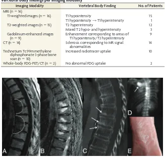
FIG 2. MR imaging, CT, and bone scan of the thoracic spine in a 74-year-old woman with back pain. Sagittal T1-weighted (A) and STIR (B) images demonstrate hypo- and hyperintensity, respec-tively, in a curvilinear or semicircular pattern (dashed line) in contiguous vertebral body segments. Note the absence of abnormal signal within the intervertebral disc spaces.C, Sagittal CT image shows associated sclerosis (arrows) corresponding to levels of abnormal increased signal on MR imaging.D, Bone scan, posteroanterior view, demonstrates focal areas of increased radiotracer uptake within thoracic vertebral bodies and the right sternoclavicular joint (arrow).E, Previously undiagnosed plantar pustulosis was evident on physical examination.
Vertebral body findings per imaging modality
Imaging Modality Vertebral Body Finding No. of Patients
MRI (n⫽16)
T1-weighted images (n⫽16) T1 hypointensity 15 T1 hypointensity3T1 hyperintensity 1 T2-weighted images (n⫽15) T2 hyperintensity 12 Mixed T2 hypo- and hyperintensity 3 Gadolinium-enhanced images
(n⫽9)
Enhancement corresponding to areas of T1 hypointensity/T2 hyperintensity
9
CT (n⫽14) Sclerosis corresponding to MR signal abnormalities
14
Technetium Tc99m methylene diphosphonate 3-phase bone scan (n⫽10)
Increased radiotracer uptake 10
[image:3.594.51.378.50.367.2]Soft Tissues
Thickening of the anterior longitudinal ligament was present in 12 patients (80%) and extended to otherwise uninvolved verte-bral bodies in 3 patients (25%). No other spinal ligaments were involved. Paraspinal soft-tissue involvement consisted of mass-like soft-tissue thickening with enhancement (Fig 4). This was observed in 6 patients (37%), with a range of 1– 6 vertebral levels involved. Masslike paraspinal soft-tissue involvement did not ex-ceed 15 mm in maximal diameter in any case (range, 2–15 mm).
Extraspinal Involvement
Seventeen of 18 patients (94%) had typical skin manifesta-tions. Ten (59%) of the 17 patients had palmoplantar
pustulo-sis (Fig 2E), 2 patients (12%) had acne conglobata, 1 patient (6%) had hidrad-enitis suppurativa, and 6 patients (35%) had nonspecific dermatoses. Two pa-tients (12%) had⬎1 skin manifestation, with 1 patient having both palmoplantar pustulosis and acne conglobata, while the other patient had both palmoplan-tar pustulosis and hidradenitis suppu-rativa. Seven of the 18 patients (39%) with SAPHO syndrome and spinal in-volvement also had concurrent sterno-clavicular involvement (Fig 2), while 4 patients (22%) had involvement of the first costovertebral joint. Four of the 18 patients (22%) had plain radio-graphs of the sacroiliac joints, with 1 patient (25%) demonstrating sclerosis and the remaining 3 patients demon-strating no involvement of the sacroil-iac joint.
Pathology/Microbiology
Twelve patients (67%) underwent biop-sy; 7 patients had a single biopsy, 3 pa-tients had 2 biopsies, and 2 papa-tients had 3 biopsies, totaling 19 biopsies. The ver-tebral body was the most commonly bi-opsied location (n⫽ 11), followed by the paravertebral soft tissues (n⫽5) and disc interspace (n⫽ 1). In 2 patients with spinal involvement, a concomitant sternal lesion was biopsied. Pathology was negative for malignancy in all biopsy specimens (n⫽19), including 2 patients (11%) with a known history of malig-nancy. Microbiologic cultures were also negative for infection in all 8 patients who had microbiologic testing performed.
Inflammatory/Infectious Markers
C-reactive protein levels were available in 13 of 18 patients (72%), with elevated lev-els in 8 patients (62%). Sedimentation rate levels were available in 16 of 18 patients (89%), with elevated levels in 6 patients (38%). White blood cell counts were available in 17 of 18 patients (94%), with all patients having normal levels.
DISCUSSION
A curvilinear or a semicircular pattern of contiguous vertebral body involvement localized to either the anterior or posterior vertebral bodies of the middle segments and adjacent anterior or posterior endplates of the surrounding vertebral bodies was found in most of our patients (63%) with SAPHO and spinal involvement. The high prevalence of this semicircular pattern of vertebral body signal alteration and enhancement may help to
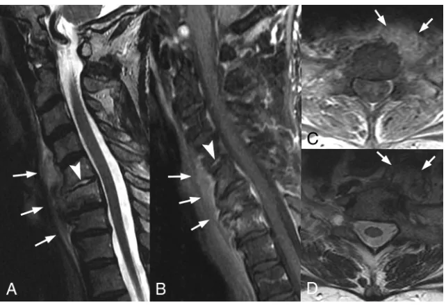
FIG 3.MR images in a 32-year-old woman with back pain. Sagittal T1-weighted fat-saturated images of the thoracic spine following administration of gadolinium demonstrate multilevel enhancement (arrows) of the spinous processes (B) and bilateral facet joints (AandC).
[image:4.594.56.379.47.216.2] [image:4.594.55.376.261.481.2]differentiate SAPHO syndrome from metastases, which tend to be randomly distributed throughout the spine. When combined with a relatively low prevalence of abnormal disc space T2-signal and enhancement, the high prevalence of the semicircular pattern may also be helpful to radiologists in cases in which spinal infec-tion is being considered. Correlainfec-tion with CT may be beneficial because sclerosis in the areas of MR signal abnormality was pres-ent in 100% of our patipres-ents, regardless of the pattern of the MR signal changes. Recognition of these findings by radiologists should prompt clinical consultation with dermatology and/or rheumatology to assess typical skin and other musculoskeletal manifestations.
Osteosclerosis of the vertebral bodies in SAPHO syndrome has been described by Leone et al15and may progress to produce
diffuse and generalized sclerosis with development of hyperosto-sis in more chronic cases. Prior case reports16-18have shown the
utility of [18F] FDG-PET/CT to differentiate active from healed chronic inflammatory lesions because PET/CT shows increased uptake only in lesions with active inflammation. The chronic os-teosclerotic changes seen in our patients are hypothesized to rep-resent a quiescent phase of the disease, which likely accounts for the normal FDG uptake in the 2 patients who had PET/CT scans in our series.
In contrast to a study performed by Laredo et al,6our patients
were more likely to have involvement at contiguous levels. In addition, while our patients had frequent involvement of verte-bral body corners, we did not identify erosion in any case. Because we are a tertiary referral center, our patients may have presented at a later clinical stage. In fact, nearly all our patients presented after other manifestations such as skin lesions or sternoclavicular in-volvement, both of which are considered highly specific, were evident.
The frequency of spondylodiscitis in SAPHO syndrome has been reported to be 9%–32%.10Prior studies have shown chronic
sterile nonspecific inflammation from intervertebral disc biopsies in patients with SAPHO syndrome, equivalent to the Andersson lesion, which has been identified in 4.5% of patients with anky-losing spondylitis.5,10Others have suggested that spondylodiscitis
is due toPropionibacterium acnes.19Although disc space
involve-ment with fluidlike signal on T2-weighted images and disc space enhancement have been previously described in patients with SAPHO syndrome,5,7our study showed that most intervertebral
discs in patients with SAPHO syndrome demonstrated the ab-sence of both fluidlike signal on T2-weighted images (69%) and disc space enhancement (78%). The absence of these findings may be helpful in distinguishing SAPHO involvement from spinal infection.
We found single-level vertebral involvement rare, with contig-uous involvement ofⱖ2 levels in nearly 90%, often withⱖ5 levels involved. This finding is in contrast to cases of spondyloarthropa-thy, in which it is uncommon to see contiguous involvement.6
Nachtigal et al7previously described the paravertebral soft
tis-sues in patients with SAPHO involvement of the spine possibly
showing abnormal signal intensity. We found similar results, with thickening and enhancement of the anterior longitudinal liga-ment present in 37% of patients with SAPHO syndrome. These findings further complicate the differential diagnosis of SAPHO syndrome versus infection but may help to differentiate it from spondyloarthropathies, which, to our knowledge, have only been reported to affect the supraspinous ligament, interspinal liga-ments, and ligamenta flava.20Additionally, in the 2 patients with
vertebral body involvement determined solely on nuclear medi-cine bone scans or plain radiographs, both demonstrated unilat-eral sacroiliac joint involvement. Depasquale et al11and Leone et
al15have both described increased sclerosis and hyperostosis,
par-ticularly at the iliac side of the joint, in patients with SAPHO. When seen in patients with features of moderate sacroiliitis, this presentation was thought to be highly suggestive of SAPHO syndrome and helped to differentiate it from other spondyloarthropathies.
Peffers et al13first described a subset of patients with
nonbac-terial osteitis involving the thoracic spine. Spinal involvement in our study had a strong female predominance and preferentially affected the thoracic spine. This is in contrast to osteomyelitis and discitis, in which males are affected twice as often as females and the lumbar spine is most commonly affected.21
MR imaging of the spine and sacroiliac joints has played a key role in patients with spondyloarthropathies and has led to im-proved understanding of the course of the disease and to an earlier diagnosis.20We suggest an imaging protocol consisting of at
least a sagittal T1-weighted turbo spin-echo sequence, sagittal fat-saturated T2-weighted turbo spin-echo sequence or STIR sequence with high resolution, and a sagittal fat-saturated gad-olinium-enhanced T1-weighted sequence for evaluation of pa-tients with suspected SAPHO syndrome, spondyloarthropa-thy, or osteomyelitis/discitis.
Our study had a number of limitations. This was a retrospec-tive review without a control group, and images were reviewed in consensus rather than independently by the 2 neuroradiologists, which might limit the reproducibility. Additionally, the number of patients was small; however, SAPHO syndrome is an uncom-mon entity, and ours is one of the largest studies to date. The MR imaging protocols were not standardized. For example, not every patient had a fat-saturated sequence, 1 patient lacked a T2-weighted sequence, and 7 of 16 patients (44%) did not receive intravenous contrast material. Variation in MR imaging field strength and imaging protocols was a relatively minor limitation because signal change was noted on precontrast T1-weighted images in all patients. Finally, biopsy and microbiologic results were not available in half of the patients, perhaps due to the presence of other highly specific anterior chest wall and/or skin manifestations.
determine whether this semicircular pattern is a unique spinal finding in patients with SAPHO syndrome.
CONCLUSIONS
In a patient with a curvilinear or semicircular pattern of contigu-ous vertebral involvement, sclerosis along ligamentcontigu-ous attach-ment sites, and the absence of intervertebral disc abnormal T2 hyperintensity and enhancement, SAPHO syndrome should be included in the differential diagnosis and clinical consultation with dermatology and/or rheumatology should be considered. This may prevent the imaging findings from being misinterpreted as discitis/osteomyelitis or metastases, with subsequent potential reduction in the number of unnecessary biopsies and delayed diagnoses.
ACKNOWLEDGMENTS
We thank Sonia Watson, PhD, for her assistance in editing the manuscript.
REFERENCES
1. Chamot AM, Benhamou CL, Kahn MF, et al. Acne-pustulosis-hy-perostosis-osteitis syndrome: results of a national survey— 85 cases [in French].Rev Rhum Mal Osteoartic1987;54:187–96Medline 2. Boutin RD, Resnick D.The SAPHO syndrome: an evolving concept for unifying several idiopathic disorders of bone and skin.AJR Am J Roentgenol1998;170:585–91CrossRef Medline
3. Kahn MF, Chamot AM.SAPHO syndrome.Rheum Dis Clin North Am1992;18:225– 46Medline
4. Maugars Y, Berthelot JM, Ducloux JM, et al.SAPHO syndrome: a followup study of 19 cases with special emphasis on enthesis in-volvement.J Rheumatol1995;22:2135– 41Medline
5. Akisue T, Yamamoto T, Marui T, et al.Lumbar spondylodiscitis in SAPHO syndrome: multimodality imaging findings.J Rheumatol
2002;29:1100 – 01Medline
6. Laredo JD, Vuillemin-Bodaghi V, Boutry N, et al.SAPHO syndrome: MR appearance of vertebral involvement. Radiology 2007;242: 825–31CrossRef Medline
7. Nachtigal A, Cardinal E, Bureau NJ, et al.Vertebral involvement in SAPHO syndrome: MRI findings.Skeletal Radiol1999;28:163– 68 CrossRef Medline
8. Perez C, Hidalgo A, Olier J, et al.MR imaging of multifocal
spondy-lodiskitis as the initial manifestations of SAPHO syndrome.AJR Am J Roentgenol1998;171:1431–32Medline
9. Tohme-Noun C, Feydy A, Belmatoug N, et al.Cervical involvement in SAPHO syndrome: imaging findings with a 10-year follow-up.
Skeletal Radiol2003;32:103– 06CrossRef Medline
10. Toussirot E, Dupond JL, Wendling D.Spondylodiscitis in SAPHO syndrome: a series of eight cases.Ann Rheum Dis1997;56:52–58 CrossRef Medline
11. Depasquale R, Kumar N, Lalam RK, et al.SAPHO: what radiologists should know.Clin Radiol2012;67:195–206CrossRef Medline 12. Hong SH, Choi JY, Lee JW, et al.MR imaging assessment of the
spine: infection or an imitation? Radiographics2009;29:599 – 612 CrossRef Medline
13. Peffers G, James SL, Stirling A, et al.Thoracic spine osteitis: a distinct clinical entity, a variant of SAPHO or late-onset non-bacterial oste-itis?Rheumatology (Oxford)2012;51:191–93CrossRef Medline 14. Benhamou CL, Chamot AM, Kahn MF.Synovitis-acne-pustulosis
hyperostosis-osteomyelitis syndrome (SAPHO): a new syndrome among the spondyloarthropathies? Clin Exp Rheumatol 1988;6: 109 –12Medline
15. Leone A, Cassar-Pullicino VN, Casale R, et al.The SAPHO syndrome revisited with an emphasis on spinal manifestations.Skeletal Radiol
2015;44:9 –24CrossRef Medline
16. Inoue K, Yamaguchi T, Ozawa H, et al.Diagnosing active inflamma-tion in the SAPHO syndrome using 18FDG-PET/CT in suspected metastatic vertebral bone tumors.Ann Nucl Med2007;21:477– 80 CrossRef Medline
17. Patel CN, Smith JT, Rankine JJ, et al.F-18 FDG PET/CT can help differentiate SAPHO syndrome from suspected metastatic bone disease.Clin Nucl Med2009;34:254 –57CrossRef Medline 18. Takeuchi K, Matsusita M, Takagishi K.A case of SAPHO
(synovitis-acne-pustulosis-hyperostosis-osteomyelitis) syndrome in which [18F]fluorodeoxyglucose positron emission tomography was use-ful for differentiating from multiple metastatic bone tumors.Mod Rheumatol2007;17:67–71CrossRef Medline
19. Gerster JC, Lagier R, Livio JJ.Propionibacterium acnes in a spondy-litis with palmoplantar pustulosis.Ann Rheum Dis1990;49:337–38 Medline
20. Sieper J, Rudwaleit M, Baraliakos X, et al.The Assessment of Spon-dyloArthritis international Society (ASAS) handbook: a guide to assess spondyloarthritis.Ann Rheum Dis2009;68(suppl 2):ii1– 44 Medline