ISSN Online: 2158-7043 ISSN Print: 2158-7027
DOI: 10.4236/jbnb.2018.91001 Dec. 18, 2017 1 Journal of Biomaterials and Nanobiotechnology
A New Biomaterial for Urinary Catheters
Roberto Santos Lima
1*, Salvador Vilar Correia Lima
2, José Lamartine De Andrade Aguiar
2,
Eziel Cavalcanti Vasconcelos Rocha
1, Flávia Cristina Morone Pinto
21Life Sciences Center, Federal University of Pernambuco (UFPE), Caruaru, Brazil
2Postgraduate Program in Surgery, Department of Surgery, Center for Health Sciences, Federal University of Pernambuco (UFPE), Recife, Brazil
Abstract
Several studies argue that an ideal biomaterial for urinary catheters is utopian. Based in literature review it seems to be true. However, research advances: the biomaterial itself, new designs, new coatings, associated drugs, etc. Once im-planted and interacting with urine, two old problems persist: encrustation and bacterial colonization. In this context, an extracellular product from bacterial synthesis on sugarcane molasses biomaterial has been studied in several expe-rimental and clinical studies. Based on its high biocompatibility, the aim of this study is to evaluate its performance in an in vivo model as an endouro-logic prosthesis implanted in the bladder of Wistar rats. We evaluate physical, chemical and biological phenomena in comparison to an already established biomaterial, polyurethane. Even though it is not a finished product, the su-garcane biopolymer presented similar performance compared to polyurethane in several analyzed parameters and has an important characteristic: low cost.
Keywords
Biomaterial, Urinary Catheters, Encrustation
1. Introduction
Since antiquity there have been urinary catheters usefulness reports. Initially they were made from papyrus or metal and were used for urinary retention relief [1] [2]. Industrialization brought the vulcanized rubber, than they becamechemically stable and flexible at different temperatures [3]. In 20th-century various synthetic
biomaterials and different applications emerged [4]. Specifically in urology, due to high using frequency and necessity to remain implanted for long term, two uri-nary catheters have been highlighted: the Foley urethral catheter and the ureteral How to cite this paper: Lima, R.S., Lima,
S.V.C., De Andrade Aguiar, J.L., Rocha, E.C.V. and Pinto, F.C.M. (2018) A New Biomaterial for Urinary Catheters. Journal of Biomaterials and Nanobiotechnology, 9, 1-12.
https://doi.org/10.4236/jbnb.2018.91001
Received: October 14, 2017 Accepted: December 15, 2017 Published: December 18, 2017
Copyright © 2018 by authors and Scientific Research Publishing Inc. This work is licensed under the Creative Commons Attribution International License (CC BY 4.0).
DOI: 10.4236/jbnb.2018.91001 2 Journal of Biomaterials and Nanobiotechnology stent, and therefore are the most studied [5]. Also, there are two major problems associated with urinary catheters: bacterial colonization (and consequent infec-tion) and encrustation. In North America, more than 100 million urinary devic-es are implanted each year, bringing immense morbidity and cost [6]. In the United States, approximately two million nosocomial infections occur annually. Forty percent are urinary tract related, and 60% of these are urinary catheters related [7]. A Foley’s catheter implanted patient has 100% chance developing bacterial colonization in 30 days term [8]. Despite existence of various biomate-rials such as latex (polyisoprene), polyethylene, polyvinylchloride (PVC), polyu-rethane, silicone, biodegradables ones, and those made from heterologous and autologous tissue, the ideal biomaterial for endourological purpose was not dis-covered [4] [9] [10]. Which characteristics does it must have? It must be resis-tant to biofilm formation and bacterial colonization; able to resist encrustation; be biologically inert; have chemical stability when in urine contact; be easy to implant and remove; not prone to migration; allow optimal flow; be sterilizable; durable; radiopaque and low cost [11].
The Sugar Cane Biopolymer (SCB) is a biodegradable biomaterial. It is a po-lysaccharide obtained by bacterial synthesis from sugarcane molasses [12]. Pre-vious studies indicate that it is biocompatible, has low toxicity and induces tissue remodeling [13] [14]. In clinical applications this biomaterial proves to be versa-tile and has been applied in different experimental studies [15]-[24]. In this text, the aim of this study was to evaluate a SCB performance when in urine con-tact comparing with a stablished biomaterial.
2. Materials and Methods
Study Design: Fifty-one male Wistar rats (Rattus norvegicus albinus), from Nutrition Department Bioterium were transferred to Experimental Surgery Nuc-leus, both of the Federal University of Pernambuco. They were 12 - 16 weeks old of age and 260 - 589 grams of weight. They were distributed in five groups:
SCB-3 group (n = 10), operated animals and SCB tubes implanted and fol-lowed up for 3 months; DJ-3 group (n = 10), operated animals and double J tubes implanted and followed up for 3 months; SCB-6 group (n = 13), animals operated and implanted with SCB tubes and followed up for 6 months; DJ-6 group(n = 9), animals operated and implanted with double J tubes and followed up for 6 months; and Control Group (9), animals operated without implanta-tion, followed for 6 months.
All animals were kept in cages with wood shavings on the ground, in an envi-ronment with temperature and humidity control, day-night cycle artificially es-tablished in 12 for 12 hours, free access to drinking water and Labina® ad libi-tum feed.
pur-DOI: 10.4236/jbnb.2018.91001 3 Journal of Biomaterials and Nanobiotechnology pose it was used a 6F polyurethane ureteral stent (Stent Ureteral Universa, Han-dle Cook®) transversely sectioned in 5 mm pieces. The choice of animals for surgery was performed at random. All tubes were sterilized by gamma radiation in the Nuclear Energy Department of Federal University of Pernambuco. Figure 1 illustrates the materials.
[image:3.595.258.489.486.563.2]Perioperative Procedures: Animals underwent 12 hours fast and were anes-thetized under Bioterium protocol that used atropine, xylazine and ketamine. During the surgical procedure receiving supplemental oxygen, the animal was positioned on the surgical table in dorsal decubitus, after abdominal trichotomy was performed antisepsis with povidone-iodine. After apposition of sterile hole drape it was performed a 4 cm length infra-umbilical incision and blunt dissec-tion until peritoneal cavity. The bladder was identified and grasped. A 6 - 7 mm incision was performed at cupula and a tube implanted in vesical lumen. In this point war performed a continuous suture with polyglactin 910 (Vicryl 6-0®). Pe-ritoneal cavity was cleaned and abdominal wall was closed with cat gut 4-0 in-terrupted suture in two layers (aponeurosis and skin). Animal was taken to indi-vidual cages with slightly elevated decubitus, remaining heated under a light source in the first hours. In first week they were evaluated daily and after eighth day, weekly. The evaluation was: consciousness level, motor activity, food and water intake and wound aspect. On euthanasia’s day animal was weighted again and received a lethal dose of intraperitoneal anesthetic. It was performed in-verted U-laparotomy. Bladder liquid content was aspirated with needle for mi-crobiological analysis than a cystectomy was performed. The bladder was opened longitudinally with introduction of scissors through the urethra. Its macroscopic appearance and content (tubes, calculations, scale, organic material) were checked and the recovered material was put into a sterile, dry tube for further analysis.
[image:3.595.207.542.593.691.2]Figure 2 shows a SCB tube implantation, bladder suture and a SCB tube visualization
Figure 1. SCB tube on the left and polyurethane tube on the right.
DOI: 10.4236/jbnb.2018.91001 4 Journal of Biomaterials and Nanobiotechnology through bladder wall on euthanasia’s time.
Histological Analysis: Bladders was fixed in 10% formalin on parchment paper to avoid contraction and folds. All material was stained with hematoxylin and eo-sin. Histopathological evaluation was performed by a single pathologist.
Microbiological Analysis: Urine samples were seeded in MacConkey medium and rich blood agar (Casoy medium with 5% defibrinated sheep’s blood) at 35˚C for 18 to 24 hours in aerophilic, and read out after 24 hours.
Chemical Analysis: Encrustation substances and stones underwent qualitative analysis with a urinary stone test often used clinically, and they underwent quan-titative analysis by Energy Dispersive X-ray Fluorescence Spectrometer EDX Se-ries EDX-720/800HS.
Analytical Procedures: Each animal was weighted in grams prior to implant surgery and before euthanasia. Macroscopic phenomena consisted of 5 categorical variables: color change, shape change, presence of organic material, encrustation and formation of calculi. These variables were expressed as frequency (proportion) in each group as well the histological categories, if there were changes or not.
Statistical Analysis: Weight in grams of each group and the loss or gain be-tween to the surgery day to euthanasia day were calculated and expressed as mean and standard deviation. Student’s t test and Tuckey test for multiple comparisons were used for comparisons between groups. Categorical variables between the groups were compared using Fisher’s exact test. It was accepted probabilities with values less than 5%. Epi Info 6.0 software was used to statistical calculations.
Ethical Procedures: This study was submitted and approved by the Animal Ethics and Research Committee of Federal University of Pernambuco, under ID number 23076.013771/2009-25.
3. Results
[image:4.595.57.542.553.680.2]All animals underwent implantation tube had weight loss, but without statistic differences between groups. Control group had statistically significant weight gain.
Table 1 shows these data.
Table 1. Parameters related to weight gain and loss.
Variables Groups (n)
SCB-3 (10) DJ-3 (10) SCB-6 (13) DJ-6 (9) Control (9) WID (g) M ± SD 515 ± 40.91 451.2 ± 64.95 424.76 ± 52.97 463.22 ± 42.92 356.0 ± 99.42 WED (g) M ± SD 495.75 ± 33.93 434.5 ± 53.46 413 ± 41.95 450.44 ± 42.13 397.57 ± 104.91
Minimum weight 449 380 350 449 260
Maximum weight 580 589 539 527 511
WED-WID = variation M ± SD −17 ± 14.2 −16.7 ± 13.7 −10.55 ± 16.3 −12.77 ± 20.0 45.71 ± 9.5 p value p < 0.001 SCB-3/DJ-3/SCB-6/DJ-6 vs control
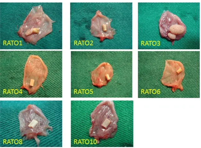
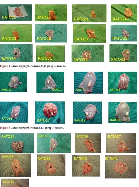
DOI: 10.4236/jbnb.2018.91001 5 Journal of Biomaterials and Nanobiotechnology There were five deaths (9.8%), all occurred in the first 24 h, with two in SCB-3 group, one in SCB-6 and one in control group. All animals underwent necropsy but no important findings. Concerning deaths there is no statistically significant findings (SCB-3 vs DJ-3 p = 0.473; SCB-6 vs DJ-6 p = 1; SCB-3 vs Control p = 0.544; SCB-6 vs Control p = 0.470). On 3 months term some findings were ob-served: almost all SCB tubes changed its color when compared to DJ tubes (7 of 8 and 0 of 10, respectively = 0.0007). Also, concerning shape changes, the SBC tubes were more likely to this alteration (4 of 9 and 0 of 10, respectively, p = 0.022). When others variable were studied (presence of organic material or cal-culi, encrustation) there is not differences between groups. On 6 months term, just the shape change variable was different, again in SCB tubes compared with DJ ones (10 of 12 and 0 de 9, respectively, p = 0.0002). The other variables were not observed differences. In control group was not found any organic material, calculi or encrustation. Figures 3-6 illustrate those macroscopic phenomena.
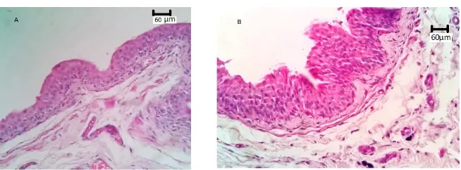
Forty six bladders were resected and processed for histological analysis. Twenty three bladders were classified as normal. When abnormal exhibited two main al-terations: Mostly in the epithelial layer (urothelium), all of them considered reac-tion from injury caused by tubes themselves, stones, and infecreac-tion; and lamina propria exhibited cells from inflammatory response. In the first ambit there was urothelium thickening, undulations, papillary growth and sometimes more spe-cific architecture such cystic cystitis. In the second, there were found inflamma-tory cells such as neutrophils, lymphocytes, plasma cells, eosinophils, and mast cells. All benign, no atypia. Comparing groups no statistic differences were found.
Figure 7 illustrates common findings in this study.
[image:5.595.213.534.469.705.2]There were six animals where urine samples microbiological growth (11.76%), with two in the DJ-3 group, three in the SCB-6 group and one in the DJ-6 group.
DOI: 10.4236/jbnb.2018.91001 6 Journal of Biomaterials and Nanobiotechnology
[image:6.595.75.528.57.665.2]Figure 4. Macroscopic phenomena. SCB group-6 months.
[image:6.595.75.524.474.708.2]Figure 5. Macroscopic phenomena. DJ group-3 months.
DOI: 10.4236/jbnb.2018.91001 7 Journal of Biomaterials and Nanobiotechnology
Figure 7. Bladder wall sections: hematoxylin and eosin stain from an optic microscope 400x. (A) Rat 4 showing preserved epithelium and lamina propria; (B) Rat 33 showing increased number of epithelial layers and undulated surface (epithelial hyperplasia).
There is no statistic difference. Isolated microorganisms were: Staphylococcus epidermidis and Staphylococcus aureus.
The physical-chemical qualitative analysis found three substances: carbonate, phosphate and ammonia in the calculi of the three animals, one from each group below: SCB-3, DJ-3 and DJ-6. Same samples underwent spectrometry revealed: phosphorus, calcium and magnesium. All samples had similar proportions of those elements pointing to higher concentration of struvite stones (magnesium ammo-nia phosphate) than calcium phosphate.
4. Discussion
blad-DOI: 10.4236/jbnb.2018.91001 8 Journal of Biomaterials and Nanobiotechnology der of foreign materials (e.g. zinc) or associations of the models [30].
Ford et al. and Carvalho Júnior et al. used methodology very similar to ours
[17] [31]. Ford evaluated performance of several 5mm squares polymers free-
floating in bladder lumen for 3 months term. Carvalho Júnior studied the per-formance of suture thread made from SCB after a transfixing suture in rats blad-der for 4 - 8 weeks. Regarding the cross-section of ureteral stents, other studies used same arrangement for encrustation evaluation and comparison [32]. Po-lyurethane stent was chosen because it is one of the most versatile biomaterials. It has composed numerous biomedical devices such as breast implants, urinary catheters, blood bags, and artificial heart. Durable, elastic, and low cost. It has great interaction with water and blood. As a ureteral stent is the most commonly used polymer in urological practice [9] [10] [33]. About physical phenomena, SCB tubes were made from thin membranes without special coating, its surface is rough, therefore deformities and discolorations were expected. That characte-ristic may block adequate flow through catheter, but this happens on others bio-degradable biomaterials. There is a study where the catheter sustained its paten-cy for only two days [10]. When the tubes were manufactured we used distilled water to mold them, so we already expected that change. One study evaluated macroscopic phenomena and among them the color change as a encrustation predictor and difficulty for the removal as well. This study was hardly criticized for its speculative character under the argument there is no evidence that change color is a good parameter for assessing encrustation and infection [34]. Certain-ly, there are differences between rats and humans life expectancy, and this aspect has to be observed in experimental studies. Our follow up time was so long. First, we based upon Ford’s study [31] that followed animals for 3 months. Also, stu-dies in humans demonstrate increasing complications catheter-related chances beyond 3 months [35]. Second, in the present study, due to the low incidence of encrustation and other phenomena observed in the three months, this time in the following groups was doubled, which gave more robustness to study. There is no study with so long follow up. Probably, this was the reason of SCB biode-gradation in one SCB-6 group animal. This fact coincides with Carvalho Júnior’s study [17]. Also there was no trace of SCB thread in one animal. There is pro-portionality between time and stone size. In a rat model for stone formation, rats received zinc implants in the bladders. Rats sacrificed from day 0 to day 7 show inlay progressive stone size 113. A clinical study evaluated time and bacterial colonization on ureteral stents. When evaluated up to 4 weeks, between 4 - 6 weeks, after 6 weeks it found colonization in 2.3%, 2.9%, and 25%, respectively [30].
DOI: 10.4236/jbnb.2018.91001 9 Journal of Biomaterials and Nanobiotechnology epidermidis.
In addition to evaluating the physico-chemical phenomena, our study eva-luated the urothelium, an important aspect of the complex called biocompatibil-ity, already pointed out in previous literature as a common lack in studies in this
area [36]. All histological changes found in this study may be considered
non-specific. Some studies show very similar results as we can see below. Ford et al. described the following histological findings in similar study: epithelial hyper-plasia, exophytic mucosal growth with deep folds (similar to cystic cystitis found), and areas with inflammatory cells such as lymphocytes, mast cells, and plamo-cytes [31]. A model to evaluate intersticial cystitis induced rat bladder inflam-mation. They found epithelial hyperplasia, acute and chronic inflammation, and the presence of mast cells in several groups [37]. Isolato et al. evaluated histolog-ical findings after rabbit prostatic urethral stenting with two different biomate-rials. They found epithelial hyperplasia and polyposis so early as one-month term [38]. Another study evaluated histologically dogs ureters after stenting. Over again, as above reposts they found epithelial hyperplasia, increased lumen, and wall thick-ening [39]. Not less important, it is malignancy potential. Spontaneous bladder tumors in Wistar rats are very rare. Cases described are usually from older male rats with a mean age of 26 months. Wistar rats live around 36 months. Both uro-thelial carcinomas and adenocarcinomas are described. Previous studies have sug-gested epithelial proliferation can induce carcinoma formation [40]. Even under long follow-up, none of ours animals presented atypia.
Chemical analysis confirmed what other studies found: mostly magnesium am-monia phosphate stones (struvite) as well as calcium phosphate (hydroxyapatite). In many studies, struvite is present in all samples collected [31] [40] [41] [42]. Calcium carbonate stone formation is not commonly induced, but it usually ap-pears in spontaneous formation. Some studies found a small fraction of carbonate stones in rats [31] [41] [42].
5. Conclusion
Despite being a prototype and unfinished biomaterial/prosthesis yet, the SCB pre-sented similar performance compared to polyurethane in several analyzed para-meters such as biofilm formation, encrustation, and infection., and it holds a re-levant attribute: low cost.
References
[1] Marino, R.A., Mooppan, U.M.M. and Kim, H. (1993) History of Urethral Catheters and Their Balloons: Drainage, Anchorage, Dilation, and Hemostasis. Journal of Endourology, 7, 89-92. https://doi.org/10.1089/end.1993.7.89
[2] Apud Beiko, D.T., et al. (2004) Urinary Tract Biomaterials. The Journal of Urology, 171, 2438-2444. https://doi.org/10.1097/01.ju.0000125001.56045.6c
DOI: 10.4236/jbnb.2018.91001 10 Journal of Biomaterials and Nanobiotechnology
[4] Beiko, D.T., et al. (2004) Urinary Tract Biomaterials. The Journal of Urology, 171, 2438-2444. https://doi.org/10.1097/01.ju.0000125001.56045.6c
[5] Foley, F.E.B. (1937) A Hemostatic Bag Catheter. The Journal of Urology, 38, 134-139.
https://doi.org/10.1016/S0022-5347(17)71935-0
[6] Jacobsen, S.M., et al. (2008) Complicated Catheter-Associated Urinary Tract Infec-tions Due to Escherichia Coli and Proteus Mirabilis. Clinical Microbiology Reviews, 21, 26-59. https://doi.org/10.1128/CMR.00019-07
[7] Jarvis, W.R. (1996) Selected Aspects of the Socioeconomic Impact of Nosocomial Infections: Morbidity, Mortality, Cost, and Prevention. Infection Control & Hospit-al Epidemiology, 17, 552-557. https://doi.org/10.2307/30141291
[8] Shaw, G.L., Choong, S.K. and Fry, C. (2005) Encrustation of Biomaterials in the Urinary Tract. Urological Research, 33, 17-22.
https://doi.org/10.1007/s00240-004-0423-9
[9] Badlani, G.H. (1997) Role of Permanent Stents. Journal of Endourology, 11, 473-475. https://doi.org/10.1089/end.1997.11.473
[10] Venkatesan, N., Shroff, S., Jayachandran, K. and Doble, M. (2010) Polymers as Ure-teral Stents. Journal of Endourology, 24, 191-198.
https://doi.org/10.1089/end.2009.0516
[11] Choong, S.K.S., Wood, S. and Whitfield, H.N. (2000) A Model to Quantify Encrus-tation on Ureteric Stents, Urethral Catheters and Polymers Intended for Urological Use. BJU International, 86, 414-421.
https://doi.org/10.1046/j.1464-410X.2000.00861.x
[12] Paterson-Beedle, M., Kennedy, J.F., Melo, F.A.D., Lloyd, L.L. and Medeiros, V.A. (2000) A Cellulosic Exopolysaccharide Produced from Sugarcane Molasses by a Zoog-loea sp. Carbohydrate Polymers, 42, 375-383.
https://doi.org/10.1016/S0144-8617(99)00179-4
[13] Pinto, F.C.M., et al. (2016) Acute Toxicity, Cytotoxicity, Genotoxicity and Antige-notoxic Effects of a Cellulosic Exopolysaccharide Obtained from Sugarcane Mo-lasses. Carbohydrate Polymers, 137, 556-560.
https://doi.org/10.1016/j.carbpol.2015.10.071
[14] De Lucena, M.T., et al. (2015) Biocompatibility and Cutaneous Reactivity of Cellu-losic Polysaccharide Film in Induced Skin Wounds in Rats. Journal of Materials Science: Materials in Medicine, 26, 82. https://doi.org/10.1007/s10856-015-5410-x
[15] Martins, A.G.S., Lima, S.V.C., Araujo, L.A.P., Vilar, F.O. and Cavalcante, N.T.P. (2013) A Wet Dressing for Hypospadias Surgery. Journal of the Brazilian Society of Urology, 39, 408-413. https://doi.org/10.1590/S1677-5538.IBJU.2013.03.15
[16] Tavares, L.H., Vilar, F.O., Aguiar, J.L.A., Paz, A.R., Melo, F.A. and Negromonte, G.R. (2014) Biopolymer Sponge for High Grade Renal Trauma: An Experimental Study in Rabbits. Open Journal of Urology, 4, 1-6.
https://doi.org/10.4236/oju.2014.41001
[17] Carvalho Junior, A.M.D., Santos, M.M., Barkokébas, B.B., Aguiar, J.L.A., Lima, S.V.C. and Dambros, M. (2012) Characterization of the Deposition of Collagen Fi-bers and Lithogenic Potential in Bladder of Rats Submitted to a Sugar Cane Biopo-lymer Graft. International Brazilian Journal of Urology, 38, 544-551.
https://doi.org/10.1590/S1677-55382012000400015
[18] Teixeira, Fernanda Mossumez Fernandes, et al. (2014) Spongy Film of Cellulosic Polysaccharide as a Dressing for Aphthous stomatitis Treatment in Rabbits. Acta Cirurgica Brasileira, 29, 231-236.
DOI: 10.4236/jbnb.2018.91001 11 Journal of Biomaterials and Nanobiotechnology
[19] Pita, P.C.C., et al. (2015) Biocompatibility of the Bacterial Cellulose Hydrogel in Subcutaneous Tissue of Rabbits. Acta Cirurgica Brasileira, 30, 296-300.
[20] Junior, C., et al. (2015) Prevention of Peritoneal Adhesion using a Bacterial Cellu-lose Hydrogel, in Experimental Study. Acta Cirurgica Brasileira, 30, 194-198.
https://doi.org/10.1590/S0102-865020150030000005
[21] Silveira, F.C.A., et al. (2016) Treatment of Tympanic Membrane Perforation using Bacterial Cellulose: A Randomized Controlled Trial. Brazilian Journal of Otorhino-laryngology, 82, 203-208.https://doi.org/10.1016/j.bjorl.2015.03.015
[22] Lima, S.V.C., Rangel, A.E.O., Aguiar, J.L.A., Sampaio, F.J.B., Cardoso, L. and Gomes, H. (2011) A New Bulking Agent to Treat Vesoureteral Reflux: An Experi-mental Study. Journal of Urology, 185, 106.
https://doi.org/10.1016/j.juro.2011.02.355
[23] Fragoso, A.S., Silva, M.B., de Melo, C.P., Aguiar, J.L.A., Rodrigues, C.G., Medeiros, P.L. and Oliveira, M.L. (2014) Dielectric Study of the Adhesion of Mesenchymal Stem Cells from Human Umbilical Cord on a Sugarcane Biopolymer. Journal of Materials Science: Materials in Medicine, 25, 229-237.
https://doi.org/10.1007/s10856-013-5056-5
[24] Medeiros Júnior, M.D., Carvalho, E.J.D.A., Catunda, I.S., Bernardino-Araújo, S. and Aguiar, J.L.A. (2013) Hydrogel of Polysaccharide of Sugarcane Molasses as Carrier of Bone Morphogenetic Protein in the Reconstruction of Critical Bone Defects in Rats. Acta Cirurgica Brasileira, 28, 233-238.
https://doi.org/10.1590/S0102-86502013000400001
[25] Morris, N.S. and Stickler, D.J. (1998) The Effect of Urease Inhibitors on the En-crustation of Urethral Catheters. Urological Research, 26, 275-279.
https://doi.org/10.1007/s002400050057
[26] Cox, A.J., Hukins, W.L. and Sutton, M. (1988) Comparison of in Vitro Encrustation on Silicone and Hydrogel-Coated Latex Catheters. BJU, 61, 156-161.
https://doi.org/10.1111/j.1464-410X.1988.tb05067.x
[27] Gorman, S.P. and Tunney, M.M. (1997) Assessment of Encrustation Behaviour on Urinary Tract Biomaterials. Journal of Biomaterials Applications, 12, 136.
https://doi.org/10.1177/088532829701200204
[28] De Boo, J. and Knight, A. (2008) Increasing the Implementation of Alternatives to Laboratory Animal Use. AATEX, 13, 109-117.
[29] Atmani, F. and Khan, S.R. (1995) Characterization of Uronic-Acid-Rich Inhibitor of Calcium Oxalate Crystallization Isolated from Rat Urine. Urological Research, 23, 95-101.https://doi.org/10.1007/BF00307939
[30] Singh, P.K., Patil, C.R., Harlalka, G.V. and Gaud, N.P. (2010) Zinc Disc Implanta-tion Model of Urinary Bladder Calculi and Humane Endpoints. Laboratory Ani-mals, 44, 226-230.https://doi.org/10.1258/la.2010.009084
[31] Ford, T.F., Parkinson, M.C., Fydelor, P.J., Ringrose, B.J. and Wickham, J.E. (1985) A Preliminary in Vivo Assessment of Acrylic Acid Graft-Copolymers in the Urinary Tract. Journal of Urology, 133, 141.https://doi.org/10.1016/S0022-5347(17)48823-9
[32] Cirioni, O., Silvestri, C., Ghiselli, R., Kamysz, W., Minardi, D., Castelli, P. and Gia-cometti, A. (2013) In Vitro and In Vivo Effects of Sub-MICs of Pexiganan and Im-ipenem on Pseudomonas aeruginosa Adhesion and Biofilm Development. Le Infe-zioni in Medicina: Rivista Periodica di Eziologia, Epidemiologia, Diagnostica, Cli-nica e Terapia delle Patologie Infettive, 21, 287-295.
Bioma-DOI: 10.4236/jbnb.2018.91001 12 Journal of Biomaterials and Nanobiotechnology terials Applications, 14, 67-90.https://doi.org/10.1177/088532829901400104
[34] Joshi, H. (2012) Re: Ureteral Stent Encrustation, Incrustation, and Coloring: Mor-bidity Related to Indwelling Times. Journal of Endourology, 26, 924-925.
https://doi.org/10.1089/end.2012.0192
[35] El-Faqih, S.R., Shamsuddin, A.B., Chakrabarti, A., Atassi, R., Kardar, A.H., Osman, M.K. and Husain, I. (1999) Polyurethane Internal Ureteral Stents in Treatment of Stone Patients: Morbidity Related to Indwelling Times. Journal of Urology, 146, 1487-1491.https://doi.org/10.1016/S0022-5347(17)38146-6
[36] Cormio, L., Talja, M., Koivusalo, A., Makisalo, H., Wolff, H. and Ruutu, M. (1995) Biocompatibility of Various Indwelling Double-J Stents. The Journal of Urology, 153, 494-496.https://doi.org/10.1097/00005392-199502000-00069
[37] Dupont, M.C., Spitsbergen, J.M., Kim, K.B., Tuttle, J.B. and Steers, W.D. (2001) Histological and Neurotrophic Changes Triggered by Varying Models of Bladder Inflammation. The Journal of Urology, 166, 1111-1118.
https://doi.org/10.1016/S0022-5347(05)65931-9
[38] Isotalo, T.M., Nuutine, J.P., Vaajanen, A., Martikainen, P.M., Laurila, M., Tormala, P. and Tammela, T.L. (2006) Biocompatibility Properties of a New Braided Biode-gradable Urethral Stent: A Comparison with a BiodeBiode-gradable Spiral and a Braided Metallic Stent in the Rabbit Urethra. BJU International, 97, 856-859.
https://doi.org/10.1111/j.1464-410X.2006.06000.x
[39] Culkin, D.J., Zitman, R., Bundrick, W.S., Goel, Y., Price, V.H., Ledbetter, S. and Venable, D.D. (1992) Anatomic, Functional, and Pathologic Changes from Internal Ureteral Stent Placement. Urology, 40, 385-390.
https://doi.org/10.1016/0090-4295(92)90397-F
[40] Dontas, I.A. and Khaldi, L. (2006) Urolithiasis and Transitional Cell Carcinoma of the Bladder in a Wistar Rat. Journal of the American Association for Laboratory Animal Science, 45, 64-67.
[41] Hukins, D.W.L., Hickey, D.S. and Kennedy, A.P. (1983) Catheter Encrustation by Struvite. BJU, 55, 304-305.https://doi.org/10.1111/j.1464-410X.1983.tb03304.x
[42] Cauda, F., Cauda, V., Fiori, C., Onida, B. and Garrone, E. (2008) Heparin Coating on Ureteral Double J Stents Prevents Encrustations: An in Vivo Case Study. Journal of Endourology, 22, 465-472. https://doi.org/10.1089/end.2007.0218