viruses
ISSN 1999-4915 www.mdpi.com/journal/viruses Review
Cetacean Morbillivirus: Current Knowledge and
Future Directions
Marie-Françoise Van Bressem 1,*, Pádraig J. Duignan 2, Ashley Banyard 3 Michelle Barbieri 4, Kathleen M Colegrove 5, Sylvain De Guise 6, Giovanni Di Guardo 7, Andrew Dobson 8,
Mariano Domingo 9, Deborah Fauquier 10, Antonio Fernandez 11, Tracey Goldstein 12, Bryan Grenfell 8,13, Kátia R. Groch 14,15, Frances Gulland 4,16, Brenda A Jensen 17, Paul D Jepson 18, Ailsa Hall 19, Thijs Kuiken 20, Sandro Mazzariol 21, Sinead E Morris 8, Ole Nielsen 22, Juan A Raga 23, Teresa K Rowles 10, Jeremy Saliki 24, Eva Sierra 11,
Nahiid Stephens 25, Brett Stone 26, Ikuko Tomo 27, Jianning Wang 28, Thomas Waltzek 29 and James FX Wellehan 30
1 Cetacean Conservation Medicine Group (CMED), Peruvian Centre for Cetacean Research
(CEPEC), Pucusana, Lima 20, Peru
2 Department of Ecosystem and Public Health, University of Calgary, Calgary, AL T2N 4Z6, Canada;
E-Mail: [email protected]
3 Wildlife Zoonoses and Vector Borne Disease Research Group, Animal and Plant Health Agency
(APHA), Weybridge, Surrey KT15 3NB, UK; E-Mail: [email protected]
4 The Marine Mammal Centre, Sausalito, CA 94965, USA;
E-Mails: [email protected] (M.B.); [email protected] (F.G.)
5 Zoological Pathology Program, College of Veterinary Medicine, University of Illinois at
Maywood, IL 60153 , USA; E-Mail: [email protected]
6 Department of Pathobiology and Veterinary Science, and Connecticut Sea Grant College Program,
University of Connecticut, Storrs, CT 06269, USA; E-Mail: [email protected]
7 Faculty of Veterinary Medicine, University of Teramo, 64100 Teramo, Italy;
E-Mail: [email protected]
8 Department of Ecology and Evolutionary Biology, Princeton University, Princeton, NJ 08544,
USA; E-Mails: [email protected] (A.D.); [email protected] (B.G.); [email protected] (S.E.M.)
9 Centre de Recerca en Sanitat Animal (CReSA), Autonomous University of Barcelona, Bellaterra,
Barcelona 08193, Spain; E-Mail: [email protected]
10 National Marine Fisheries Service, Marine Mammal Health and Stranding Response Program,
Silver Spring, MD 20910, USA; E-Mails: [email protected] (D.F.); [email protected] (T.K.R.)
11 Department of Veterinary Pathology, Institute of Animal Health, Veterinary School,
Universidad de Las Palmas de Gran Canaria, Las Palmas 35413, Spain; E-Mails: [email protected] (A.F.); [email protected] (E.S.)
12 One Health Institute School of Veterinary Medicine University of California, Davis, CA 95616,
USA; E-Mail: [email protected]
13 Fogarty International Center, National Institutes of Health, Bethesda, MD 20892, USA
14 Department of Pathology, School of Veterinary Medicine and Animal Sciences, University of São
Paulo, São Paulo 05508-207, Brazil; E-Mail: [email protected]
15Instituto Baleia Jubarte (Humpback Whale Institute), Caravelas, Bahia 45900-000, Brazil
16 Marine Mammal Commission, 4340 East-West Highway, Bethesda, MD 20814, USA
17 Department of Natural Sciences, Hawai`i Pacific University, Kaneohe, HI 96744, USA;
E-Mail: [email protected]
18 Institute of Zoology, Regent’s Park, London NW1 4RY, UK; E-Mail: [email protected] 19 Sea Mammal Research Unit, Scottish Oceans Institute, University of St. Andrews,
St. Andrews KY16 8LB, UK; E-Mail: [email protected]
20 Department of Viroscience, Erasmus MC, Rotterdam 3015 CN, The Netherlands;
E-Mail: [email protected]
21 Department of Comparative Biomedicine and Food Science, University of Padua, Padua 35020,
Italy; E-Mail: [email protected]
22 Department of Fisheries and Oceans Canada, Central and Arctic Region, 501 University Crescent,
Winnipeg, MB R3T 2N6 , Canada; E-Mail: [email protected]
23 Marine Zoology Unit, Cavanilles Institute of Biodiversity and Evolutionary Biology,
University of Valencia, Valencia 22085, Spain; E-Mail: [email protected]
24 Athens Veterinary Diagnostic Laboratory, College of Veterinary Medicine, University of Georgia,
Athens, GA GA 30602 , USA; E-Mail: [email protected]
25 School of Veterinary and Life Sciences, Murdoch University, Perth 6150, Western Australia,
Australia; E-Mail: [email protected]
26 QML Vetnostics, Metroplex on Gateway, Murarrie, Queensland 4172, Australia;
E-Mail: [email protected]
27 South Australian Museum, North Terrace, Adelaide 5000, South Australia, Australia;
E-Mail: [email protected]
28 Commonwealth Scientific and Industrial Research Organisation (CSIRO), East Geelong,
Victoria 3220, Australia; E-Mail: [email protected]
29 Department of Infectious Diseases and Pathology, College of Veterinary Medicine,
University of Florida, Gainesville, FL 32611, USA; E-Mail: [email protected]
30 Department of Small Animal Clinical Sciences, College of Veterinary Medicine,
University of Florida, Gainesville, FL 32611, USA; E-Mail: [email protected]
* Author to whom correspondence should be addressed; E-Mail: [email protected]; Tel.: +49-30-53051397.
Received: 7 November 2014; in revised form: 2 December 2014 / Accepted: 16 December 2014 / Published: 22 December 2014
Abstract: We review the molecular and epidemiological characteristics of cetacean morbillivirus (CeMV) and the diagnosis and pathogenesis of associated disease, with six different strains detected in cetaceans worldwide. CeMV has caused epidemics with high mortality in odontocetes in Europe, the USA and Australia. It represents a distinct species within the Morbillivirus genus. Although most CeMV strains are phylogenetically closely related, recent data indicate that morbilliviruses recovered from Indo-Pacific bottlenose dolphins (Tursiops aduncus), from Western Australia, and a Guiana dolphin (Sotalia guianensis), from Brazil, are divergent. The signaling lymphocyte activation molecule (SLAM) cell receptor for CeMV has been characterized in cetaceans. It shares higher amino acid identity with the ruminant SLAM than with the receptors of carnivores or humans, reflecting the evolutionary history of these mammalian taxa. In Delphinidae, three amino acid substitutions may result in a higher affinity for the virus. Infection is diagnosed by histology, immunohistochemistry, virus isolation, RT-PCR, and serology. Classical CeMV-associated lesions include bronchointerstitial pneumonia, encephalitis, syncytia, and lymphoid depletion associated with immunosuppression. Cetaceans that survive the acute disease may develop fatal secondary infections and chronic encephalitis. Endemically infected, gregarious odontocetes probably serve as reservoirs and vectors. Transmission likely occurs through the inhalation of aerosolized virus but mother to fetus transmission was also reported.
Keywords: cetacean morbillivirus; epidemics; mass stranding; SLAM; phylogeny; pathogenesis; diagnosis; endemic infections
1. Introduction
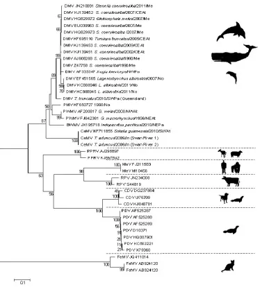
Figure 1. Cetacean species in which the six CeMV strains were isolated or detected by RT-PCR. (A) Common bottlenose dolphin (Tursiops truncatus), Fraser Island, Australia, 2010 (© E. Pearce); (B) Indo-Pacific bottlenose dolphin (Tursiops aduncus), Swan River, Perth, Australia, 2009 (© N. Stephens); (C) Harbour porpoise (Phocoena phocoena), Kent, UK, 2005 (© R. Deaville); (D) Long-finned pilot whale (Globicephala melas), Alicante, Spain, 2007 (© A.J. Raga); (E) Striped dolphin (Stenella coeruleoalba), Valencia, Spain, 2007 (© A.J. Raga); (F) Emaciated calf Guiana dolphin (Sotalia guianensis), Guriri, Espirito Santo, Brazil 2010 (© K. Groch); (G) Longman’s beaked whale (Indopacetus pacificus), Hawaii, US, March 2010 (© K. West, Hawaii Pacific University, NOAA Permit number 932-1905).
2. Antigenic and Molecular Characteristics of CeMV
Very little is known about the three new CeMV related strains recently detected in odontocetes from Hawaii, Brazil, and Australia [5–7]. However, recent sequencing data of the P gene of the isolates recovered from two T. aduncus from the west coast of Australia (Indian Ocean) and from a S. guianensis from Brazil suggest that they differ significantly from the DMV, PMV and PWMV strains [6,7] and may represent another CeMV lineage (Figure 2). The beaked whale morbillivirus (BWMV) clusters with the ‘old’ CeMV lineage and should be considered as a new strain of this lineage (Figure 2). Sequences from a fragment of the P gene revealed that it has 86% similarity to DMV and 84% similarity to PWMV [5]. We propose to use the terminology CeMV-1 for the ‘old’ lineage that includes DMV, PMV, PWMV and BWMV and CeMV-2 for the ‘new’ lineage that includes the T. aduncus and S. guianensis morbilliviruses until the taxonomy of these viruses is further explored.
The close genetic relationship between cetacean and ruminant morbilliviruses has led to the suggestion that they may have a common ancestor [7,16]. Closely related to the hippopotamus (Hippopotamus amphibius), cetaceans belong to the clade Cetartiodactyla [36,37]. As several species of this clade are susceptible to RPV and PPRV [38,39], it is possible that a host jump occurred between a cetacean and another member of the Cetartiodactyla, and that ecological isolation led to distinct virus species. The presence of similar host proteins and cell receptors in cetaceans and artiodactyls may favour cross-species transmission [9,13,40]. However, further studies are needed to confirm this hypothesis.
3. Mechanisms of Cellular Entry and Receptors
The H glycoprotein is responsible for virus attachment to the host cell membrane and for cellular entry. The F glycoprotein causes fusion with the host cell membrane and, together with the M protein, invokes cell-to-cell fusion [20,41]. H and F interact with cellular receptors that allow virus entry and determine host susceptibility, tissue tropism and viral pathogenesis [12,42]. The signaling lymphocyte activation molecule (SLAM or CD150) and the poliovirus like receptor 4 (PVLR4 or nectin 4) have both been recently identified as the major receptors for wild-type morbilliviruses in immune and polarized epithelial cells, respectively [9,13,42–45]. Besides, CD147, a transmembrane glycoprotein that belongs to the immunoglobulin family and is present on a variety of cells including neuronal and endothelial cells, and the membrane bound form of heparin binding epithelial growth factor have been suggested to function as entry receptors for MeV and PDV, respectively [42,46]. Most morbilliviruses, including MV, CDV, PDV, PPRV, and RPV use the SLAM of their respective host species as a receptor [42,43,47,48].
residues) than between the Pacific white-sided dolphin (Lagenorhynchus obliquidens) and the spotted seal (Phoca largha) SLAM (21 amino acid residues) [9,13], as would be expected based on the host relationships. Among the nine cetacean families examined, variations were found between six amino acid residues, with charge alterations for four of them [13]. Interestingly, three residue substitutions (G68, H90 and H130) that introduced charge alteration and possible change in viral affinity were observed in the SLAM of the Delphinidae, while these residues were mostly conserved in the receptor of the other cetacean families [13]. As morbillivirus mass mortalities have mostly been detected in the Delphinidae, it is possible that their SLAMs have a higher affinity for CeMV resulting in increased viral infectivity and dissemination [13]. Among the Delphinidae, only T. truncatus, T. aduncus and, S. coeruleoalba had variation at position 130 [13] and during CeMV outbreaks mass die-offs were overwhelmingly dominated by these species [7,51,52]. The only other odontocete that presented this H130Q variation was P. phocoena, a species that was affected by morbillivirus infection in 1988–1990 [53,54]. Further studies are needed to confirm if the SLAM of dolphins, porpoises and whales is indeed the immune cell receptor for CeMV and should investigate whether alternate potential receptors, such as nectin4 and CD147, are present on the cells of these mammals.
4. Diagnosis
Though virus isolation remains the gold standard for definitive diagnosis, it is challenging when dealing with stranded cetacean carcasses. RT-PCR followed by sequencing has proven very helpful for obtaining rapid confirmation of CeMV infection, to differentiate between PMV and DMV and to identify new strains [4–7,16,20,55]. Histology and immunohistochemistry have provided further confirmation of the disease and insights into its pathogenesis and have permitted differentiation between systemic disease and localized chronic infection of the central nervous system (CNS) [56–60]. Serological studies have also been useful for studying CeMV epidemiology, to assess the immune status of populations before and after an outbreak and to predict the occurrence of new epidemics [21,23,24,26,61–65].
4.1. Histology and Immunohistochemistry
4.2. Virus Isolation
The isolation of DMV and PMV has been achieved using homogenates of lung tissue from S. coeruleoalba and P. phocoena inoculated onto monolayers of African green monkey kidney (Vero) cells following standard methodologies [1,3,22]. Primary canine kidney epithelial cell cultures, bovine foetal lung cells and T. truncatus peripheral blood mononuclear cells have also proved useful for isolation of CeMV directly or after co-cultivation with Vero cells [1,17,21]. Primary culture of kidney cells derived from diseased P. phocoena permitted direct virus isolation [22]. Repeated passages of the inoculated cell cultures and, consequently, several weeks are typically needed before virus growth can be detected [17,22]. Recently, Vero cells expressing the canine SLAM (Vero.DogSLAMtag cells) were shown to reduce the time necessary for PDV isolation from weeks to days [72]. These cells were also successfully used to grow stocks of PMV and DMV initially passaged on Vero cells and to isolate DMV from the brain of a G. melas stranded in Valencia during the 2006–2008 epidemic [27,55,67]. More recently, they proved useful to isolate CeMV from fresh tissues as part of the investigation into the T. truncatus morbillivirus outbreak along the eastern Atlantic coast of USA in 2013 [71]. Virus isolation has the added benefit of providing antigen necessary to carry out serological testing, as described in the serology section below. It may also provide genomic material for more complete phylogenetic analysis.
4.3. Serology
Virus neutralization (VN) tests, plaque reduction (PR) assays and indirect enzyme-linked immunosorbent assays (iELISAs) are the main platforms used to detect antibodies against CeMV. The iELISA allows the detection of antibodies directed against the N, P, F and H CeMV proteins [73] whereas only antibodies to the surface glycoproteins (H and F) are detected by the VN and PR assays [16]. Morbilliviruses are antigenically closely related and may cross-neutralize one another. However, serum raised against one morbillivirus will neutralize the homologous virus at a higher titer than it will heterologous morbilliviruses [63,74]. Thus, when working with cetaceans it is very important to use CeMV strains in the serological tests to avoid false negatives.
The VN test is highly sensitive and very specific and is considered the most reliable assay for the detection of CeMV antibodies [74]. Antibody titers are expressed as the reciprocal of the highest dilution of sera that completely neutralizes cytopathic effects. Titers of 1:16 or higher are considered to be indicative of exposure to CeMV, although higher thresholds can be used to reduce the likelihood of false positives. A more conservative interpretation is recommended when either new host species or new geographic areas are under investigation. A PR assay was developed to allow detection of antibodies in hemolyzed sera [78,79]. In this test, titers are expressed as the reciprocal of the highest dilution that gave 80% reduction in the number of plaques compared to the negative control [79]. Although Vero cells are most commonly utilized in these tests, use of Vero. DogSLAMtag cells, which allows for improved virus replication and permits reduction of incubation time from nine days to four days ([72]; Saliki, unpublished observations) may be a more robust and cost-effective alternative.
4.4. Reverse Transcription Polymerase Chain Reaction
5. Pathology and Pathogenesis of CeMV Infection
Most morbilliviruses are lymphotropic and epitheliotropic [12]. After initial replication in the lymphoid tissues, the virus is disseminated by infected lymphocytes through the lymphatic system and spreads to epithelial cells [12,83–86]. Histology and immunohistochemistry data indicate that CeMV-associated pathology resembles that commonly seen in other morbillivirus infections in animal and human hosts [54].
[image:11.595.108.490.201.475.2]5.1. Acute, Systemic Disease
Acutely fatal CeMV infection is generally associated with severe multifocal to diffuse interstitial broncho-pneumonia characterized by necrosis of type I pneumocytes and bronchiolar epithelial cells, interstitial oedema, type II pneumocyte hyperplasia, and formation of large syncytia in the alveolar and bronchiolar lumina. Intracytoplasmic and intranuclear inclusion bodies can be noted and are sometimes numerous in respiratory epithelia, bronchiolar gland epithelia and the syncytial cells. Generalized lymphoid depletion with germinal center necrosis is usually present and syncytial cells (Warthin-Finkeldey type) are often prominent in lymphoid tissues (Figure 3A). There may be evidence of viral replication (inclusion bodies) in epithelia and neural cells of other body systems (Figure 3B,C). Multifocal non-suppurative encephalitis may also be present (Figure 3D). Therefore, there may be strong IHC staining in the lungs, lymphoid organs (Figure 3A) and other tissues that is variable in extent between individual cases [7,54,57,59,60,68,87].
5.2. Sub-Acute Systemic Disease
Animals that survive the acute stage of infection may succumb to opportunistic infections (Toxoplama gondii, herpesviruses, bacteria such as Photobacterium damselae, and fungi) as a consequence of the profound immunosuppression. This typical pattern has been commonly seen in odontocetes that died during outbreaks of CeMV in Europe, South America, the USA and Australia [6,7,32,57–60,68,88–91]. While some of the lesions typical of acute infection may no longer be present or be largely obscured by the inflammatory response to the opportunistic pathogens, non-suppurative demyelinating meningoencephalitis (Figure 3D), often focally distributed, is a feature of sub-acute infection. Colonization of the brain by opportunistic mycotic pathogens (e.g., Aspergillus spp.) is also common [7,59]. IHC and RT-PCR are useful for confirmation of the diagnosis of morbilliviral infection in these cases.
5.3. Chronic Systemic Infection
Animals may survive the acute and sub-acute manifestations of infection but succumb sometime later to the secondary infections acquired as a result of viral immunosuppression, or from complications of CNS infection. Typically these animals are in poor body condition at the time of death and the proximate cause of death may be multifactorial. Invariably there are no or few lesions directly attributable to CeMV but viral antigen may be detectable by IHC in some lymph nodes and lungs [57] and viral RNA may be amplified by RT-PCR [57,92].
survived the acute phase of the infection but died following profound immunosuppression and secondary infections [7]. If the pathogenesis of CeMV is similar to that of MV [93], cetaceans that survived acute and sub-acute infection could show prolonged RNA persistence in the blood and lymphoid organs and could be RT-PCR positive in the absence of typical morbillivirus lesions. The concurrent use of histology, IHC and molecular techniques is recommended to further explore the pathogenesis of chronic systemic infections.
5.4. Chronic, Localized CeMV Encephalitis
Cetaceans that have cleared and resolved DMV systemic infection may develop a CNS form that is characterized by the presence of lesions and virus only in the brain [33,56,66,94]. This CNS form was consistently observed in S. coeruleoalba after the two epidemics in the Mediterranean Sea [56,94]. By contrast with the sub-acute cerebral CeMV infection, cytoplasmic or nuclear eosinophilic inclusions were only occasionally detected and syncytial cells were not observed in the CNS form. Many neuronal processes showed immunostaining for CeMV, and some areas had massive accumulation of CeMV-antigen, while contiguous zones of the brain had almost no staining. This suggests that the presence of CeMV was more the result of cell-to-cell spreading of infection rather than of a multifocal infection indicative of blood-borne infection. The CNS form appears to share histological characteristics with subacute sclerosing panencephalitis (SSPE) and old dog encephalitis (ODE), chronic latent localized infections that affect humans and dogs, respectively, and are caused by defective forms of MV and CDV, respectively [95–97]. As in SSPE and ODE, the CNS lesions were localized predominantly in the cerebral cortex, subcortical white matter, and the thalamus, while the cerebellum was mostly spared. In the three conditions perivascular cuffing, diffuse gliosis, and glial nodules with neurophagia were the most prominent changes [94]. Focal malacia was not detected [94,96,98–100]. Demyelination was less prominent in dolphins with the exclusively CNS presentation and in dogs with ODE than is seen in the meningoencephalitis of CeMV or CDV, respectively [56,66,94,101,102]. As in the human and canine presentations, antigen and viral RNA could be detected in dolphin brains but the virus proved difficult to isolate [101]. The mechanism for this is unknown but delayed clearance of antigen and RNA from the CNS may be related to reduced immune surveillance in an immuno-privileged site [99,100]. RT-PCR studies on the brain of S. coeruleoalba chronically affected by CeMV suggest that the sequence of the P gene is different in these cases [56], but further research is needed. The role of CD147 and other cell receptors in the pathogenesis of this form of the disease should be further examined [103].
seems greater than that seen within other morbillivirus species. This is unexpected for a virus that induces lifelong immunity and high cross-protection between congeneric viruses in long-lived hosts. It is possible that CNS persistence plays a role in the maintenance of strains in an ocean basin, although, as a dead end infection, it is unlikely to contribute to virus transmission to other cetaceans.
Although the CNS form has been mostly described in Mediterranean S. coeruleoalba, a similar presentation was described in a few other sporadic cases, namely a mature L. obliquidens stranded on the coast of Miyasaki, Japan, in March 1998 [106], in a juvenile white-beaked dolphin (Lagenorhynchus albirostris) that beached on the island of Ameland, the Netherlands, in June 2011 and died in a rehabilitation center six months later [34], in four S. coeruleoalba stranded along the coasts of the Canary Islands in 2002–2011 [33], and in several T. truncatus from North America (Colegrove, pers. observation). The CNS form was not detected in P. phocoena from the North Sea and Northeastern (NE) Atlantic, although the brains from relatively few animals were examined [107]. Current data would suggest that of the three known strains of CeMV, the DMV variant is the only one associated with the chronic CNS presentation. However, it is also the most prevalent variant detected in nature, and much more research would be required before conclusions could be drawn.
5.5. Subclinical Infection
The pathogenesis and clinical course of morbillivirus infections in cetaceans are poorly understood as there are no comparable laboratory studies to those on CDV in dogs and MV in primates [85,108]. Acute and subacute systemic presentations and chronic CNS infection causing death have been documented, as described in preceding sections. However, the existence and nature of subclinical infection remains speculative. Between 1995–1997, a series of DMV seropositive (> 1:50) D. delphis beached along the southern California coast [75]. One of the six dolphins survived and developed very high titers (1:720) against DMV while in rehabilitation. The other five were euthanized and, at necropsy, none had classical morbillivirus lesions. However, one had a mild lymphocytic meningoencephalitis and its brain was positive for morbillivirus RNA by RT-PCR, as described in Mediterranean S. coeruleoalba. Morbilliviral RNA was also detected in the spleen and heart of two other dolphins without histological lesions [75]. Whether this actually represents subclinical infection in D. delphis or an atypical viral strain/host presentation in the eastern Pacific is unknown.
Similarly, a low prevalence of serum antibodies in apparently healthy live-captured T. truncatus from the Indian River Lagoon, on Florida’s Atlantic coast, without a prior increase in mortalities in the population was suggestive of virus circulation in the absence of an epidemic and thus, of subclinical infection [64]. However, this is a complex system with documented evidence of repeated CeMV epidemics over a period of at least 1982 to 2013 [25,57,72]. Thus, a better understanding of CeMV pathogenesis and immunity in dolphins is required before we can adequately interpret findings from field investigations.
5.6. Immune Function and CeMV Infections
mitogen-induced T cell proliferation along with an increase in lysozyme concentrations and a marginally significant increase in monocyte phagocytosis, along with a marginally significant decrease in the numbers of CD4+ T lymphocytes in T. truncatus that had antibody titers ≥ 1:8 against morbillivirus, suggestive of previous exposure to the virus, compared to animals with lower or no titers. They found no effects on neutrophil phagocytosis. While the timing of the morbillivirus infection (active infection, chronic infection, resolved past infection) cannot be determined from titers alone, it is clear that there is an association between modulation of immune functions and previous exposure to morbillivirus infection in T. truncatus, as observed in other species. Further studies are needed.
5.7. CeMV Transmission 5.7.1. Horizontal Transmission
Morbillivirus transmission is thought to occur mostly after the inhalation of aerosolized virus shed by infected individuals [113]. This horizontal transmission is likely to occur among cetaceans too and to be favored by a gregarious behavior and a high density of cetaceans [67,114]. Transmission by inhalation of expired blowhole droplets possibly occurs during breathing in a synchronized fashion when large numbers of tightly grouped cetaceans are travelling and feeding together or are engaged in social activities [20,115,116].
5.7.2. Evidence for Vertical Transmission
Morbillivirus antigen was detected in the mammary gland of T. truncatus from the US Atlantic coast epidemic in 1987–1988 and of S. coeuruleoalba from the Mediterranean Sea outbreak in 1990 [59,87] and in the penile and preputial epithelium of a P. phocoena from the North Sea [107]. Furthermore, a testicular fibroma collected in a short-beaked common dolphin (Delphinus delphis ponticus) from the Black Sea was positive for morbillivirus RNA by RT-PCR [117]. The first evidence that vertical transmission may occur was the detection of morbilliviral RNA in the brain, lung, spleen, lymph node, and liver from the seven-month fetus of a DMV-infected G. melas stranded in the Balearic Islands in 2007 [88]. These data suggest that CeMV infected females may transmit the infection to their fetuses and neonates in utero and during lactation, respectively. In utero transmission has been reported for MV in humans. The effects on the fetus depend on the stage of pregnancy and include abortion, in utero death or premature birth [118]. When MV infects pregnant women in the peri-natal period, neonates are at risk of congenital measles and have a higher risk of developing early and fulminant SSPE because of the incomplete transfer of protective antibodies [96,119]. A similar situation may have happened in a S. coeruleoalba calf stranded in Italy in November 2009 with a CNS infection [120] and in a neonate sperm whale (Physeter macrocephalus) washed ashore in Oahu, Hawaii in May of 20l1 [121].
6. Outbreaks of Disease and Epidemiology
However, the persistence of morbilliviruses in relatively small (possibly multispecies) host metapopulations remains an important unsolved problem in disease ecology [122]. Newborn individuals typically have maternal immunity if their mothers had previously been infected. After some months, this immunity is lost and the young individuals are fully susceptible to infection [123,124].
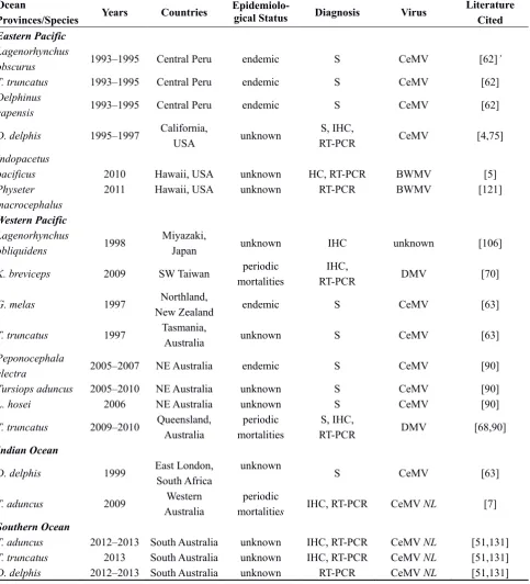
[image:16.595.59.537.420.715.2]CeMV infection has been detected using various techniques in several species of odontocetes and mysticetes worldwide (Table 1). DMV is the strain most commonly observed in cetaceans from the Northern Hemisphere, followed by PMV and PWMV (Table 1). Serological studies strongly suggest that CeMV is endemic in gregarious odontocete species in the North Atlantic and, possibly in the Southwestern Atlantic and in the South Pacific [23–25,61,63]. Pilot whale (Globicephala spp.), dusky dolphin (Lagenorhynchus obscurus), Fraser’s dolphin (Lagenodelphis hosei) and melon-headed whale (Peponocephala electra) populations had high prevalences of DMV-seropositives and may be reservoirs and vectors of the infection to susceptible species [23–25,62,63,90]. In the absence of, or decrease in, herd immunity, outbreaks of lethal disease may occur in susceptible species, as has repeatedly been observed in Europe, the Americas, and Australia since the late 1980s.
Table 1. CeMV infection in odontocetes and mysticetes worldwide. Abbreviations are: VI = virus isolation, IHC = immunohistochemistry, S = serology, RT-PCR = reverse-transcriptase polymerase chain reaction, PMV = porpoise morbillivirus, CeMV = cetacean morbillivirus, DMV = dolphin morbillivirus, PWMV = pilot whale morbillivirus and CeMV, NL = new lineage of CeMV.
Ocean
Provinces/Species Years Countries
Epidemiolo-gical Status Diagnosis Virus
Literature Cited
Eastern Atlantic & North Sea
Phocoena phocoena 1988–1990 N. Ireland, UK,
Netherlands
periodic mortalities
VI, IHC, S,
RT-PCR PMV
[1,22,53], [54,61,63]
Delphinus delphis 1988–1990 UK, Netherlands unknown S CeMV [22,61,63]
Lagenorhynchus albirostris 1988–1990, 2007, 2011 Germany, Netherlands periodic mortalities S, IHC,
RT-PCR DMV
[22,34,61, 125]
Balaenoptera physalus 1983 Iceland unknown S CeMV [17]
B. physalus 1997–1998 Belgium, France periodic
mortalities IHC unknown [126]
Tursiops truncatus 1999 Kent, UK unknown S CeMV [63]
Globicephala macrorhynchus T. truncatus 1996 2005 Canary Islands Canary Islands periodic mortalities periodic mortalities RT-PCR IHC, RT-PCR PWM DMV [30] [69]
S. coeruleoalba 2002–2011 Canary Islands periodic
mortalities IHQ, RT-PCR DMV [33]
D. delphis 2007 Canary Islands periodic
Table 1. Cont.
Ocean
Provinces/Species Years Countries
Epidemiolo-gical Status Diagnosis Virus
Literature Cited
Mediterranean Sea
S. coeruleoalba 1990–1992 Spain, France, Italy,
Greece epidemic
VI, IHC, S,
RT-PCR DMV
[2,3,21,58, 127]
S. coeruleoalba 2006–2008 Spain, France, Italy epidemic IHC, RT-PCR DMV [32,66,67]
T. truncatus 1994; 2007–
2008, 2011
Israel, Spain, France, Italy
periodic mortalities
IHC,
RT-PCR, S DMV
[32,63,66, 128]
D. delphis 1990 Italy unknown S CeMV [21]
Globicephala melas 2006–2007 Spain, France epidemic IHQ, RT-PCR DMV [88]
Grampus griseus 1997, 1999 Valencia, Spain unknown S CeMV [63]
Balaenoptera
acutorostrata 1993 Tuscany, Italy unknown S unknown [58]
B. physalus 2011 Tuscany, Italy periodic
mortalities RT-PCR DMV [89]
Northwestern Atlantic
T. truncatus 1982 Florida, USA epidemic S, IHC CeMV [25,129]
T. truncatus 1987–1988 East coast USA epidemic IHC,
RT-PCR CeMV [57,92]
T. truncatus 1993–1994 Gulf of Mexico,
USA epidemic IHC, RT-PCR CeMV [25,57,92]
T. truncatus 2003–2007 Florida, USA unknown S, IHC CeMV [64,65]
T. truncatus 2013–2014 East coast USA epidemic IHC, RT-PCR DMV [71,130]
T. truncatus 1992–1994 East coast USA endemic S CeMV [25]
G. melas 1982–1993 Northeast coast USA endemic S CeMV [23]
G. macrorhynchus 1986–1994 Florida, USA endemic S CeMV [23]
G. melas late nineties New Jersey, USA periodic
mortalities
IHC,
RT-PCR PWM [4]
S. coeruleoalba 1991–1993 Northeast coast USA unknown S CeMV [24]
Stenella frontalis 1993 Northeast coast USA unknown S CeMV [24]
D. delphis 1980–1994 Northeast coast USA possibly
endemic S CeMV [24]
Lagenorhynchus
acutus 1985–1993 Northeast coast USA unknown S CeMV [24]
Kogia breviceps 1983–1991 Southeast coast USA unknown S CeMV [24]
Feresa attenuata 1983 Southeast coast USA unknown S CeMV [24]
Pseudorca crassidens 1982–1988 Southeast coast USA possibly
endemic S CeMV [24]
Lagenodelphis hosei 1994 Gulf of Mexico,
USA
possibly
endemic S CeMV [24]
P. phocoena 1993–1994 East coast, Canada unknown S CeMV [24]
Southwestern Atlantic
L. hosei 1999 Puerto Madryn,
Argentina unknown S CeMV [63]
L. hosei 1999 Rio de Janeiro,
Brazil unknown S CeMV [63]
Sotalia guianensis 2010 Espirito Santo, Brazil unknown IHC,
Table 1. Cont.
Ocean
Provinces/Species Years Countries
Epidemiolo-gical Status Diagnosis Virus
Literature Cited
Eastern Pacific
Lagenorhynchus
obscurus 1993–1995 Central Peru endemic S CeMV [62]’
T. truncatus 1993–1995 Central Peru endemic S CeMV [62]
Delphinus
capensis 1993–1995 Central Peru endemic S CeMV [62]
D. delphis 1995–1997 California,
USA unknown
S, IHC,
RT-PCR CeMV [4,75]
Indopacetus pacificus Physeter macrocephalus 2010 2011 Hawaii, USA Hawaii, USA unknown unknown HC, RT-PCR RT-PCR BWMV BWMV [5] [121] Western Pacific Lagenorhynchus
obliquidens 1998
Miyazaki,
Japan unknown IHC unknown [106]
K. breviceps 2009 SW Taiwan periodic
mortalities
IHC,
RT-PCR DMV [70]
G. melas 1997 Northland,
New Zealand endemic S CeMV [63]
T. truncatus 1997 Tasmania,
Australia unknown S CeMV [63]
Peponocephala
electra 2005–2007 NE Australia endemic S CeMV [90]
Tursiops aduncus 2005–2010 NE Australia unknown S CeMV [90]
L. hosei 2006 NE Australia unknown S CeMV [90]
T. truncatus 2009–2010 Queensland,
Australia
periodic mortalities
S, IHC,
RT-PCR DMV [68,90]
Indian Ocean D. delphis T. aduncus 1999 2009 East London, South Africa Western Australia unknown periodic mortalities S IHC, RT-PCR CeMV CeMV NL [63] [7] Southern Ocean
T. aduncus 2012–2013 South Australia unknown IHC, RT-PCR CeMV NL [51,131]
T. truncatus D. delphis 2013 2012–2013 South Australia South Australia unknown unknown IHC, RT-PCR RT-PCR CeMV NL CeMV NL [51,131] [51,131] 6.1. Europe
6.1.1. North Sea, NE Atlantic, and CE Atlantic
DMV-seropositivity was declining over time and that only adult porpoises and dolphins were positive in 1997–1999. This suggested that the virus had not persisted as an endemic infection in these populations [61,63]. Similarly, with the exception of a P. phocoena with systemic morbillivirus infection beached in Kent, UK, in late 1990, systemic morbilliviral disease was not detected in any porpoise that stranded along the coasts of Belgium, northern France, England and Germany in 1990–2000 [132–135]. Though the number of P. phocoena in the North Sea and adjacent waters was theoretically large enough to sustain an endemic infection (341,366 individual [95% confidence interval = 260,000–449,000] in 1994 [136]), their solitary behavior likely did not favor morbillivirus transmission and maintenance in this population. The presence of high titers of DMV antibodies in the serum of a juvenile G. melas collected in the English Channel in 1996 suggested that this species could be involved in the maintenance of the virus in the NE Atlantic [61]. However, further serological surveys and molecular investigations are needed to understand the ecology of CeMV in this ocean basin.
Recently, CeMV infection was detected in Delphinidae from the CE Atlantic Ocean. A virus closely related to the PWMV strain was detected by RT-PCR in the brain of a G. macrorhynchus stranded in Tenerife, Canary Islands, Spain, in 1996 [30]. In addition, an IHC and RT-PCR retrospective survey showed that DMV caused chronic CNS disease in S. coeruleoalba and D. delphis washed ashore in the Canary Islands in the period 2002-2011 [33]. Finally, a systemic DMV infection was observed in a T. truncatus stranded in Lanzarote, Canary Islands, in 2005 [69]. Thus, at least two strains of CeMV are circulating in cetaceans from this ocean province.
6.1.2. Mediterranean Sea
beginning of July and then a sharp increase in mid-August [67]. The outbreak extended to France and Italy during the following months, also affecting T. truncatus [32,138]. Mostly juveniles were affected during this mortality event, likely because adults were still protected by immunity acquired during the 1990–1992 epidemic [32,67]. The virus strains amplified by RT-PCR from tissues of S. coeruleoabalba, G. melas and T. truncatus were similar to those isolated during the 1990–1992 epidemic but not identical ([27,31,32,67,88], this paper). An estimated 200 striped dolphins died in the western Mediterranean but the total number of deaths remains unknown [67]. As well as the deaths caused by the acute infection, there were also several cases, ultimately lethal, of a chronic CNS form of infection in 1991–1994 and 2008–2011 in the western Mediterranean and in 2009–2011 in the Eastern Mediterranean [56,66,94]. In the Western Mediterranean chronic morbillivirus encephalitis represented the most common single cause of stranding and death in mature S. coeruleoalba in the years following a DMV epizootic [56]. These data suggest that the second DMV outbreak may also have had a negative impact on the Mediterranean S. coeruleoalba population, though to a lesser extent than the previous one. Little is known about the impacts of the outbreak on populations of the other cetacean species affected. However, Wierucka et al. [139] found that the 2006–2008 DMV epidemic lowered the survival rate of some clusters (groups of individuals that associate with each other more often than with others) of G. melas (from 0.919 (95% CI: 0.854−0.956) to 0.547 (95% CI: 0.185−0.866)) in the Alboran Sea and Gulf of Vera.
The S. coeruleoalba population density in the Gulf of Valencia (0.49 dolphin/km2) was again close
to the maximum reported for this species in the Western Mediterranean in 2001–2003 [104,140]. This high population density, with a large proportion of susceptible individuals, likely favored viral transmission and permitted the start of a new epidemic when DMV was reintroduced into the population [67]. As both the 1990–1992 and 2006–2007 DMV epidemics started close to, or in, the Gibraltar Strait, it was suggested that DMV endemically infected cetaceans, possibly G. melas transmitted the infection to S. coeruleoalba with which they occasionally form mixed groups (Raga et al. pers. observations). The recent detection of DMV strains in S. coeruleoalba from the CE Atlantic Ocean that are almost identical to the Mediterranean strains [33] indicates that this population could also transmit the virus to the Mediterranean S. coeruleoalba through occasional contacts in the Strait of Gibraltar. The finding of systemic morbillivirus infection in two adult S. coeruleoalba stranded on the southwestern (Atlantic) coast of Spain, close to Gibraltar in 2011 and 2012 [141] further indicates that this Strait plays an important role in the epidemiology of CeMV. Environmental factors (higher sea-surface temperatures and limited prey availability), as well as fisheries interactions, inbreeding, migration, and high contaminant loads may synergistically interact to increase the severity of the disease and favor transmission between species [127,142–145]. When CeMV herd immunity significantly decreases in Mediterranean S. coeuruleoalba, the population will again be at risk for an epidemic. Serological surveys are needed to determine the current immune status of these dolphins and facilitate development of predictive epidemiological models.
the proximate cause of the event is still under investigation (Di Guardo and Mazzariol, pers. observations.).
6.1.3. Black Sea
Two D. delphis ponticus that stranded during an outbreak of mortality in Crimea in August and September 1994 had broncho-pneumonia, syncytia and lymphoid depletion [117]. Morbillivirus antigen was observed by IHC in the lungs, cerebrum, spleen and lymph nodes. However, morbillivirus RNA could only be detected in a formalin-fixed sample of a testicular fibroma by RT-PCR. There was no evidence of morbillivirus in the frozen tissues using either virus isolation or an antigen capture ELISA that had proven useful during other mortality events [3,22,117]. As virus isolation was negative and sequencing of the PCR products was not performed, it is unclear which morbillivirus caused the death of these dolphins. The last S. coeruleoalba reported to die of acute DMV infection in Greek waters was found in the spring of 1992, two years before the D. delphis ponticus mortality in the Black Sea. S. coeruleoalba are not known to enter the Turkish Strait Systems (Bosphorus, Marmara Sea and Dardanelles) where D. delphis ponticus are commonly seen, and are absent from the Black Sea [147]. D. delphis ponticus have also not been reported in Aegean waters [147]. Thus, a link between the 1990–1992 morbillivirus outbreak in Mediterranean S. coeruleoalba and the morbillivirus infection in the two D. delphis ponticus stranded in Crimea is unclear. Whether the morbillivirus was in fact CeMV, originating in the Mediterranean, or another morbillivirus should be further examined.
6.2. North America
There have been several die-offs in coastal T. truncatus populations from the Gulf of Mexico and the Atlantic coast of the US since 1982 [25,57,80,92,130,148].
6.2.1. Atlantic Coast
From January to May of 1982, 43 carcasses were recovered in the Indian River Lagoon System (IRL), Florida, among a community estimated at 211 individuals [129]. Serological data indicated that this outbreak was likely due to a morbillivirus infection and contact with endemically infected species such as offshore T. truncatus was hypothesized to be the source of infection for the event [25]. Further serological studies performed on samples collected from 2003–2007 indicated that IRL dolphins born after the 1982 mortality had antibodies to a DMV-like virus, indicating exposure and infection, though no outbreaks or associated deaths were documented after 1982 [64].
and false killer whales (Pseudorca crassidens)) in which CeMV is endemic [23–25] may have been the sources of infection for the 1987–88 and 2013–2014 outbreaks. Seasonal overlap between resident coastal T. truncatus stocks at certain times of the year, and migration of the coastal migratory stock, may have favored transmission of the disease down the coast [149]. A serological survey performed on samples collected from live capture-released coastal and estuarine T. truncatus along the east coast of the US in 1999–2004 indicated that the seroprevalence decreased over the years of the study, suggesting that CeMV did not persist as an endemic infection in these populations, as had been predicted [25,26]. Therefore, population immunity likely continued to decrease over time leading to increased numbers of susceptible individuals and resulting in the 2013–2014 epidemic. The role of environmental and anthropogenic factors in this mortality and the population impacts is being investigated [71].
6.2.2. Gulf of Mexico
In 1993–1994, CeMV caused another outbreak of mortality, this time in a population of T. truncatus from the Gulf of Mexico, spanning from Florida (Panama City) to Texas [92,150]. A total of 171 specimens were retrieved from the entire Texas coast in March and April 1994 [151]. About a quarter of 34 dolphins sampled in Matagorda Bay in 1992 had CeMV antibodies, indicating that this community/population had been exposed to the virus before the 1993–1994 outbreak occurred [25].
6.2.3. North Pacific
Morbillivirus infection was detected by RT-PCR in a juvenile male I. pacificus stranded at Hana, Maui in March 2010, following traumatic maxillary and mandibular bone fractures [5]. The whale had chronic encephalitis and was also concurrently infected by an alphaherpesvirus [5]. Though morbillivirus RNA was detected by RT-PCR in samples of the lungs, spleen, thymus, and lymph nodes the juvenile did not have any typical morbillivirus lesions in these organs [5]. This may reflect prolonged persistence of morbilliviral RNA following acute infection, as described for MV [93] but further analyses are necessary to confirm this hypothesis. The virus represents a new strain of CeMV-1, tentatively named beaked whale morbillivirus. BWMV was also detected by RT-PCR in the tracheobronchial lymph node and spleen of a neonate P. macrocephalus beached on the island of Oahu, Hawaii, in May 2011. However, typical morbillivirus lesions were not detected in this individual [121].
Six of 18 D. delphis that stranded along the coast of California from August 1995 through August 1997 had serum antibodies against DMV. Morbilliviral RNA was detected in the normal spleen and heart of two seropositive dolphins that did not show any typical morbillivirus lesions and in the brain of a third dolphin that suffered mild lymphocytic meningoencephalitis [75]. Together, these data indicate that CeMV strains are circulating in the North Pacific.
6.3. South America
depletion, interstitial pneumonia, and meningoencephalitis [6]. The S. guianensis community off Guriri may be related to the Abrolhos Bank population that concentrates around the estuaries of Caravelas (estimated at 57–124 individuals) and Doce rivers [152,153]. Though S. guianensis have not been observed mixing with other cetacean species in this region, it is sympatric with the rough-toothed dolphin (Steno bredanensis), T. truncatus, the humpback whale (Megaptera novaeangliae) and the southern right whale (Eubalaena australis) [154,155]. Interactions between these species may have resulted in the infection of the S. guianensis calf. Preliminary IHC studies suggested that morbilliviruses have infected other cetacean species along the Brazilian coast [156].
6.4. Asia and Australasia 6.4.1. Asia
CeMV infection was detected by serology, IHC and RT-PCR in a stranded L. obliquidens from Japan, in a pygmy sperm whale (Kogia breviceps) beached in Taiwan and in a captive T. aduncus from Taiwan [63,70,106]. The L. obliquens was diagnosed with a chronic persistent morbillivirus encephalitis while the K. breviceps had a systemic, acute infection [70,106]. Partial sequence of the P gene of the K. breviceps virus had 97.6% similarity with DMV. The T. aduncus had very high titers against DMV likely acquired after an infection developed while still in the wild [63]. Though these data indicate that CeMV is present in odontocetes from the Northwest Pacific, mass mortalities were not reported in this ocean basin. Further investigations are necessary to determine the distribution of virus, the identity of strains and susceptibility of hosts in this region.
6.4.2. Australasia
7. Conclusions
Significant progress in our understanding of the epidemiology, molecular biology and pathogenesis of CeMV have been made since PMV and DMV were first detected in small odontocetes in European waters in 1988–1992. Large herds of gregarious species were found to be the likely reservoirs and sources of CeMV infection to susceptible species in the Atlantic and Pacific Oceans [23–25,61–63,90]. New species and lineages of CeMV have been recently discovered [5–7]. Several techniques have been developed to optimize the diagnosis of CeMV infection, to differentiate the strains and to reduce the possibility of cross-contamination [16,20,55]. Serological assessment may enable prediction of future outbreaks [157]. The development of Next Generation Sequencing technologies has greatly enhanced the detection and genetic characterization across all forms of life [158]. To date, such technologies have not yet been applied to morbillivirus infections of aquatic mammals although they recently enabled an assessment of the evolution of ruminant morbilliviruses [159] and their application to cetacean morbilliviruses may allow a greater understanding of their evolution. Such studies may, where sampling permits, enable the use of genetic data to trace transmission routes between cetacean species and indicate key interactions between species that could lead to significant outbreak events. Standard sampling and preservation protocols should be used during suspected morbillivirus outbreaks and complete genomes of CeMV strains and lineages should be sequenced [19]. The recent discoveries of several new morbilli-related viruses in bats [160], as well a potentially novel feline morbillivirus representing a basal divergence in the genus [11], are likely to lead to a revision of the phylogeny and understanding of the evolution of morbilliviruses. Identification of the SLAM cell receptor in several cetacean species [9,13] represents a major step in our understanding of the pathogenesis of CeMV infection, especially with regard to susceptibility and transmission to non-classical hosts, such as pinnipeds [9,13,161,162]. Further studies should confirm whether the SLAM cell receptor is indeed the primary immune receptor for CeMV, as is the case for other studied morbilliviruses, and should look for the nectin4 epithelial cell receptor and other cell receptors in cetaceans. Further studies are also warranted to delineate the host responses to CeMV strains and lineages, and the factors that determine the outcome of infection in cetaceans. Mathematical models should be developed to examine the long-term dynamic consequences of the epidemics on odontocete populations and to predict the risk of epidemics, as has been done for PDV in harbor seals (Phoca vitulina) [157,163]. The concurrent use of the different diagnostic techniques in the context of an integrative approach that includes epidemiological parameters, life history of the affected species and environmental parameters should provide a better and more complete picture of the ecology and evolution of CeMV.
Acknowledgements
Author Contributions
M-F Van Bressem designed paper outlines and Figures 1 and 3, did the main writing and literature review; P. Duignan helped to design the ms outlines, contributed to the writing of the introduction, diagnosis, pathology and epidemiology sections, reviewed and improved earlier drafts, made many helpful suggestions, facilitated access to the T. aduncus morbillivirus and helped with literature search; A. Banyard build the phylogenetic tree (Figure 2), contributed to the virology section and conclusions, made useful comments and reviewed several drafts of the manuscript; F. Gulland organized the marine mammal morbillivirus workshop in Princeton, USA in August 2014, contributed to draw the paper outlines, made helpful suggestions and reviewed various drafts of the manuscript; K. Colegrove contributed to the epidemiology and pathology sections, reviewed various drafts of the manuscript and provided unpublished information; D. Fauquier and T. Goldstein contributed to the epidemiology section, reviewed several drafts of the manuscript and provided unpublished information; O. Nielsen contributed to the receptor, virology and diagnosis sections and reviewed drafts of the manuscript; S. De Guise wrote the paragraph ‘Immune function and CeMV infections’, reviewed drafts of the manuscript and provided unpublished information; J. Saliki contributed to the diagnosis and virology sections, made helpful suggestions and reviewed drafts of the manuscript; G. Di Guardo, M. Domingo, S. Mazzariol, A. Fernandez, and E. Sierra contributed to the pathology sections and to Figures 1 and 3, reviewed drafts of the manuscript and made useful comments; B. Stone, I. Tomo and N. Stephens contributed to the epidemiology and virology sections and to Figures 1 and 3, made useful comments and allowed access the Australian dolphin morbilliviruses; K. Groch contributed to the epidemiology and virology sections, greatly helped with the figures, formatting of the manuscript and literature, reviewed drafts of the manuscript and provided unpublished data; T. Raga contributed to the introduction and epidemiology section and Figure 1, made helpful suggestions and reviewed drafts of the manuscript; J. Wellehan contributed to the introduction, receptors and virology sections and conclusions, reviewed an advanced version of the manuscript and made helpful comments; T. Waltzek reviewed an advanced draft of the manuscript and made useful comments; J. Wang provided the sequences of the Australian T. aduncus morbilliviruses, reviewed an advanced draft of the manuscript and made useful comments; A. Hall reviewed an advanced draft of the manuscript and made useful comments; B. Grenfell organized the marine mammal morbillivirus workshop in Princeton in August 2014, contributed to the epidemiology section and reviewed an advanced draft of the manuscript; A. Dobson made useful comments on the manuscript and reviewed an advanced draft of the manuscript; P. Jepson reviewed an earlier version of the manuscript and provided Figure 1C.; B. Jensen provided Figure 1G and made useful comments; T.K. Rowles organized the marine mammal morbillivirus workshop in Princeton in August 2014, revised drafts of the manuscript and made useful suggestions; M. Barbieri and S.E. Morris participated to the workshop, facilitated access to CeMV literature and made constructive comments; T. Kuiken provided literature.
Conflicts of Interest
References and Notes
1. McCullough, S.J.; McNeilly, F.; Allan, G.M.; Kennedy, S.; Smyth, J.A.; Cosby, S.L.; McQuaid, S.; Rima, B.K. Isolation and characterisation of a porpoise morbillivirus. Arch. Virol. 1991, 118, 247–252.
2. Domingo, M.; Ferrer, L.; Pumarola, M.; Marco, A.; Plana, J.; Kennedy, S.; McAliskey, M.; Rima, B.K. Morbillivirus in dolphins. Nature 1990, 348, 21.
3. Van Bressem, M.-F.; Visser, I.K.G.; van de Bildt, M.W.; Teppema, J.S.; Raga, J.A.; Osterhaus, A.D.M.E. Morbillivirus infection in Mediterranean striped dolphins (Stenella coeruleoalba). Vet Rec. 1991, 129, 471–472.
4. Taubenberger, J.K.; Tsai, M.M.; Atkin, T.J.; Fanning, T.G.; Krafft, A.E.; Moeller, R.B.; Kodsi, S.E.; Mense, M.G.; Lipscomb, T.P. Molecular genetic evidence of a novel morbillivirus in a long-finned pilot whale (Globicephala melas). Emerg. Infect. Dis. 2000, 6, 42–45.
5. West, K.L.; Sanchez, S.; Rotstein, D.; Robertson, K.M.; Dennison, S.; Levine, G.; Davis, N.; Schofield, D.; Potter, C.W.; Jensen, B. A Longman’s beaked whale (Indopacetus pacificus) strands in Maui, Hawaii, with first case of morbillivirus in the central Pacific. Mar. Mamm. Sci. 2013, 29, 767–776.
6. Groch, K.R.; Colosio, A.C.; Marcondes, M.C.; Zucca, D.; Díaz-Delgado, J.; Niemeyer, C.; Marigo, J., Brandão, P.E., Fernández, A.; Luiz Catão-Dias, J. Novel cetacean morbillivirus in Guiana dolphin, Brazil. Emerg. Infect. Dis. 2014, 20, 511–513.
7. Stephens., N.; Duignan, P.J.; Wang, J.; Bingham, J.; Finn, H.; Bejder, L.; Patterson, A.P.; Holyoake, C. Cetacean morbillivirus in coastal Indo-Pacific bottlenose dolphins, Western Australia. Emerg. Infect. Dis. 2014, 20, 666–670.
8. Barrett, T. Morbillivirus infections, with special emphasis on morbilliviruses of carnivores. Vet. Microbiol. 1999, 69, 3–13.
9. Ohishi, K.; Suzuki, R.; Maruyama, T. Host-Virus Specificity of the Morbillivirus Receptor, SLAM, in Marine Mammals: Risk Assessment of infection based on three-dimensional models. In New Approaches to the Study of Marine Mammals; Romero, A., Keith, E.O., Eds.; InTech: Rijeka, Croatia, 2012; pp. 183–204.
10. Hall, A.J. Morbilliviruses in marine mammals. Trends Microbiol. 1995, 3, 4–9.
11. Woo, P.C.; Lau, S.K.; Wong, B.H.; Fan, R.Y.; Wong, A.Y.; Zhang, A.J.; Wu, Y.; Choi, G.K.; Li, K.S.; Hui, J.; et al. Feline morbillivirus, a previously undescribed paramyxovirus associated with tubulointerstitial nephritis in domestic cats. Proc. Natl. Acad. Sci. USA 2012, 109, 5435–5440. 12. Delpeut, S.; Noyce, R.S.; Richardson, C.D. The tumor-associated marker, PVRL4 (nectin-4),
is the epithelial receptor for morbilliviruses. Viruses 2014, 6, 2268–2286.
13. Shimizu, Y.; Ohishi, K.; Suzuki, R.; Tajima, Y.; Yamada, T.; Kakizoe, Y.; Bando, T.; Fujise, Y.; Taru, H.; Murayama, T.; et al. Amino acid sequence variations of signaling lymphocyte activation molecule and mortality caused by morbillivirus infection in cetaceans. Microbiol. Immunol. 2013, 57, 624–632.
15. Osterhaus, A.D.M.E.; de Swart, R.L.; Vos, H.W.; Ross, P.S.; Kenter, M.J.H.; Barrett, T. Morbillivirus infections of aquatic mammals: Newly identified members of the genus. Vet. Microb. 1995, 44, 219–227.
16. Barrett, T.; Visser, I.K.G.; Mamaev, L.; Goatley, L.; van Bressem, M.-F.; Osterhaus, A.D.M.E. Dolphin and porpoise morbilliviruses are genetically distinct from phocine distemper virus. Virology 1993, 193, 1010–1012.
17. Blixenkrone-Möller, M.; Bolt, G.; Gottschalk, E.; Kenter, M. Comparative analysis of the gene encoding the nucleocapsid protein of dolphin morbillivirus reveals its distant evolutionary relationship to measles virus and ruminant morbilliviruses. J. Gen. Virol. 1994, 75, 2829–2834. 18. Bolt, G.; Blixenkrone-Möller, M.; Gottschalk, E.; Wishaupt, R.G.A.; Welsh, M.J.; Earle, P.J.A.;
Rima, B.K. Nucleotide and deduced amino acid sequences of the matrix (M) and fusion (F) protein genes of cetacean morbilliviruses isolated isolated from a porpoise and a dolphin. Virus Res. 1994, 34, 291–304.
19. Rima, B.K.; Collin, A.M.; Earle, J.A. Completion of the sequence of a cetacean morbillivirus and comparative analysis of the complete genome sequences of four morbilliviruses. Virus Genes. 2005, 30, 113–119.
20. Banyard, A.C.; Grant, R.J.; Romero, C.H.; Barrett, T. Sequence of the nucleocapsid gene and genome and antigenome promoters for an isolate of porpoise morbillivirus. Virus Res. 2008, 132, 213–219.
21. Van Bressem, M.-F.; Visser, I.K.G.; de Swart, R.L.; Örvell, C.; Stanzani, L.; Androukaki, E.; Siakavara, K.; Osterhaus, A.D.M.E. Dolphin morbillivirus in different parts of the Mediterranean Sea. Arch. Virol. 1993, 129, 235–242.
22. Visser, I.K.G.; van Bressem, M.-F.; de Swart, R.L.; van de Bildt, M.W.G.; Vos, H.W.; van der Heijden, R.W.J.; Saliki, J.T.; Örvell, C.; Kitching, P.; Kuiken, T.; et al. Characterization of morbilliviruses isolated from dolphins and porpoises in Europe. J. Gen. Virol. 1993, 74, 631–641. 23. Duignan, P.J.; House, C.; Geraci, J.R.; Early, G.; Copland, H.G.; Walsh, M.T.; Bossart, G.D.;
Cray, C.; Sadove, S.; St. Aubin, D.J.; et al. Morbillivirus infection in two species of pilot whales (Globicephala sp.) from the western Atlantic. Mar. Mamm. Sci. 1995, 11, 150–162.
24. Duignan, P.J.; House, C.; Geraci, J.R.; Duffy, N.; Rima, B.K.; Walsh, M.T.; Early, G.; St Aubin, D.J.; Sadove, S.; Koopman, H.; et al. Morbillivirus infection in cetaceans of the western Atlantic. Vet. Microbiol. 1995, 44, 241–249.
25. Duignan, P.J.; House, C.; Ode11, D.K.; Wells, R.S.; Hansen, W.; Walsh, M.T.; St. Aubin, D.J.; Rima, B.K.; Geraci, J.R. Morbillivirus in bottlenose dolphins: Evidence for recurrent epizootics in the western Atlantic and Gulf of Mexico. Mar. Mamm. Sci. 1996, 12, 499–515.
26. Rowles, T.R.; Schwacke, L.S.; Wells, R.S.; Saliki, J.T.; Hansen, L.; Hohn, A.; Townsend, F.; Sayre, R.A.; Hall, A.J. Evidence of susceptibility to morbillivirus infection in cetaceans from the United States. Mar. Mamm. Sci. 2011, 27, 1–19.
27. Banyard, A.C.; Tiwari, A.; Barrett, T. Morbillivirus infection in pilot whales: Strict protein requirement drives genetic conservation. Arch. Virol. 2011, 156, 1853–1839.
29. King, A.M.Q.; Adams, M.J.; Carstens, E.B.; Lefkowitz, E.J. Virus Taxonomy: Classification and Nomenclature of Viruses: Ninth Report of the International Committee on Taxonomy of Viruses; Elsevier/Academic Press: London, UK, 2011.
30. Bellière, E.N.; Esperón, F.; Fernández, A.; Arbelo, M.; Muñoz, M.J.; Sánchez-Vizcaíno, J.M. Phylogenetic analysis of a new Cetacean morbillivirus from a short-finned pilot whale stranded in the Canary Islands. Res. Vet. Sci. 2011, 90, 324–328.
31. Bellière, E.N.; Esperón, F.; Sánchez-Vizcaíno, J.M. Genetic comparison among dolphin morbillivirus in the 1990–1992 and 2006–2008 Mediterranean outbreaks. Infect. Genet. Evol. 2011, 11, 1913–1920.
32. Keck, N.; Kwiatek, O.; Dhermain, F.; Dupraz, F.; Boulet, H.; Danes, C.; Laprie, C.; Perrin, A.; Godenir, J.; Micout, L.; et al. Resurgence of Morbillivirus infection in Mediterranean dolphins off the French coast. Vet. Rec. 2010, 166, 654–655.
33. Sierra, E.; Sánchez, S.; Saliki, J.T.; Blas-Machado, U.; Arbelo, M.; Zucca, D.; Fernández A. Retrospective study of etiologic agents associated with nonsuppurative meningoencephalitis in stranded cetaceans in the Canary Islands. J. Clin. Microbiol. 2014, 52, 2390–2397.
34. Van Elk, C.E.; van de Bildt, M.W.; Jauniaux, T.; Hiemstra, S.; van Run, P.R.; Foster, G.; Meerbeek, J.; Osterhaus, A.D.M.E.; Kuiken, T. Is Dolphin Morbillivirus Virulent for White-Beaked Dolphins (Lagenorhynchus albirostris)? Vet. Pathol. 2014, doi:10.1177/0300985813516643.
35. Tamura, K.; Peterson, D.; Peterson, N.; Stecher, G.; Nei, M.; Kumar, S. MEGA5: Molecular evolutionary genetics analysis using maximum likelihood, evolutionary distance, and maximum parsimony methods. Mol. Biol. Evol. 2011, 28, 2731–2739.
36. Milinkovitch, M.C.; Thewissen, J.G.M. Even-toed fingerprints on whale ancestry. Nature 1997, 388, 622–624.
37. Nikaido, M.; Rooney, A.P.; Okada, N. Phylogenetic relationships among cetartiodactyls based on insertions of short and long interpersed elements: Hippopotamuses are the closest extant relatives of whales. Proc. Natl. Acad. Sci. USA 1999, 96, 10261–10266.
38. Barrett, T.; Rossiter, P.B. Rinderpest: The disease and its impact on humans and animals. Adv. Virus Res. 1999, 53, 89–110.
39. Kumar, N.; Maherchandani, S.; Kashyap, S.K.; Singh, S.V.; Sharma, S.; Chaubey, K.K.; Ly, H. Peste des petits ruminants virus infection of small ruminants: A comprehensive review. Vet. Med. Int. 2014, 6, 2287–2327.
40. Nollens, H.H.; Ruiz, C.; Walsh, M.T.; Gulland, F.M.D.; Bossart, G.; Jensen, E.E.; McBain, J.F.; Wellehan, J.F.X. Cross- reactivity of immunoglobulin G of whales and dolphins correlates with evolutionary distance of cytochrome B genes. Clin. Vacc. Immunol. 2008, 15, 1547–1554. 41. Wild, T.F.; Malvoisin, E.; Buckland, R. Measles virus: Both the haemagglutinin and fusion
glycoproteins are required for fusion. J. Gen. Virol. 1991, 72, 439–442.
42. Melia, M.M.; Earle, J.P.; Abdullah, H.; Reaney, K.; Tangy, F.; Cosby, S.L. Use of SLAM and PVRL4 and identification of pro-HB-EGF as cell entry receptors for wild type phocine distemper virus. PLoS One 2014, 9, e106281.
44. Muhlebach, M.D.; Mateo, M.; Sinn, P.L.; Prufer, S.; Uhlig, K.M.; Leonard, V.H.; Navaratnarajah, C.K.; Frenzke, M.; Wong, X.X.; Sawatsky, B.; et al. Adherens junction protein nectin-4 is the epithelial receptor for measles virus. Nature 2011, 480, 530–533.
45. Noyce, R.S.; Bondre, D.G.; Ha, M.N.; Lin, L.T.; Sisson, G.; Tsao, M.S.; Richardson, C.D. Tumor cell marker PVRL4 (nectin 4) is an epithelial cell receptor for measles virus. PLoS Pathog. 2011, 7, e1002240.
46. Watanabe, A.; Yoneda, M.; Ikeda, F.; Terao-Muto, Y.; Sato, H.; Kai, C. CD147/EMMPRIN acts as a functional entry receptor for measles virus on epithelial cells. J. Virol. 2010, 84, 4183–4193 47. Baron, M.D. Wildtype rinderpest virus uses SLAM (CD150) as its receptor. J. Gen. Virol. 2005,
86, 1753–1787.
48. Adombi, C.M.; Lelenta, M.; Lamien, C.E.; Shamaki, D.; Koffi, Y.M.; Traoré, A.; Silber, R.; Couacy-Hymann, E.; Bodjo, S.C.; Djaman, J.A.; et al. Monkey CV1 cell line expressing the sheep-goat SLAM protein: A highly sensitive cell line for the isolation of peste des petits ruminants virus from pathological specimens. J. Virol. Methods 2011, 173, 306–313.
49. Ono, N.; Tatsuo, H.; Tanaka, K.; Minagawa, H.; Yanagi, Y. V domain of human SLAM (CDw150)
is essential for its function as a measles virus receptor. J. Virol. 2001, 75, 1594–1600.
50. Bieringer, M.; Han, J.W.; Kendl, S.; Khosravi, M.; Plattet, P.; Schneider-Schaulies, J. Experimental adaptation of wild-type canine distemper virus (CDV) to the human entry receptor CD150. PLoS One 2013, 8, e57488.
51. Kemper, M.C.; Woolford, L.; Tomo, I.; Dickason, C.; Bastianello, S.; Gibbs, S.; Kelly, D.; Wang, J.; Bingham, J. Abnormally high dolphin mortalities linked to Morbillivirus in South Australia. In Proceedings of the 20 Biennial Conference on the Biology of Marine Mammals, Dunedin, New Zealand, 9–13 December 2013.
52. Van Bressem, M.-F.; Raga, J.-A.; di Guardo, G.; Jepson, P.D.; Duignan, P.J.; Siebert, U.; Barrett, T.; Santos, M.C.; Moreno, I.B.; Siciliano, S.; et al. Emerging infectious diseases in cetaceans worldwide and the possible role of environmental stressors. Dis. Aquat. Org. 2009, 86, 143–157.
53. Kennedy, S.; Smyth, J.A.; Cush, P.F.; McCullough, S.J.; Allan, G.M.; McQuaid, S. Viral distemper now found in porpoises. Nature 1988, 336, 21.
54. Kennedy, S.; Smyth, J.A.; Cush, P.F.; McAliskey, M.; McCullough, S.J.; Rima, B.K. Histopathologic and immunocytochemical studies of distemper in harbor porpoises. Vet. Pathol. 1991, 28, 1–7.
55. Grant, R.J.; Banyard, A.C.; Barrett, T.; Saliki, J.T.; Romero, C.H. Real-time RT-PCR assays for the rapid and differential detection of dolphin and porpoise morbilliviruses. J. Virol. Methods. 2009, 156, 117–123.
56. Soto, S.; Alba, A.; Ganges, L.; Vidal, E.; Raga, J.A.; Alegre, F.; González, B.; Medina, P.; Zorrilla, I.; Martínez, J. Post-epizootic chronic dolphin morbillivirus infection in Mediterranean striped dolphins Stenella coeruleoalba. Dis. Aquat. Organ. 2011, 96, 187–194.