ISSN Online: 2160-5629 ISSN Print: 2160-5440
DOI: 10.4236/oju.2019.910018 Oct. 17, 2019 153 Open Journal of Urology
Evaluation of Outcome of Ureteroscopic
Pneumatic Lithotripsy in Single Lower
Ureteric Calculus and Its Association with
CT Parameters
Rahul Bhatia, Tariq Hameed*, Pawan Lal, Lovenish Bains, Gaurish Sawant, Haraesh Maranna
Department of Surgery, Maulana Azad Medical College, New Delhi, India
Abstract
Objective: To evaluate the outcome of ureteroscopic pneumatic lithotripsy in single lower ureteric calculus and correlate its success with different CT pa-rameters like HU, size of calculus and hydrnephrosis, if present. Patients and Methods: This study was conducted from October 2017 to March 2019 in Department of General Surgery, Maulana Azad Medical College, New Delhi. 30 patients (out of which 6 were excluded due to spontaneous passage of cal-culus), with single lower ureteric calculus were chosen and the outcome of URSL was compared with respect to CT parameters of Size, HU and Hydro-nephrosis and intra-operative clearance of calculus. Results: Success rate of URSL in single lower ureteric calculus was found to be 75%. Lower HU (774.12 ± 212.85) was associated with higher success rate. Similarly smaller size of calculus (9 ± 2.1) mm was associated with success group. Patients with gross hydronephrosis had a poor outcome of URSL. Lower urinary tract in-fection (8.33%) was the most common complication. Conclusion: Patients with small size calculus, low HU and absence of hydronephrosis have a better outcome of URSL.
Keywords
Computed Tomography (CT), Ureteroscopic Lithotripsy (URSL), Hounsfield Unit (HU), Hydronephrosis (HDN)
1. Introduction
Ureteral stones are common (5% - 12%) urologic condition [1]. They are com-monly made up of Calcium salts, while others are also made of other compounds like triple phosphates, cysteine and uric acid [2]. Due to this variability in its
How to cite this paper: Bhatia, R., Ha-meed, T., Lal, P., Bains, L., Sawant, G. and Maranna, H. (2019) Evaluation of Outcome of Ureteroscopic Pneumatic Lithotripsy in Single Lower Ureteric Calculus and Its Association with CT Parameters. Open Journal of Urology, 9, 153-161.
https://doi.org/10.4236/oju.2019.910018
Received: September 22, 2019 Accepted: October 14, 2019 Published: October 17, 2019
Copyright © 2019 by author(s) and Scientific Research Publishing Inc. This work is licensed under the Creative Commons Attribution International License (CC BY 4.0).
DOI: 10.4236/oju.2019.910018 154 Open Journal of Urology
composition, the stones themselves can occur in various shapes, sizes and hard-ness. The stone may lie at different anatomical positions within the ureter, hence they are also classified as upper, middle and lower ureteric stone. Lower ureteric calculus (distal ureter + UVJ) is most common, more than 60% of all ureteric calculus [3].
Non Contrast Computed Tomography (NCCT) is accepted as one of the best imaging modality for detection and evaluation of ureteric calculus with sensitiv-ity as high as 96% [4]. It is the imaging modality of choice during acute renal colic. Other than the anatomical details, we can rule out other pathologies that may mimic renal colic [5]. CT also helps in knowing about the nature or the hardness of the stone measured in terms of Hounsfield unit (HU) [6] [7]. In terms of position it tells the exact distance of the stone from PUJ/VUJ.
Ureteroscopic lithotripsy (URSL) is a good intervention modality for treat-ment of ureteric stones [8]. Its outcome is affected by size, position and nature of stones. In our study we are evaluating the outcome of URSL in lower ureteric calculus and its association with CT parameters. The parameters used are di-mensions and HU of stone which depict the nature and size of stone.
2. Material and Methods
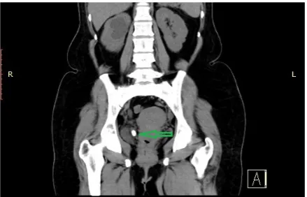
The study was conducted in department of General Surgery, Maulana Azad Medical College and Lok Nayak hospital, New Delhi from October 2017 to March 2019. It was a prospective study. A total of 30 patients were taken for study, which was done for a period of 1.5 years. Inclusion criteria was all adults (>18 years) with diagnosis of single lower ureteric calculus on X-Ray KUB (Figure 1). Exclusion criteria were presence of ureteral injury/stricture, urinary tract infection, single kidney, renal failure. Out of the 30 patients, 6 were ex-cluded due to spontaneous passage of stone. Remaining 24 patients underwent unenhanced helical computed tomography scan (Figure 2 & Figure 3). The im-ages were reviewed at a Picture Archiving and Communication System (PACS) workstation.
The size of the stone was measured using the dimension tool of Radiant DICOM software. The longest transverse diameter of stone was taken as the size of stone. Other stone factor used was height of stone in form number of axial cuts images of stone (slice thickness 5 mm). The Hounsfield units (HU) for each stone were calculated for the cross-section with the largest diameter and the secondary sign like hydronephrosis was graded between 0 - 3. All the CT studies were reviewed by radiologist who was blindfolded to pre and postoperative CT parameters.
Hydronephrosis was graded as follows: 0 = absent, 1 = prominence of the intra-renal pelvis or mild ureteral dilatation, 2 = dilatation of the intra-renal calices or mod-erate ureteral dilatation, and 3 = severe dilatation of the collecting system [9].
DOI: 10.4236/oju.2019.910018 155 Open Journal of Urology
[image:3.595.265.484.252.388.2]Figure 1. X-Ray KUB showing right lower ureteric calculus.
Figure 2. CT KUB showing right lower ureteric calculus.
Figure 3. CT KUB with calculus in right lower ureter.
Statistical analysis was performed to evaluate the association between the outcome of URSL and patient’s age and gender. We also evaluated the associa-tion between the outcome of URSL and CT parameters of the stone like size of the stone, Hounsfield unit. We also included hydronephrosis as secondary sign, duration and complication of the procedure as statistical parameters in our study.
[image:3.595.264.484.416.557.2]DOI: 10.4236/oju.2019.910018 156 Open Journal of Urology
Kolmogorov-Smirnov test. If the normality was rejected then non parametric test was used. Analysis was done using Statistical Package for Social Sciences (SPSS) version 21.0. A p value of less than 0.05 was considered significant.
3. Results
Data from 24 patients with single lower ureteric stone were analyzed. Outcome was classified as successful or failure, based on intra-operative clearance of stone and post operative CT findings. In our study 18 patients had complete removal of stones and 6 patients had failed outcome of URSL. The overall rate of treat-ment success was 75% (n = 18) and the failure rate was 25% (n = 6). The out-come of URSL was correlated with duration of the procedure and CT parameters such as size of stone (diameter and height), Hounsfield unit and presence of hy-dronephrosis. In group 1 (success group) the mean duration of URSL was 25.56 ± 6.49 minutes and mean Hounsfield unit of stone was 774.12 ± 212.85 HU. In group 2 (failure group) the mean duration of URSL was 40.75 ± 4.95 minutes and mean Hounsfield unit of stone was 1193.12 ± 327.51 HU (Table 1).
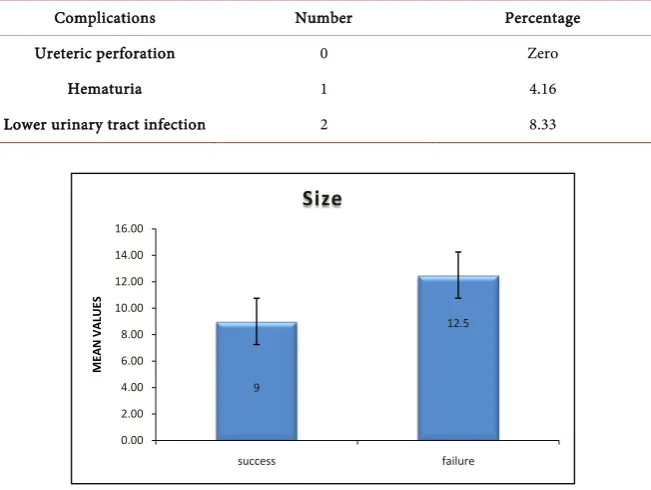
Out of 24 patients, 18 had positive outcome of URSL with mean size of stone 9 ± 2.1 mm and 6 had negative outcome with mean size of stone 12.5 ± 3.3 mm. The p value for stone size in relation to outcome was <0.0004 which was statis-tically significant (Figure 4).
In our study population the shortest duration of URSL was 15 min and the longest duration was 48 min. Out of 24 patients, 18 had positive outcome of URSL with mean duration of procedure 25.56 ± 6.49 and 6 had negative out-come with mean duration of 40.75 ± 4.95. The p value for procedure duration was <0.0001 which was statistically significant (Figure 5).
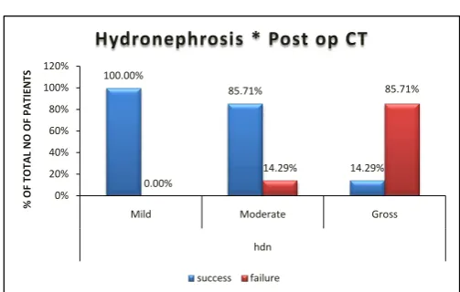
Out of total sample size of the study 58.33% of patients had moderate hydro-nephrosis (HDN), 29.17% had gross HDN and 12.50% had mild HDN. Patients with gross HDN had a poor outcome of URSL (Figure 6 & Figure 7).
The patient’s sex, age, height, weight, BMI and previous stone history did not differ significantly between group 1 and group 2 (Table 2).
The most common complication seen was lower urinary tract infection, which accounted for 8.33%. Second most common complication was hematuria which accounted for 4.16%. There was no evidence of any other known complication including ureteric perforation (Table 3).
4. Discussion
The success rate of URSL depends on the location of stone in the ureter, size of the stone, the use of surgical instruments including the ureteroscope, and the operator’s surgical technique.
The stone size factors that were included in the study were longest diameter, transverse diameter, and height by CT axial cuts. Most studies have focused on the longest stone diameter for measuring stone size. However, Abdelrahim et al.
DOI: 10.4236/oju.2019.910018 157 Open Journal of Urology
Table 1. Comparison of outcome of URSL in respect to duration of procedure, size and
HU of calculus and Hydronephrosis.
Success (Group 1)
Sample size 18 Failure (Group 2) Sample size 6 P value
Duration (minutes) <0.0001
Mean ± SD 25.56 ± 6.49 40.75 ± 4.95
HU 0.001
Mean ± SD 774.12 ± 212.85 1193.12 ± 327.51
Size (mm) 0.004
Mean ± SD 9 ± 2.1 12.5 ± 3.3
Hydronephrosis
[image:5.595.208.540.296.431.2](grade 0/1/2/3) 0/3/13/2 0/0/1/5 0.001
Table 2. Comparison of various parameters in success and failure group.
Parameters Success (group 1) Failure (group 2) p value
Sex (m/f) 14/4 5/1 ˃0.05
Age (years) 26.75 ± 6.2 26.80 ± 6.1 >0.05
Height (cm) 160.66 ± 9.3 160.56 ± 9.6 >0.05
Weight (kg) 65.3 ± 8.7 66.1 ± 8.3 >0.05
BMI (kg/m2) 25.2 ± 3.4 24.6 ± 3.2 >0.05
Previous stone history
[image:5.595.212.538.462.707.2](present/not) 6/13 2/4 >0.05
Table 3. Complications after URSL.
Complications Number Percentage
Ureteric perforation 0 Zero
Hematuria 1 4.16
Lower urinary tract infection 2 8.33
Figure 4. size of stone with outcome of URSL.
9
12.5
0.00 2.00 4.00 6.00 8.00 10.00 12.00 14.00 16.00
success failure
ME
AN
V
AL
U
ES
DOI: 10.4236/oju.2019.910018 158 Open Journal of Urology
[image:6.595.246.499.255.405.2]Figure 5. Duration with outcome of URSL.
Figure 6. Distribution of Hydronephrosis.
Figure 7. Hydronephrosis and outcome of URSL.
There is still controversy whether the secondary signs can predict outcome in patients with ureteral stones or not. In a report on the incidence rates of second-ary signs and their influences on patient management, Ege et al. [11] identified hydroureter, hydronephrosis, periureteral edema, and unilateral renal enlarge-ment as the most reliable signs for endoscopic removal of stones. Takahashi et al. [12] reported that the severity of perinephric edema correlates with the probability of stone expulsion spontaneously. He also suggested that the degree
25.56 40.75 0.00 10.00 20.00 30.00 40.00 50.00 60.00 success failure ME AN V AL U ES Duration 12.50% 58.33% 29.17% Hydronephrosis
Mild Moderate Gross
100.00% 85.71% 14.29% 0.00% 14.29% 85.71% 0% 20% 40% 60% 80% 100% 120%
Mild Moderate Gross
hdn % OF T OT AL N O OF P AT IE N TS
Hydronephrosis * Post op CT
[image:6.595.247.499.429.589.2]DOI: 10.4236/oju.2019.910018 159 Open Journal of Urology
of edema indicated the seriousness of ureteral obstruction. Seitz et al. [13] re-ported that the presence of secondary signs before URSL did not correlate with the preoperative stone-free rate. In our study 7 out of 24 patients had gross HDN and among them 6 had failed outcome of URSL. Patients with gross HDN had poor outcome of URSL. This was because patients with gross HDN has longer duration of stone disease which resulted in impaction of stone and thus resulting in failure of URSL.
In our study the stone with higher HU had a negative outcome as compared to stones with a low HU. The result was in concordance with the study done by Magnunson et al. [14] who showed that Hounsfield unit reflects the stone’s composition and hardness and correlates with the rate of successful ESWL.
Our study compares well with other world literature in terms of outcome of URSL. In our study the overall success rate was 75% and complication rate of 16.67%. Knispel et al. had a success rate of 73.7% in his series of 135 patients who were treated with lithocast [15] Shrestha et al. had a success rate of 88.5% in 52 patients with lower ureteric calculi when treated with pneumatic lithotripter
[16]. Oktay et al. and Leidi et al. also showed the success rate of 91% in lower ureteric calculi [17] [18]. Another study done by Salman et al. compared pneu-matic lithotripsy and laser lithotripsy. Out of 50 patients taken for pneupneu-matic li-thotripsy, 41 patients had successful outcome giving a success rate of 82% [19]. In our study we were not able to include the later complications such as stricture formation, which has been documented in literature, due to difficulties in the further follow up.
5. Conclusions
Size of the stone and Hounsfield units of the stone are the factors which directly influence the outcome of URSL; the bigger the stone or the higher the HU of stone, the less the chances of complete removal of stone. Gross hydronephrosis is associated with poor outcome of URSL.
This study also points towards higher incidence of ureteric stones in males as compared to females with mean age of presentation between 20 - 40 years. We found that ureteroscopy is safe with minimal complications in the management of lower ureteric calculus.
Ethical Clearance
Study was approved by institutional ethics committee.
Conflicts of Interest
The authors declare no conflicts of interest regarding the publication of this pa-per.
References
DOI: 10.4236/oju.2019.910018 160 Open Journal of Urology
Treatment. Postgraduate Medical Journal, 83, 469-472. https://doi.org/10.1136/pgmj.2006.055913
[2] Bhat, A., Singh, V., Bhat, M., Kumar, V. and Bhat, A. (2018) Spectrum of Urinary Stone Composition in Northwestern Rajasthan Using Fourier Transform Infrared Spectroscopy. Indian Journal of Urology, 34, 144-148.
https://doi.org/10.4103/iju.IJU_363_16
[3] Song, H.-J., Cho, S.-T. and Kim, K.-K. (2010) Investigation of the Location of the Ureteral Stone and Diameter of the Ureter in Patients with Renal Colic. Korean Journal of Urology, 51, 198-201.https://doi.org/10.4111/kju.2010.51.3.198
[4] Worster, A., Preyra, I., Weaver, B. andHaines, T. (2002) The Accuracy of Noncon-trast Helical Computed Tomography versus Intravenous Pyelography in the Diag-nosis of Suspected Acute Urolithiasis: A Meta-Analysis. Annals of Emergency Med-icine, 40, 280-286.
[5] Ahmad, N.A., Ather, M.H. and Rees, J. (2003) Incidental Diagnosis of Disease on Un-Enhanced Helical Computed Tomography Performed for Ureteric Colic. BMC Urology, 3, 2-6.
[6] Mostafavi, M.R., Ernst, R.D. and Saltzman, B. (1998) Accurate Determination of Chemical Composition of Urinary Calculi by Spiral Computerized Tomography.
Journal of Urology, 159, 673-675.https://doi.org/10.1016/S0022-5347(01)63698-X
[7] Motley, G., Dalrymple, N., Keesling, C., Fischer, J. and Harmon, W. (2001) Houns-field Unit Density in the Determination of Urinary Stone Composition. Urology, 58, 170-173.https://doi.org/10.1016/S0090-4295(01)01115-3
[8] Etafy, M., Morsi, G.A.M., Beshir, M.S.M., etal. (2013) Management of Lower Ure-teric Stones: A Prospective Study. Central European Journal of Urology, 66, 456-462. https://doi.org/10.5173/ceju.2013.04.art19
[9] Kim, S.Y., Kim, M.J., Yoon, C.S., Lee, M.S., Han, K.H. and Lee, M.J. (2013) Com-parison of the Reliability of Two Hydronephrosis Grading Systems: The Society for Foetal Urology Grading System vs. the Onen Grading System. Clinical Radiology, 68, e484-e490.https://doi.org/10.1016/j.crad.2013.03.023
[10] Abdelrahim, A.F., Abdelmaguid, A., Abuzeid, H., Amin, M., El Mousa, S. and Ab-delrahim, F. (2008) Rigid Ureteroscopy for Ureteral Stones: Factors Associated with Intraoperative Adverse Events. Journal of Endourology, 22, 277-280.
https://doi.org/10.1089/end.2007.0072
[11] Ege, G., Akman, H., Kuzucu, K. and Yildiz, S. (2003) Acute Ureterolithiasis: Inci-dence of Secondary Signs on Unenhanced Helical CT and Influence on Patient Management. Clinical Radiology, 58, 990-994.
https://doi.org/10.1016/S0009-9260(03)00294-0
[12] Takahashi, N., Kawashima, A., Ernst, R.D., Boridy, I.C., Goldman, S.M., Benson, G.S. and Sandler, C.M. (1998) Ureterolithiasis: Can Clinical Outcome Be Predicted with Unenhanced Helical CT? Radiology, 208, 97-102.
https://doi.org/10.1148/radiology.208.1.9646798
[13] Seitz, C., Memarsadeghi, M., Fajkovic, H. and Tanovic, E. (2008) Secondary Signs of Non-Enhanced CT Prior to Laser Ureterolithotripsy: Is Treatment Outcome Pre-dictable? Journal of Endourology, 22, 415-418.
https://doi.org/10.1089/end.2007.0248
[14] Magnuson, W.J., Tomera, K.M. and Lance, R.S. (2005) Hounsfield Unit Density Accurately Predicts ESWL Success. Alaska Medicine, 47, 6-9.
DOI: 10.4236/oju.2019.910018 161 Open Journal of Urology
Patients. JournalofEndourology, 12, 513-515.
https://doi.org/10.1089/end.1998.12.513
[16] Shrestha, B., Karki, D. and Baidya, J. (1970) The Outcome of Pneumatic Lithotripsy for the Management of Ureteric Calculi. Kathmandu University Medical Journal, 6, 355-360.https://doi.org/10.3126/kumj.v6i3.1711
[17] Leidi, G.L., Berti, G.L., Canclini, L., etal. (1997) Ureteroscopy and Stone Lithotripsy with Lithoclast: Personal Experience. Archivio Italiano di Urologia e Andrologia, 69, 181-183.
[18] Oktay, B., Yavascaoglu, I., Simsek, U. and Ozyurt, M. (1997) Intracorporeal Pneu-matic Lithotripsy for Ureteral and Vesical Calculi. Scandinavian Journal of Urology and Nephrology, 31, 333-336.https://doi.org/10.3109/00365599709030615