ISSN Online: 2158-2882 ISSN Print: 2158-284X
Echocardiographic and Clinical Evaluation
of Rheumatic Mitral Stenosis in
Younger and Elderly Patients
Ramakrishna C. D.
*, Placid Sebastian Kanattu
Department of Cardiology, Pariyaram Medical College, Kannur, India
Abstract
Background: Rheumatic heart disease (RHD) is common form of heart dis-ease among population, especially in developing countries like India. Mitral stenosis (MS) is majorly caused by rheumatic heart disease with mitral com-missural adhesion, fibrosis and calcification of the chordae tendineae. The aim of present study was clinical and echocardiographic evaluation for mitral stenosis in RHD patients with different age group. Methods: This was a re-trospective, nonrandomized, and single-centre study in which 203 consecutive patients presented rheumatic mitral stenosis. All the patients were divided into different age group viz. <40 years, 40 to 65 years and >65 years. Cardio-vascular examination and echocardiography were done in each patient. Mitral valve area (MVA), mitral valve gradient (MVG) and left atrial (LA) diameter were assessed by echocardiography. Mitral valve score was recorded to analyse the degenerative changes in mitral valve structure. Results: A total of 203 pa-tients (133 females) were enrolled and divided into three age groups. Papa-tients with age above 65 years were considered as elderly and those patients with age below 40 years were considered as younger. Echocardiographic assessment showed mean 4.7 and 4.9 cm LA diameter, 0.92 and 0.86 cm2 MVA and 11.2 and 9.7 mm Hg MVG in younger and elderly patients respectively. Total mi-tral valve score has shown significant (p < 0.001) difference between younger and elder patients. Moreover, calcification and subvalvular thickening score with >2 had shown significant difference (p < 0.001) between younger and elderly patients. Conclusion: Present study provides unique contemporary data on characteristics and management of patients with rheumatic mitral stenosis. Majority of elderly patients are unsuitable for percutaneous com-missurotomy due to degenerative changes in mitral valve structure.
Keywords
Percutaneous Commissurotomy, Rheumatic Heart Disease, Rheumatic Mitral Stenosis
How to cite this paper: Ramakrishna C.D. and Kanattu, P.S. (2017) Echocardiogra- phic and Clinical Evaluation of Rheumatic Mitral Stenosis in Younger and Elderly Patients. International Journal of Clinical Medicine, 8, 128-135.
https://doi.org/10.4236/ijcm.2017.83012
Received: January 17, 2017 Accepted: March 12, 2017 Published: March 15, 2017
Copyright © 2017 by authors and Scientific Research Publishing Inc. This work is licensed under the Creative Commons Attribution International License (CC BY 4.0).
1. Introduction
Mitral stenosis (MS) causes an obstruction to blood flow from the left atrium to left ventricle. As a result, there is an increase in pressure within the left atrium, pulmonary vasculature, and right side of the heart, while the left ventricle is un-affected in isolated MS. Nearly all cases of MS are caused by rheumatic heart disease with mitral commissural adhesion; thickened, immobile mitral valve leaflets; and fibrosis, thickening, shortening, fusion, and calcification of the chordae tendineae. Infrequent causes of MS include mitral annular calcification and congenital mitral stenosis [1]. MS is highly prevalent in developing coun-tries because of its association with the prevalence of rheumatic fever but is in-creasingly being identified in an unusual form in developed countries [2].
Two-thirds of the world’s population live in developing countries with a high prevalence of rheumatic fever or rheumatic heart disease (RHD), resulting in a large population with mitral stenosis. In a survey of rheumatic fever in India [2], the mean age of presentation was 15 years, and two-thirds of the participants had signs of mitral stenosis, of whom half had limiting symptoms. Up to 30 mil-lion school children and young adults have chronic RHD worldwide, and nearly a third of these also have mitral stenosis [3].
Echocardiography is used to diagnose and judge stage of disease, assess mitral regurgitation, exclude that mimic mitral stenosis, and provide information about suitability for percutaneous balloon valvuloplasty (PBV). Both valve area and gradient can be accurately measured, but several measurements with more than one method are often needed to accurately estimate haemodynamics of the mi-tral valve. The most reliable method to calculate valve area is planimetry with 2D echocardiography cross-section images [4].
Even though the prevalence of mitral stenosis is high in India, the age-specific clinical and anatomical characteristics of the disease are not well studied among Indian patients. The present study was therefore undertaken with clinical and echocardiographic evaluation for Mitral Stenosis in RHD patients who attending the cardiology services at Pariyaram Medical College, Kannur, Kerala.
2. Methods
2.1. Study Design and Patients Population
A total of 203 consecutive patients presenting rheumatic mitral stenosis at the department of cardiology, Pariyaram Medical College, Kannur, Kerala from January 2012 to May 2014 were included in the present study. All the patients were divided into different age group. However, patients with grade > 2 mitral regurgitation, more than mild lesion of the other valves and history of previous surgical commissurotomy either percutaneous or surgical were excluded from this study.
planimetry and pressure half time method [5]. To analyse the effect of mitral valve structure due to degenerative changes, we scored the echocardiographic study of each patient for: (a) leaflet mobility, (b) leaflet thickening, (c) subvalvar thickening, and (d) calcification. Table 1 shows the scoring system [6]. This study was approved by the institutional review board (IRB) and all patients signed the written informed consent.
2.2. Statistical Analysis
Patient baseline characteristics were presented as frequency and percentages. Differences between two point estimates were determined to be statistically sig-nificant at the 0.05 level using two-sided significance tests (z-tests).
3. Results
3.1. Baseline Demographic Characteristics
[image:3.595.205.541.380.734.2]A total of 203 patients were divided into three age groups viz. <40 years (68 pa-tients), 40 to 65 years (78 patients) and >65 years (57 patients). The mean age was 52 years (17 to 90 years). Patients with age above 65 years were considered
Table 1. Scoring of mitral valve characteristics from the echocardiographic examination
[6].
Variable (Scores) Leaflet mobility
1 Highly mobile
2 Reduced mobility
3 Basal leaflet motion only
4 Minimal motion
Valve thickening
1 Near normal (4 - 5 mm)
2 Thickened tips
3 Entire leaflet thickened (5 - 8 mm) 4 Marked leaflet thickening (>8 - 10 mm)
Calcification
1 Single area of brightness
2 Scattered areas at leaflet margins 3 Brightness extends to mid leaflets
4 Extensive leaflet brightness
Subvalvular thickening
1 Minimal chordal thickening
2 Chordal thickening up to 1/3
3 Distal third of chordae thickening
as elderly and those patients with age below 40 years were considered as younger.
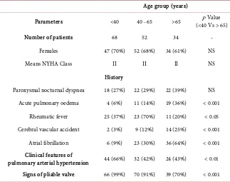
The past history of acute pulmonary oedema, rheumatic fever and atrial fib-rillation were present in 19 (36%), 11 (20%) and 36 (64%) for elder patients group while youngest group present with acute pulmonary oedema 4 (6%), rheumatic fever 25 (37%) and atrial fibrillation 6 (9%) respectively. Cerebrovas-cular accident (CVA) due to infraction was present in 14 (25%) of elderly com-pared to 2 (3%) of younger patients (Table 2).
3.2. Echocardiographic Examination
[image:4.595.214.537.326.581.2]
Assessment of echocardiography showed mean left atrial (LA) diameter of 4.7 cm, mitral valve area (MVA) of 0.92 cm2 and mitral valve gradient (MVG) of 11.2 mm of Hg in younger patients, while mean LA diameter (4.9 cm), MVA (0.86 cm2) and MVG (9.7 mm Hg) were present in elderly patients (Table 3).
Table 2. Baseline characteristics of three age groups of patients with rheumatic mitral
stenosis.
Age group (years)
Parameters <40 40 - 65 >65 (<40 Vs > 65) p Value
Number of patients 68 52 34 -
Females 47 (70%) 52 (68%) 34 (61%) NS
Means NYHA Class II II II NS
History
Paroxysmal nocturnal dyspnea 18 (27%) 22 (29%) 22 (39%) NS Acute pulmonary oedema 4 (6%) 11 (14%) 19 (36%) < 0.001
Rheumatic fever 25 (37%) 23 (70%) 11 (20%) < 0.05 Cerebral vascular accident 2 (3%) 9 (12%) 14 (25%) < 0.001
Atrial fibrillation 6 (9%) 23 (30%) 36 (64%) < 0.001 Clinical features of
pulmonary arterial hypertension 44 (66%) 32 (42%) 24 (43%) < 0.01 Signs of pliable valve 66 (99%) 70 (91%) 39 (70%) < 0.001 Values are expressed as n (%). NYHA, New York Heart Association; Significance at p < 0.05 calculated us-ing a z-test comparus-ing <40 years (younger) with >65 years (Elderly) patients; NS: Non-significant.
Table 3. Comparison of left atrial (LA) size, mitral valve area (MVA) and mitral valve
gradient (MVG) of three age groups of patients with rheumatic mitral stenosis.
Age group (years)
Parameters <40 40 - 65 <65
LA, mean (cm) 4.7 4.5 4.9
MVA, mean (cm2) 0.92 0.9 0.86
[image:4.595.209.540.651.735.2]3.3. Mitral Valve Score
According to classification of mitral valve score as depicted in Table 1, the score of less than 8 was present in 88%, 54% and 23% of younger (<40 years), 40 to 65 year old and elder (>65 years) patients. However, 23% of elder patients had mi-tral valve score greater than 11 and only 2% patients had mimi-tral valve score greater than 11 (Figure 1).
Higher calcification and subvalvular score (>2) was present in 68% and 61% in elderly patients respectively. Though, only 3% and 13% patients with younger population present with higher calcification and subvalvular thickening, respec-tively (Figure 2).
4. Discussion
[image:5.595.262.486.405.515.2]Mitral valve disease is a common cause of morbidity and mortality in patient over age of 65. Physical findings and natural history of rheumatic mitral stenosis may differ in older and younger patients. In addition, symptoms of mitral steno-sis may be masked or exacerbated by coexistent coronary artery disease, pulmo-nary disease, hypertension, and other systemic disorders that commonly occur in older adults [7]. According to previous published reports for the treatment of mitral stenosis, the mean age of the patients ranged from 15 to 56 years. One third of the patients who undergo percutaneous mitral valvotomy are >65 years old [8] [9]. In the present single center study that excluded the patients who had
Figure 1. Comparison of mitral valve score (<8, between 9 to 11 and >11) of three age
groups of patients with rheumatic mitral stenosis. †The P < 0.001 calculatedusing a z-test comparing <40 years (younger) with >65 years (Elderly) patients.
Figure 2. Calcification score and subvascular score having >2 of three age groups of
[image:5.595.268.481.571.692.2]undergone surgical or percutaneous commissurotomy, 28% were above the age of 65 years, the oldest being 90 years of age. Only two patients (3%) were below 18 years. Two thirds of patients were females. Mean duration of dominant symptom of dyspnoea on exertion was 4.9 years that is comparable to younger patients which indicates that the disease may remain latent for many decades and may be participated by arrhythmias like atrial fibrillation which is signifi-cantly higher in elderly patients. Incidence of acute pulmonary edema was sig-nificantly higher in elderly patients (36%) than younger group (6%). Difference could be due to multiple factors like higher incidence of atrial fibrillation, lower atrial compliance, presence of diastolic dysfunction and other co morbid condi-tions like systemic arterial hypertension or coronary artery disease. This is con-trary to earlier observations that acute pulmonary oedema occurs in the early stages of the disease in the younger patients. Average age of patients with acute pulmonary oedema in Wood’s series was 32 years [10].
History of rheumatic fever is present in 60% of cases [10]. In our study 37% of younger patients gave history of rheumatic fever where as only 20% of elderly group could remember about the occurrence of rheumatic fever in childhood. Incidence of cerebrovascular accident was higher among elderly group than younger (25% vs 3%) probably due to higher incidence of atrial fibrillation. Trial fibrillation is the most common complication (40%) of mitral stenosis [10]. Ag-ing is reported as an independent risk factor for the genesis of atrial fibrillation in mitral stenosis [11]. Moreover, Previous study showed patients who devel-oped AF were older and had higher mitral echo score but equal mitral valve area and left atrial size [12]. According to our study, AF was significantly more common in elderly group (64% Vs 9%, P =< 0.001). Tandon et al. (2010) found more severe pulmonary vascular changes in younger patient [13]. These results have support to Sinha et al. (1997) findings that mean pulmonary arterial pres-sure and pulmonary vascular resistance are greater in juvenile patients with mi-tral stenosis when compared with adults [14]. In our study also clinical evidences of pulmonary arterial hypertension, electrocardiographic evidence of right ven-tricular hypertrophy, radiology evidence of main pulmonary artery dilatation; electrocardiographic feature of right ventricular dilatation (RVIDd > 2.6 cm) and pulmonary arterial systolic of more than >50 mmHg were significantly more common among younger age group.
higher mitral valve degenerative changes. After balloon dilatation the younger patients achieved a greater increase in valve area. Moreover, procedural success was higher in the younger group with a greater increase in mitral valve area but complications were similar in both groups. In our study among elderly group, approximately 80% of patients were not suitable for closed mitral valvotomy or percutaneous commissurotomy. Complications of balloon valvotomy were more common in the older patients [16].
5. Conclusion
In older patients there is a greater tendency for valve degenerative change in ad-dition to commissural fusion, and such patients often have co-morbidities. Ma-jority of younger patients (88%) have Wilkin’s score of <8, while most of elderly patients (77%) have score more than 9 despite having similar mean mitral valve area and mean mitral gradient. Thus, in older patients, improvement may be made by palliative treatment for those unsuitable for surgery.
References
[1] Chandrashekhar, Y., Westaby, S. and Narula, J. (2009) Mitral Stenosis. The Lancet, 374, 1271-1283.https://doi.org/10.1016/S0140-6736(09)60994-6
[2] Padmavati, S. (2001) Rheumatic Fever and Rheumatic Heart Disease in India at the Turn of the Century. Indian Heart Journal, 53, 35-37.
[3] Waller, B.F., Howard, J. and Fess, S. (1994) Pathology of Mitral Valve Stenosis and Pure Mitral Regurgitation—Part I. Clinical Cardiology, 17, 330-336.
https://doi.org/10.1002/clc.4960170611
[4] Messika-Zeitoun, D., Fung Yiu, S., Cormier, B., Iung, B., Scott, C., Vahanian, A., Tajik, A.J. and Enriquez-Sarano, M. (2003) Sequential Assessment of Mitral Valve Area during Diastole Using Colour M-Mode Flow Convergence Analysis: New In-sights into Mitral Stenosis Physiology. European Heart Journal, 24, 1244-1253.
https://doi.org/10.1016/S0195-668X(03)00208-2
[5] Kang, W.S., Choi, J.W., Kang, J.E., Chung, J.W. and Kim, S.H. (2013) Determina-tion of Mitral Valve Area with Echocardiography, Using Intra-Operative 3-Dimen- sional versus Intra- & Post-Operative Pressure Half-Time Technique in Mitral Valve Repair Surgery. Journal of Cardiothoracic Surgery, 8, 98.
https://doi.org/10.1186/1749-8090-8-98
[6] Wilkins, G.T., Weyman, A.E., Abascal, V.M., Block, P.C. and Palacios, I.F. (1988) Percutaneous Balloon Dilatation of the Mitral Valve: An Analysis of Echocardio-graphic Variables Related to Outcome and the Mechanism of Dilatation. British Heart Journal, 60, 299-308.https://doi.org/10.1136/hrt.60.4.299
[7] Segal, B.L. (2003) Valvular Heart Disease, Part 2. Mitral Valve Disease in Older Adults. Geriatrics, 58, 26-31.
[8] Skagen, K., Hansen, J.F. and Olesen, K.H. (1978) Closed Mitral Valvulotomy after the Age of Fifty. Scandinavian Journal of Thoracic and Cardiovascular Surgery, 12, 85-89.https://doi.org/10.3109/14017437809100354
Medical Journal, 1, 1051-1063.https://doi.org/10.1136/bmj.1.4870.1051
[11] Diker, E., Aydogdu, S., Ozdemir, M., Kural, T., Polat, K., Cehreli, S., Erdogan, A. and Goksel, S. (1996) Prevalence and Predictors of Atrial Fibrillation in Rheumatic Valvular Heart Disease. American Journal of Cardiology, 77, 96-98.
https://doi.org/10.1016/S0002-9149(97)89145-X
[12] Eid Fawzy, M., Shoukri, M., Al Sergani, H., Fadel, B., Eldali, A., Al Amri, M. and Canver, C.C. (2006) Favorable Effect of Balloon Mitral Valvuloplasty on the Inci-dence of Atrial Fibrillation in Patients with Severe Mitral Stenosis. Catheterization and Cardiovascular Interventions: Official Journal of the Society for Cardiac Angi-ography & Interventions, 68, 536-541.https://doi.org/10.1002/ccd.20770
[13] Tandon, H.D. and Kasturi, J. (1975) Pulmonary Vascular Changes Associated with Isolated Mitral Stenosis in India. British Heart Journal, 37, 26-36.
https://doi.org/10.1136/hrt.37.1.26
[14] Sinha, N., Kapoor, A., Kumar, A.S., Shahi, M., Radhakrishnan, S., Shrivastava, S. and Goel, P.K. (1997) Immediate and Follow Up Results of Inoue Balloon Mitral Valvotomy in Juvenile Rheumatic Mitral Stenosis. The Journal of Heart Valve Dis-ease, 6, 599-603.
[15] Fawzy, M.E., Stefadouros, M.A., Hegazy, H., Shaer, F.E., Chaudhary, M.A. and Fad-ley, F.A. (2005) Long Term Clinical and Echocardiographic Results of Mitral Bal-loon Valvotomy in Children and Adolescents. Heart, 91, 743-748.
https://doi.org/10.1136/hrt.2004.040766
[16] Shaw, T.R., Sutaria, N. and Prendergast, B. (2003) Clinical and Haemodynamic Pro-files of Young, Middle Aged and Elderly Patients with Mitral Stenosis Undergoing Mitral Balloon Valvotomy. Heart, 89, 1430-1436.
https://doi.org/10.1136/heart.89.12.1430
Submit or recommend next manuscript to SCIRP and we will provide best service for you:
Accepting pre-submission inquiries through Email, Facebook, LinkedIn, Twitter, etc. A wide selection of journals (inclusive of 9 subjects, more than 200 journals)
Providing 24-hour high-quality service User-friendly online submission system Fair and swift peer-review system
Efficient typesetting and proofreading procedure
Display of the result of downloads and visits, as well as the number of cited articles Maximum dissemination of your research work
Submit your manuscript at: http://papersubmission.scirp.org/
![Table 1. Scoring of mitral valve characteristics from the echocardiographic examination [6]](https://thumb-us.123doks.com/thumbv2/123dok_us/7776180.719460/3.595.205.541.380.734/table-scoring-mitral-valve-characteristics-echocardiographic-examination.webp)