EFFECTIVENESS and VALIDITY OF SONOGRAPHIC EVALUATION IN ACUTE SCROTUM
*
Kumar Avinash, Lakhkar Bhushita, Hittalamani Iranna Mallappa and Ballur Holebasu
Department of Radiology, Shri B.M. Patil
ARTICLE INFO ABSTRACT
Background:
can be treated with medical management, but some conditions like torsion
require immediate surgical intervention to treat the patient. Ultrasound with colour Doppler is an important non
Aims:
To study cases of acute scrotum with ultrasound appearances of various diseases.
To correlat
To find out limitations of sonography and color Doppler for evaluation of acute scrotum. Settings and Design:
Methods and Material:
sonographic evaluation were taken for study from November 2015 to April 2017. vascularity of both the testes
Statistical analysis used: Results:
of epididymo
confirmed by surgical exploration or follow up on the medical line of treatment Conclusions:
Copyright © 2018, Kumar Avinash et al. This is an open
use, distribution, and reproduction in any medium, provided
INTRODUCTION
The clinical features of acute scrotum are usually pain or scrotal swelling or both occurring simultaneously. In maximum cases, conservative or non-surgical approach can be followed but if there is spermatic cord torsion, immediate surgical intervention has to be done so that testis can be preserved. Ultrasound with colour Doppler provides an important modality option to differentiate between these causes noninvasively and help in proper management
2013). The common etiological factors for acute pain in scrotum are epididymo-orchitis and spermatic cord torsion. It is very difficult to differentiate these conditions solely based on physical examination or laboratory tests and results may not be accurate in 50% of patients (Mueller
surgical intervention is done in torsion within six hours, the testis can be saved in approximately 80 to 100 percent cases.
ISSN: 0975-833X
Article History:
Received 18th March, 2018
Received in revised form 25th April, 2018
Accepted 29th May, 2018
Published online 30th June, 2018
Citation: Kumar Avinash, Lakhkar Bhushita, Hittalamani Iranna Mallappa and Ballur Holebasu
organizations in agricultural development”, International
Key words:
Acute scrotum,
Ultrasound and colour Doppler, Testicular torsion,
Epididymo-orchitis,
Incarcerated hernia, Varicocele.
*Corresponding author
RESEARCH ARTICLE
VALIDITY OF SONOGRAPHIC EVALUATION IN ACUTE SCROTUM
A STUDY OF 50 CASES
Kumar Avinash, Lakhkar Bhushita, Hittalamani Iranna Mallappa and Ballur Holebasu
Department of Radiology, Shri B.M. Patil Medical College Hospital and Research
Vijaypur, Karnataka- 586103
ABSTRACT
Background: Acute scrotum has different etiological factors. Some factors like epididymo can be treated with medical management, but some conditions like torsion
require immediate surgical intervention to treat the patient. Ultrasound with colour Doppler is an important non-invasive tool to rule out these aetiologies for acute scrotum.
To study cases of acute scrotum with ultrasound and color Doppler and note the diagnostic appearances of various diseases.
To correlate sonographic findings with clinical follow up and
To find out limitations of sonography and color Doppler for evaluation of acute scrotum. Settings and Design: This is a cross section type of study performed in our department of
Methods and Material: Patients of acute scrotal pain referred to Department of Radiology for sonographic evaluation were taken for study from November 2015 to April 2017.
vascularity of both the testes and epididymis were evaluated.
Statistical analysis used: Data was analyzed using diagram, mean ± SD, proportions.
Results: In our study of 50 patients presenting with acute scrotum, 5 cases were of torsion, 36 cases of epididymo-orchitis, 2 cases of incarcerated hernia and 7 cases of varicocele. The diagnosis was confirmed by surgical exploration or follow up on the medical line of treatment
Conclusions: Ultrasound with colour Doppler is an excellent modality for diagnosis of acute scrotum.
open access article distributed under the Creative Commons Attribution provided the original work is properly cited.
The clinical features of acute scrotum are usually pain or scrotal swelling or both occurring simultaneously. In surgical approach can be tic cord torsion, immediate surgical intervention has to be done so that testis can be preserved. Ultrasound with colour Doppler provides an important modality option to differentiate between these help in proper management (Yusuf, The common etiological factors for acute pain in spermatic cord torsion. It is very difficult to differentiate these conditions solely based or laboratory tests and results may not
et al., 1988). If
surgical intervention is done in torsion within six hours, the testis can be saved in approximately 80 to 100 percent cases.
If more than twelve hours are taken for surgical procedure, only twenty percent chance of testicular preservation will be there (Hricak, 1983). Hence, it is important and urgent to differentiate between torsion and inflammatory pathologies like epididymo-orchitis to decide further course of action. Sonography is cost effective and
radiation exposure and the examination is relatively of shorter duration. Scrotum is superficial structure so it can be easily examined by high resolution sonography. Utilization of gray scale sonography with color Doppler pr
required anatomical details along with vascularity, thereby providing the prompt diagnosis.
MATERIALS AND METHODS
Patients with complaint of acute scrotal pain who came to Shri B.M. Patil medical college, Hospital and Research centre,
International Journal of Current Research
Vol. 10, Issue, 06, pp.70548-70552, June, 2018
Kumar Avinash, Lakhkar Bhushita, Hittalamani Iranna Mallappa and Ballur Holebasu, 2018.
International Journal of Current Research, 10, (06), 70548-70552.
VALIDITY OF SONOGRAPHIC EVALUATION IN ACUTE SCROTUM –
Kumar Avinash, Lakhkar Bhushita, Hittalamani Iranna Mallappa and Ballur Holebasu
Research centre,
Acute scrotum has different etiological factors. Some factors like epididymo-orchitis can be treated with medical management, but some conditions like torsion and incarcerated hernia require immediate surgical intervention to treat the patient. Ultrasound with colour Doppler is an
invasive tool to rule out these aetiologies for acute scrotum.
color Doppler and note the diagnostic
and post operative diagnosis. To find out limitations of sonography and color Doppler for evaluation of acute scrotum.
This is a cross section type of study performed in our department of radiology. Patients of acute scrotal pain referred to Department of Radiology for sonographic evaluation were taken for study from November 2015 to April 2017. Echotexture and
Data was analyzed using diagram, mean ± SD, proportions.
In our study of 50 patients presenting with acute scrotum, 5 cases were of torsion, 36 cases erated hernia and 7 cases of varicocele. The diagnosis was confirmed by surgical exploration or follow up on the medical line of treatment
Ultrasound with colour Doppler is an excellent modality for diagnosis of acute scrotum.
ribution License, which permits unrestricted
If more than twelve hours are taken for surgical procedure, ent chance of testicular preservation will be Hence, it is important and urgent to differentiate between torsion and inflammatory pathologies orchitis to decide further course of action. and widely available. There is no radiation exposure and the examination is relatively of shorter duration. Scrotum is superficial structure so it can be easily examined by high resolution sonography. Utilization of gray scale sonography with color Doppler provides relevant and required anatomical details along with vascularity, thereby providing the prompt diagnosis.
MATERIALS AND METHODS
Patients with complaint of acute scrotal pain who came to Shri B.M. Patil medical college, Hospital and Research centre,
INTERNATIONAL JOURNAL OF CURRENT RESEARCH
Vijaypur, Karnataka and were referred to Department of Radiology for ultrasound evaluation have been included in our study. For sonographic examination, patient was asked to lie down in supine position with a rolled sheet placed between legs to provide scrotal support and penis was displaced superiorly. A high-frequency probe (7.5–15 MHz) was utilized for scanning in transverse as well as sagittal planes. Lower frequency transducer (5-7MHz) was used in cases of significantly enlarged scrotal sac. Echotexture and vascularity of both testis and epididymis were noted. Color Doppler imaging was used to identify vascularity in the scrotal contents (Brian Gorman, 2011).The machines used in the study were Siemens Acuson X700 and Philips HD11-XE. The duration of study was from November 2015 to April 2017.
Inclusion criteria: Patients with complaint of acute scrotal
pain.
Exclusion criteria: Patients with trauma to scrotum.
Type of study: Cross sectional study.
No. of Cases: Fifty
Statistical analysis: Datawasanalysed using diagram, mean ±
SD, proportions.
RESULTS
A study involving fifty patients of acute scrotum was done. The presenting symptoms were pain, erythema and scrotum. The age group of patients ranged from 2 years to 80 years. Echotexture of affected testis was homogenous in 66% cases, heterogeneous in 24% and predominantly hypoechoic heterogeneous echotexture in 10% cases. Vascularity of testis was increased in 64%, normal in 26% and nil in 10% cases. Epididymis echotexture was heterogeneous in 68% while homogenous in 32% cases. Increased vascularity of epididymis was found in 72% cases while normal in 8% cases. In 10% cases prominent venous channels was the finding. In 10% cases variable vascularity of epididymis was noted due to whirlpool sign.
DISCUSSION
Our study comprises of fifty patients referred to Department of Radiology for evaluation of acute scrotum over a period of 18 months. A study of similar sample size was done by A M Agrawal et al. (2014). Testicular torsion or spermatic cord torsion refers to the twisting of spermatic cord. In 2010, Korkes et al. described the incidence of torsion as 1.4 out of 100 000 men (Korkes, 2012).In our study of 50 patients, 5 cases (10%) were diagnosed positive for torsion. The echotexture was hypoechoic heterogeneous in 4 cases out of 5 while one case showed heterogeneous echotexture as compared to opposite side. There was absent vascularity in affected testis in all the cases. In all the five cases there was heterogeneous echogenicity of the epididymis. Also, whirlpool sign was noted in all the cases, in which there was snail shaped or whorled appearance of spermatic cord affecting the epididymis as well. The part proximal to this whorled appearance was showing vascularity, while there was absence of vascularity distal to it (Figure 1-4). This was the typical feature present in our study and also the most characteristic one as well.
The diagnosis was confirmed on surgical exploration in all the five cases. In a study by Vijayaraghavan, whirlpool sign with no vascularity in distal part was present in torsion in the 61 out of 221 cases. This finding was consistent as we found whirlpool sign in all our cases (Vijayaraghavan, 2006). However in other 4 cases of Vijayaraghavan, there was distal vascularity present. Such cases were diagnosed with incomplete torsion. In these 4 cases, there were focal areas of hypoechogenicity with absent vascularity. These were the regions of segmental infarction in the testis. In our study, there was no such case of partial presence of vascularity in the testis or in the distal part of spermatic cord. A snail shaped mass was described by Kalfa et al. in 199 out of 208 patients of torsion. Furthermore, if linear spermatic cord was present, it indicated other causes of other acute scrotum instead of torsion (Kalfa et al., 2007).Yagil et al found 18 cases of torsion which showed absent intratesticular vascularity and few cases were of hypovascular testis (Yagil et al., 2010).Cassar et al described that for arterial flow to disappear, minimum of 540° twisting is required. If twisting or rotation is less than 360°, there may be presence of arterial flow with obstruction of venous flow (Cassar et al., 2008).Hence, apart from absent intaratesticular vascularity, there can be finding of hypovascular testis as well. S A Rizvi et al. found 4 cases of torsion with similar findings (Rizvi et al., 2011). A M Agrawal found 8 cases of torsion out of 50 cases with similar findings of absent testicular flow. There was sensitivity and specificity of 100%, which was also observed in our study (Agrawal et al., 2014). The infectious or inflammatory changes present in epididymis constitute the epididymitis. The urinary tract infection is commonly associated in such cases (Knight et al., 1984).In our study of 50 patients, 36 cases (72%) were diagnosed for epididymo-orchitis and epididymitis. Out of these 4 cases were diagnosed for epididymitis only.
There was homogenous normal echotexture of testis in 24 cases. Heterogeneous echogenicity of testis was noted in 11 cases while 1 case showed hypoechoic heterogeneous echotexture. Testicular vascularity was characteristically increased in 32 cases while in remaining 4 cases it was normal. In these 4 cases the diagnosis of epididymitis was confirmed. The epididymal echotexture was heterogeneous in majority 29 cases and homogenous in 7 cases. Increased epididymal vascularity was present in all 36 cases. In our study, one case which was diagnosed for epididymo-orchitis was later on confirmed for varicocele. Rest all the cases were confirmed positive with sonographic diagnosis by taking follow up of medical management administered. In our study, increased vascularity of testis and epididymis were the major criteria for the diagnosis of epididymo-orchitis and epididymitis. Also, the echotexture was a non-specific finding. Similar findings have been suggested by other authors. Vijayaraghavan opined increased vascularity of testis or epididymis or both as the main diagnostic feature (Vijayaraghavan, 2006).D’Andrea et al and Thinyu et al also confirmed the samefinding (D’Andrea
et al., 2013; Thinyu et al., 2009). A M Agrawal quoted
Table 1. Association of diagnosis and testis echotexture
Testis Echotexture Epididymo-Orchitis Obstructed Hernia Torsion Varicocele p value
N % N % N % N %
Heterogenous 11 30.6 0 0.0 1 20.0 0 0.0 <0.001
Homogenous 24 66.7 2 100.0 0 0.0 7 100.0
Hypoechoic & Heterogenous 1 2.8 0 0.0 4 80.0 0 0.0
[image:3.595.89.505.175.235.2]Total 36 100.0 2 100.0 5 100.0 7 100.0
Table 2. Association of diagnosis and testis vascularity
Testis Vascularity Epididymo-Orchitis Obstructed hernia Torsion Varicocele p value
N % N % N % N %
Increased 32 88.9 0 0.0 0 0.0 0 0.0 <0.001
Normal 4 11.1 2 100.0 0 0.0 7 100.0
Nil 0 0.0 0 0.0 5 100.0 0 0.0
Total 36 100.0 2 100.0 5 100.0 7 100.0
Table 3. Association of diagnosis and epididymis echotexture
Epididymis echotexture Epididymo-orchitis Obstructed hernia Torsion Varicocele p value
N % N % N % N %
Heterogenous 29 80.6 0 0.0 5 100.0 0 0.0 <0.001
Homogenous 7 19.4 2 100.0 0 0.0 7 100.0
[image:3.595.59.533.353.576.2]Total 36 100.0 2 100.0 5 100.0 7 100.0
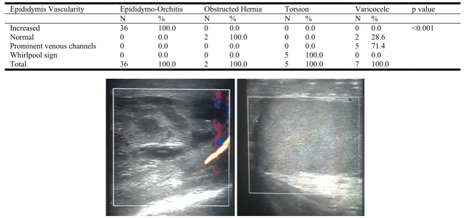
Table 4. Association of diagnosis and epididymis vascularity
Epididymis Vascularity Epididymo-Orchitis Obstructed Hernia Torsion Varicocele p value
N % N % N % N %
Increased 36 100.0 0 0.0 0 0.0 0 0.0 <0.001
Normal 0 0.0 2 100.0 0 0.0 2 28.6
Prominent venous channels 0 0.0 0 0.0 0 0.0 5 71.4
Whirlpool sign 0 0.0 0 0.0 5 100.0 0 0.0
[image:3.595.129.468.603.753.2]Total 36 100.0 2 100.0 5 100.0 7 100.0
Figure 1 A. Whirlpool Sign with twisting vessels and Figure no.1 B. Absence of vascularity in testis.
Figure no.2 A: On color Doppler image, increased epididymal vascularity Figure no. 2B: On color Doppler image, increased testicular vascularity
Figure no.3A Bowel loop seen in the Inguinoscrotal region.
Figure no.3 B Bowel loop seen in the Inguinoscrotal region without vascularity, however vascularity is seen in pampiniform
plexus.
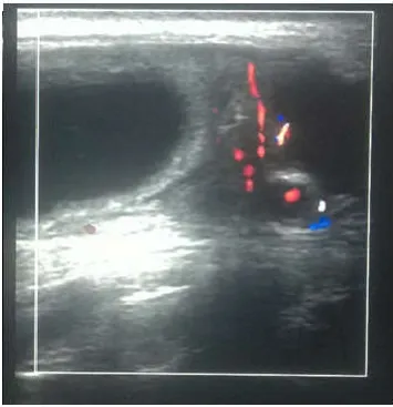
Figure no. 4A & B Colour Doppler showing dilated pampoiniform plexus veins
Figure No 4. Varicocele
Testicular echotexture and vascularity were normal in both of the cases. Similarly epididymis was homogenous with maintained vascularity in both cases. The diagnostic feature was a dilated akinetic bowel loop present in the inguino-scrotal region. There were internal echoes and fluid within bowel loop with no significant peristalsis on real time ultrasound. Also, omentum was noted as significantly echogenic area. On color Doppler, no significant vascularity in the wall of bowel loop appreciated. Similar findings were in the previous studies. Ogata et al diagnosed strangulated hernia in 35 out of 39 patients with akinetic loop which was dilated (Ogata et al., 1994). Celestino et al. also indicated the same feature of dilated akinetic loop of intestine for strangulated hernia (Aso et al., 2005). A M Agrawalet al found 1 case of obstructive inguino-scrotal hernia. Non-peristaltic or akinetic bowel loop in the scrotum was the characteristic feature for strangulation of hernia (Agrawal et al., 2014). Varicoceles correspond to dilatation of the pampiniform plexus predominantly due to ineffective valves of veins. There were 7 cases of varicocele in our study with all patients above the age of 20 years. There was positive history of infertility in 2 patients. Testicular echotexture and vascularity both were normal in all the 7 cases. Epididymis was homogenous in all the cases, but there were significantly prominent multiple anechoic vascular channels on the gray scale which demonstrated venous flow on colour Doppler study.
This characteristic feature of venous flow with reflux on valsalva maneuver was the diagnostic feature in our study. Same feature was found in previous studies. Yagil et al. found 107 cases of varicocele out of 620 with predominant finding of dilated venous channels (Yagil et al., 2010). S A Rizvi et al
diagnosed 14 cases out of 122 patients with positive findingof anechoic serpiginous areas multiple in number with increased flow in venous channels on valsalva (Rizvi et al., 2011). D’Andrea et al. described varicocele in 18 out of 125 cases. There were venous channels which appeared anechoic, tubular and tortuous on gray scale sonography. There was increased flow on color Doppler (D’Andrea et al., 2013). It was observed in our study that sometimes it was difficult to appreciate the change in size and the reflux. The reason was lack of patient complying with the instruction or due to lack of coordination between the patient, probe and the timing. To overcome this difficulty, multiple attempts were taken for demonstration of flow and reflux.
Limitation found in our study: The relatively less common
causes of acute scrotum like fournier’s gangrene, idiopathic scrotal wall oedema and neoplasms are not encountered in our study. This may be due to regional variation.
Conclusion
Acute scrotum is an emergency condition in which early decision is required for better prognosis. Immediate surgical intervention is required for torsion cases and incarcerated hernias. Ultrasound with color Doppler can be confidently used to diagnose them.
Conflicts of interest: Nil
Funding: Nil
REFERENCES
management. Journal of family medicine and primary care. Oct;3(4):409.
Aso C, Enríquez G, Fité M, Torán N, Piró C, Piqueras J, Lucaya J. 2005. Gray-scale and color Doppler sonography of scrotal disorders in children: an update. Radiographics. Sep; 25(5):1197-214.
Brian Gorman. 2011. The scrotum. In: Rumack CM, Wilson SR, Charboneau JW, eds. Diagnostic ultrasound.4th ed. Philadelphia: Elsevier Mosby.
Cassar S, Bhatt S, Paltiel HJ, Dogra VS. 2008. Role of spectral Doppler sonography in the evaluation of partial testicular torsion. J Ultrasound Med., 27:1629–38
D’Andrea A, Coppolino F, Cesarano E, Russo A, Cappabianca S, Genovese EA, Fonio P, Macarini L. 2013. US in the assessment of acute scrotum. Critical ultrasound journal. Dec 1; 5(S1):S8.
Hricak H, Lue T, Filly RA, et al. 1983. Experimental study of the sonographic diagnosis of testicular torsion. J
Ultrasound Med., 2:349-356.
Kalfa N, Veyrac C, Lopez M, Lopez C, Maurel A, Kaselas C, Sibai S, Arena F, Vaos G, Bréaud J, Merrot T. 2007. Multicenter assessment of ultrasound of the spermatic cord in children with acute scrotum. The Journal of urology. Jan 31; 177(1):297-301.
Knight PJ, Vassy LE. 1984. The diagnosis and treatment of the acute scrotum in children and adolescents. Annals of
surgery. Nov; 200(5):664.
Korkes F, Cabral PR, Alves CD, Savioli ML, Pompeo AC. 2012. Testicular torsion and weather conditions: analysis of
21,289 cases in Brazil. International braz jurol.
Apr;38(2):222-9.
Kulacoglu H, Kulah B, Hatipoglu S, Coskun F. 2000. Incarcerated direct inguinal hernias: a three-year series at a large volume teaching hospital. Hernia. Sep 29; 4(3):145-7. Mueller DL, Amundson GM, Rubin SZ, et al. 1988. Acute
scrotal abnormalities in children: diagnosis by combined sonography and scintigraphy. AJR Am J Roentgenol.,
150:643-646.
Ogata M, Imai S, Hosotani R, Aoyama H, Hayashi M, Ishikawa TE. 1994. Abdominal ultrasonography for the diagnosis of strangulation in small bowel obstruction.
British journal of surgery. Mar 1; 81(3):421-4.
Rizvi SA, Ahmad I, Siddiqui MA, Zaheer S, Ahmad K. 2011. Role of color Doppler ultrasonography in evaluation of scrotal swellings: pattern of disease in 120 patients with review of literature. Urology journal. Jan 1; 8(1):60. Stedman’s medical dictionary, 26th ed. Baltimore, MD:
Lippincott Williams and Wilkins, 1995: 788–790
Thinyu S, Muttarak M. 2009. Role of ultrasonography in diagnosis of scrotal disorders: a review of 110 cases. Biomedical imaging and intervention journal. Jan; 5(1). Vijayaraghavan SB. 2006. Sonographic differential diagnosis
of acute scrotum. Journal of ultrasound in medicine. May 1; 25(5):563-74.
Yagil Y, Naroditsky I, Milhem J, Leiba R, Leiderman M,
Badaan S, Gaitini D. 2010. Role of Doppler
ultrasonography in the triage of acute scrotum in the emergency department. Journal of Ultrasound in Medicine. Jan 1; 29(1):11-21.
Yusuf GT, Sidhu PS. 2013. A review of ultrasound imaging in scrotal emergencies. J Ultrasound., 16:171–8.