Original Article
MiR-150 inhibits hypoxia-induced autophagy in oral
squamous carcinoma by negatively regulating BNIP3
Yan-Jun Cui1, Chao Liu2, Fa-Wei Pang1, Guang-Ping Liu1, Jian-Hua Zhou1, Xiao-Ming Shi2, Sheng-Yun Huang2,
Dong-Sheng Zhang1,2
1Department of Oral and Maxillofacial Surgery, School of Stomatology, Shandong University, Jinan, China; 2 Depart-ment of Oral and Maxillofacial Surgery, Shandong Provincial Hospital Affiliated to Shandong University, Jinan, Shandong, China
Received December 14, 2016; Accepted December 27, 2016;Epub March 1, 2017; Published March 15, 2017
Abstract: Oral squamous cell carcinoma (OSCC) is a malignant solid tumor characterized by hypoxia. Autophagy is considered to play a protective role in cancer cells under hypoxia. A variety of microRNAs (miRNAs) that play a critical role in tumorigenesis have been identified. However, the underlying mechanisms of miR-150 in OSCC development remain unclear. In this study, we observed decreased downregulated expression of miR-150 in association with increased expression of BNIP3 in CAL-27 cells under hypoxia. Hypoxia induced the initiation of the BNIP3-mediated autophagic process, and overexpression of miR-150 inhibited hypoxia-induced autophagy. In addition, we demon-strated that miR-150 inhibited autophagy by negatively regulating the expression of the BNIP3 gene. Collectively, our study identified miR-150 as a key modulator of autophagy by inhibiting BNIP3 in OSCC cells.
Keywords:miR-150, autophagy, oral squamous carcinoma, BNIP3
Introduction
Oral squamous cell carcinoma (OSCC) is the most common type of cancer in the oral cavity, with an estimated global incidence of more than 500,000 annually, including 300,000 newly diagnosed cases and an annual mortality of approximately 145,000 [1, 2]. Because most OSCCs are diagnosed in the advanced stage, patients frequently develop a second primary cancer, local recurrence, and distant metasta-sis after treatment failure [3]. At present, the treatment modalities for OSCC involve radical surgical resection with adjuvant chemotherapy, radiotherapy, or both. In spite of great improve-ments in the prevention of oral cancer, little improvement in the relative 5-year survival rate of patients with OSCC has been observed in the past several decades [4]. Hence, the under-standing of the molecular mechanisms related to the pathogenesis and progression of this dis-ease could allow to establish new and more effective therapeutic strategies to improve the survival and quality of life of patients.
Autophagy is a lysosomal degradation pathway whereby degrades long-lived cellular
macro-molecules and organelles [5, 6], and maintains intracellular homeostasis and prolongs cell sur-vival under stress. Autophagy is up-regulated in response to stress conditions such as nutrient deprivation, growth factor depletion, and hypox-ia [7-9]. Emerging evidence suggested that the autophagy is related to a variety of pathological disorders including cancer, neurodegenerative and cardiovascular diseases [10-12]. In cancer cells, autophagy appears to play a role at mul-tiple levels of tumor development and may have a protective role in carcinogenesis [13], and the elucidation of the exact role of autophagy at dif-ferent stages of cancer progression and in treatment responsiveness is a complex and
challenging task. Further uncovering the role of autophagy in OSCC might lead to the identifica -tion of novel strategies for OSCC treatment. Recently, a novel class of endogenous small non-coding gene regulatory RNAs, termed
microRNA (miRNAs), has gained significant
Accumulating evidence shows that miRNAs play important roles in the regulation of most cellular processes, including differentiation, proliferation, apoptosis, metabolism and autophagy [17-20]. Deregulation of miRNAs is
closely linked to the pathological mechanisms
of many human diseases, including cancer [21]. Several studies have also shown that miR-150 is frequently downregulated in pancreatic cancer, esophageal cancer, colorectal cancer, and liver cancer [22-25], suggesting a tumor-suppressive role of miR-150 in human tumori-genesis. However, the biological functions and mechanisms of miRNA-150 in OSCC are unclear.
Materials and methods
Reagents and antibodies
Human miR-150 mimic and mimic negative controls (miR-NC) were purchased from Ribobio (Guangzhou, China). MiR-150 inhibitor and inhibitor negative control (miR-NC) were also purchased from Ribobio (Guangzhou, China). Transfection of miRNA was performed with a riboFECTTM Transfection Kit (Ribobio, Guang- zhou, China) according to the manufacturer’s protocol. The following primary antibodies were used for Western blot: anti-BNIP3 polyclonal antibody, HIF-1 polyclonal antibody, anti-LC3B polyclonal antibody, anti-Beclin1 mono-clonal antibody, anti-Atg5 monomono-clonal antibody, anti-p62 monoclonal antibody, and anti-actin polyclonal antibody (Abcam, Cambridge, MA, USA). Fluorescein isothiocyanate-labeled goat anti-rabbit IgG antibody Invitrogen (Thermo
Fisher Scientific, Inc., Waltham, MA, USA) was used as a fluorescent secondary antibody.
Alexa Fluor 488-labeled goat anti-rabbit IgG antibody (Beijing ComWin Biotech Co., Ltd., Beijing, China) was used as the secondary anti-body for western blot.
Cell culture and transfection
The OSCC cell line CAL-27 was obtained from Ninth People’s Hospital, Shanghai Second Medical University. All cells were cultured in
Dulbecco’s modified eagle medium (DMEM)
(Invitrogen, Carlsbad, CA, USA) supplemented with 10% fetal bovine serum (Thermo Fisher,
Waltham, MA, USA), 100 μM penicillin, and 100 μM streptomycin (Invitrogen, Carlsbad, CA, USA) in a humidified 5% CO2 environment at
37°C. Hypoxic conditions were achieved with a gas-controlled chamber (Thermo Electron Corp., Marietta, OH, USA) maintained at 1% O2, 94% N2, and 5% CO2 at 37°C. A total of 1 × 10⁵
CAL-27 cells were seeded on six-well plates,
and after reaching 60-70% confluence, the
cells were transfected with miR-150 mimic (and mimic negative control) or inhibitor (and inhibi-tor negative control) at concentrations ranging from 50 to 100 nM using a riboFECTTM Transfection Kit (Ribobio, Guangzhou, China) according to the manufacturer’s instructions. The cells were washed 16 h after transfection, replenished with complete medium, and har-vested at 24 or 48 h after transfection for sub-sequent analysis. Quantitative real-time poly-merase chain reaction (qRT-PCR) was used to
verify the efficiency of transfection.
RNA extraction and qRT-PCR
Total RNA was extracted from harvested cells
using TRIzol reagent (Takara Biotechnology,
Co., Ltd., Dalian, China) according to the manu-facturer’s instructions. The concentration and purity of RNA were determined spectrophoto-metrically using a NanoDrop ND-1000 (NanoDrop Technologies, Wilmington, DE, USA). The ratio of A260:A280 was used to indicate the purity of the total RNA. A two-step qRT-PCR was performed using a PrimeScript™ RT
reagent kit (Takara Biotechnology, Co., Ltd.,
Dalian, China). The total RNA was converted
into cDNA using a PrimeScript™ RT reagent kit (Takara Biotechnology, Co., Ltd., Dalian, China). Then, significantly up-regulated or down-regu
-lated miRNAs were quantified by real-time PCR using SYBRRPremix Ex Taq TM (Takara
Biotechnology, Co., Ltd., Dalian, China). PCR was performed in a real-time PCR system (Roche Light Cycler 480 Germany) as follows: 95°C for 3 min, 35 cycles of 95°C for 5 sec, 60°C for 20 sec and 72°C for 30 sec, and then 94°C for 1 min and 60°C for 1 min, with the addition of a cycle for every 0.5°C. The relative expression levels of BNIP3 and miR-150 were respectively normalized to GAPDH and U6. The
following gene-specific primers were used:
Three independent experiments were per-formed in triplicate.
Western blot analysis
were washed twice with cold phosphate-buff-ered saline (PBS) and lysed on ice in RIPA buf-fer (Shennong Bocai Biotechnology Co., Ltd., Shanghai, China). After incubation on ice for 30 min, the lysates were cleared by centrifugation at 14,000 g for 15 min at 4°C, and the protein concentrations were determined using BCA
protein assay kits (Shennong Bocai
Biotech-nology Co., Ltd., Shanghai, China). Equal am- ounts of total protein were loaded and sepa-rated by 10-15% sodium dodecyl sulfate-poly-acrylamide gel electrophoresis (Shennong Bocai Biotechnology Co., Ltd., Shanghai, China) and transferred to PVDF membranes (Shennong Bocai Biotechnology Co., Ltd., Shanghai, China).
After blocking with 5% nonfat milk in phos -phate-buffered saline, the membranes were
incubated with specific primary antibodies
overnight at 4°C, followed by incubation with secondary antibodies labeled with HRP for 60 min. After washing three times with TBST, the proteins were visualized using an Alpha Imager 2200 system (Alpha Innotech Corporation, San
Leandro, CA, USA). The bands of the specific proteins were quantified after normalization to
the density of actin using Image J instrument software.
Immunofluorescence microscopy
A total of 2 × 10⁵ CAL-27 cells were seeded in
12-well plates with 14-mm-diameter cover-slips. After incubation for at least 24 h, the cells were treated with miR-150 mimic (and mimic negative control), miR-150 inhibitor (and inhibi-tor negative control) for 48 h under hypoxia (1% O2) or normoxia (20% O2). The cells were then
fixed in 4% paraformaldehyde (Beijing ComWin
Biotech Co., Ltd., Beijing, China) for 20 min and permeabilized in 0.2% Triton X-100 (Beijing ComWin Biotech Co., Ltd., Beijing, China) for 10 min. After washing three times with PBS, the
slices were blocked with goat serum for 1 h at
room temperature (RT), then incubated with rabbit polyclonal anti-BNIP3 antibody (1:100 diluted in PBS) at 4°C overnight. The next day, the cells were washed three times with PBS and incubated for 1 h with goat anti-rabbit immunoglobulin (Ig) antibody conjugated with
fluorescein isothiocyanate (FITC; Beijing
Com-Win Biotech Co., Ltd., Beijing, China) (1:400 diluted in PBS) for 1 h at room temperature in
the dark. Nuclei were counterstained with DAPI
(BeijingComWin Biotech Co., Ltd., Beijing,
China). After washing with PBS, cells were mounted with antifade mounting media (BeijingComWin Biotech Co., Ltd., Beijing, China), and signals were visualized under a
Nikon-E600 fluorescence microscope (TE2000; Nikon Corporation, Tokyo, Japan). Images were
recorded using RSImage software.
Transmission electron microscopy
The CAL-27 cells (1 × 10⁶) were seeded in 6-well
plates. After reaching 70-80% confluence, the
cells were transfected with miR-150 mimic (and mimic negative control), miR-150 inhibitor (and inhibitor negative control) using the riboFECTTM Transfection Kit (Ribobio, Guangzhou, China) according to the manufacturer’s protocol, fol-lowed by culture for 48 h under hypoxia (1% O2) or normoxia (20% O2). The cells were then fixed
with 2% glutaraldehyde in 0.1 M Sorensen buf-fer (pH 7.3; Beijing Chemical Industry Group, Co., Ltd., Beijing, China) for 1 h at 4°C and
post-fixed in 1% osmium tetroxide in 0.1 M cacodyl -ate buffer for 1 h at room temperature. The cells were then dehydrated in solutions of etha-nol (30-90%), embedded in epon resin (Beijing Chemical Industry Group, Co., Ltd., Beijing, China) and sliced with a UC7 microtome (Leica,
Wetzlar, Germany) to a 70-nm thickness and
placed on uncoated copper grids. Sections were subsequently counterstained with 4% ura-nyl acetate (Beijing Chemical Industry Group, Co., Ltd., Beijing, China) and visualized using a JEM-1200EX Transmission Electron Microscope
(JEOL, Ltd., Tokyo, Japan).
Statistical analysis
Differences between groups were analyzed using a Student’s t-test. All data were present-ed as the mean ± standard deviation from the three independent experiments and analyzed using SPSS software, version 19.0 (IBM SPSS,
Armonk, NY, USA). P<0.05 was considered as
statistically significant difference.
Results
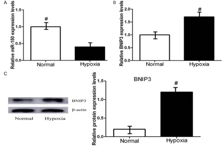
Expression level of miR-150 and BNIP3 in hypoxia-induced CAL-27 cells
To determine the expression levels of miR-150
and BNIP3 in CAL-27 cells, we first measured
nor-moxia (20% O2) by qRT-PCR. As shown in Figure 1A, miR-150 levels were decreased significant -ly following culture in hypoxic conditions com-pared with normal cell lines. Figure 1B shows that the expression of BNIP3 in OSCC CAL-27
cells was significantly increased under hypoxia
compared with normoxia. We further examined the protein level of BNIP3 in CAL-27 cells by Western blot, which revealed that the BNIP3 protein level was higher in CAL-27 cells than in normal cell lines (Figure 1C). These results demonstrate that hypoxia may decrease the expression of miRNA-150 and increase the expression of BNIP3.
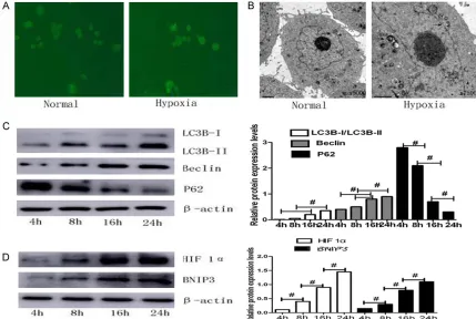
Hypoxia induces autophagy in CAL-27 cells
One of the physiological responses of hypoxia is the induction of autophagy [28]. To investi-gate whether autophagy could be induced by hypoxia in OSCC cell lines, CAL-27 cells were incubated under hypoxic conditions for 4, 8, 16, and 24 h. When autophagy is activated, the
LC3-I protein localized in the cytoplasm is cleaved, lipidated, and inserted as LC3-II into autophagosome membranes [29], providing a means of observing autophagy induction. As shown in Figure 2A, a diffuse distribution of
green fluorescence and weak punctate dots
were observed under normoxia, whereas a cor-nucopia of LC3 punctate dots in the cytoplasm
was observed under hypoxic conditions by fluo -rescence microscopy. Transmission electron microscopy analysis revealed an increase in double-membrane-bound vacuoles, where cytoplasmic material and/or membrane vesi-cles were encapsulated in vacuoles, the gold standard for determination of autophagy in CAL-27 cells incubated at 1% O2 compared with 20% O2 (Figure 2B, black arrows). During
autophagy, a series of autophagy genes associ-ated with the formation of autophagosomes
are highly activated [30]. To confirm
[image:4.612.86.519.74.359.2]hypoxia-induced autophagy, western blot analyses were performed to measure the expression of LC3, Atg5, Beclin1 and P62 at the protein levels. We
observed that hypoxia increased the expres-sion of LC3, Atg5, and Beclin1 in a time-depen-dent manner (Figure 2C). As HIF-1 is the key
protein responsible for cellular adaptation to low oxygen conditions, we examined whether HIF-1 was involved in the hypoxia-induced autophagy. Our results also showed that cells subjected to hypoxia presented increased pro-tein levels of HIF-1 and BNIP3 in hypoxia-treat-ed CAL-27 cells (Figure 2D). These results sug-gest that hypoxia could induce the presence of the double membrane of autophagic vesicles and expression of autophagy-related proteins,
including BNIP3, after 24 h of hypoxia. Taken
together, these results demonstrate that autophagy might be induced during oxygen hypoxic stress and that initiation of the autoph-agic process is mediated through HIF-1 signal-ing in CAL-27 cells.
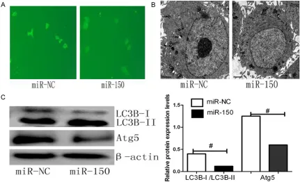
miR-150 inhibits hypoxia-induced autophagy in CAL-27 cells
Emerging evidence indicates that miRNA regu-lates autophagy by targeting autophagy-related genes [31-33]. To investigate if miR-150 modu-lates autophagy under hypoxia, we transfected CAL-27 cells with miR-150 mimic or negative control (miR-NC) under hypoxia for 48 h. Our data showed that the number of LC3 punctate
dots formed per cell significantly decreased in
cells transfected with miR-150 mimic (Figure 3A). In addition, the number of autophagolyso-somes was lower after miR-150 treatment than miR-NC treatment, as observed by transmis-sion electron microscopy (Figure 3B). The inhib-itory effects of miR-150 on autophagy were
also confirmed by western blot analysis. Consistent with the other markers of autopha
[image:5.612.91.520.73.361.2]gy inhibition, we observed that following treat-ment with miR-150 mimic, the LC3B-II/LC3B-I ratio was attenuated, and polyubiquitin-binding protein p62 levels were increased after overex-pression of miR-150 (Figure 3C). These results indicated that endogenous miR-150 contrib-utes to the limitation of autophagic responses in CAL-27 cells and that miR-150 may be a novel inhibitor of autophagy.
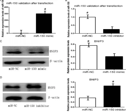
miR-150 negatively regulates the BNIP3 pro-tein in CAL-27 cells under hypoxia
Because precise modulation of BNIP3 protein levels is essential for tumorigenesis, we hypoth-esized that BNIP3 is regulated through a miR-mediated mechanism. Since bioinformatics analysis indicated that miR-150 is inversely cor-related correlation with BNIP3 in OSCC, we next
verified whether miR-150 can regulate endoge -nous BNIP3 expression under hypoxia. CAL-27 cells were transiently transfected with miR-150 mimic or inhibitor, and the expression of BNIP3 was examined. Figure 4A, 4B validation
miR-150 after transfected with miR-miR-150 mimic or inhibitor. Western blot analysis revealed that the protein expression levels of BNIP3 were clearly decreased by the miR-150 mimic com-pared with the negative control after 48 h (Figure 4C). By contrast, BNIP3 protein levels
were significantly increased in miR-150 inhibi -tor-transfected cells compared with negative control-transfected cells under hypoxia (Figure 4D). Taken together, we can conclude that
miR-150 negatively regulates the protein expres-sion of BNIP3 in CAL-27 cells under hypoxia. Discussion
A variety of miRNAs have been identified to play
[image:6.612.91.522.71.333.2]a critical role in tumorigenesis, and important regulatory functions of miRNAs in several bio-logical processes associated with tumor have been described [34]. However, the underlying mechanisms of miRNAs in OSCC development have not been fully elucidated. To understand the impact and molecular mechanisms under-lying miRNA function, we searched for target
genes that might be regulated by miR-150. We demonstrated here that hypoxia resulted in decreased expression of miR-150 in CAL-27 cells in association with increased expression of BNIP3. Forced overexpression of miR-150 inhibited autophagy of CAL-27 cells. Additionally, the upregulation of miR-150 negatively regulat-ed the protein levels of BNIP3. These results
suggest that miR-150 is a key modulator of
autophagy by inhibiting BNIP3 in OSCC cells. MiRNAs play vital roles by regulating the expres-sion of target genes via the degradation or translational inhibition of their target mRNAs [35-37]. A growing body of evidence now sug-gests that miRNAs act either as oncogenes or
tumor suppressors in a variety of cancers [38, 39]. To determine the role of miR-150 in OSCC, we searched for physiological targets using
bio-informatics analysis. Bcl-2 nineteen-kilodalton
interacting protein (BNIP3), a BH3-only
mem-ber of the Bcl-2 family, was identified as one of
the targets of miR-150. BNIP3 is increased in many cancers, such as gastric adenocarcino-mas, breast cancer, non-small cell lung cancer, and primary prostate tumors [40-43]. During the past decades, a series of studies have sug-gested that increased expression of BNIP3 might induce cell death through multiple path-ways including apoptosis, necrosis and autoph-agy, depending on cell lines and conditions
[image:7.612.95.518.77.454.2]that overexpression of BNIP3 is not sufficient to induce cell death and defines a role for
BNIP3 in promoting autophagy in response to hypoxia [47]. BNIP3 competes with Beclin1-Bcl2 and Beclin1-Bcl-xl complexes, resulting in release of Beclin1 and thus enhancing autoph-agy [48, 49]. BNIP3 induces autophautoph-agy by interacting with RheB, resulting in mTOR inhibi-tion [50]. Moreover, BNIP3 could be involved in autophagosome-lysosome fusion in later stag-es of autophagy [51, 52]. Here, we found that
the expression of BNIP3 was significantly
increased in CAL-27 cells under hypoxia expo-sure compared to normoxia. Transfection of miR-150 mimic decreased the protein levels of BNIP3 in CAL-27 cells under hypoxia exposure, whereas administration of the miR-150 inhibi-tor increased BNIP3 protein levels compared with the negative control. These results sug-gest that BNIP3 protein levels are inversely associated with miR-150 expression in CAL-27 cells, which suggest that miR-150 might be an important factor in OSCC cells through directly targeting BNIP3. Further studies exploring the role BNIP3 as a novel target in therapeutic applications are needed.
Recently, the role of miRNAs in tumorigenesis has been widely investigated, and their impor-tant regulatory functions in several biological processes associated with tumors have been
described [34]. Among known miRNAs with sig
-nificantly decreased expression levels, the
highly conserved miRNA miR-150 attracted our attention. Decreased levels of miR-150 have been observed in esophageal carcinoma, colorectal cancer, pancreatic cancer, chronic
lymphocytic leukemia, and liver cancer [23-25,
53, 54], whereas miR-150 is upregulated in gastric cancer, breast cancer, and lung cancer [55-57]. Aberrant expression of miR-150 is associated with cancer development and pro-gression through regulating oncogenes or tumor suppressor genes [58-60]. Based on the literature, we explored the biological function of miR-150 expression in OSCC. Our results revealed decreased expression of miR-150 in CAL-27 cells under hypoxic conditions com-pared to normal conditions. Forced overexpres-sion of miR-150 inhibited autophagy of CAL-27
cells. Our findings provide evidence suggesting
that miR-150 plays an important role in modu-lating autophagosome formation in OSCC cells.
Hence, identifying the specific involvement of
miR-150 in oral squamous cell carcinogenesis will help expand our understanding of OSCC and aid the development of new targets for diagnosis and therapy.
In summary, our study suggests that miR-150 exerts its roles in OSCC through negative regu-lation of the protein levels of BNIP3, and over-expression of miR-150 inhibited autophagy in
CAL-27 cells in vitro. These findings suggest
that miR-150 may be a potential novel predic-tor and therapeutic target for future
develop-ment of specific therapeutic interventions in
OSCC. However, further studies with in vivo
observations are required to confirm our
results.
Acknowledgements
We thank all the staff members in laboratory
center for the technical help and critical read-ing of the manuscript. This study was support-ed by grants from the National Natural Science Foundation of China (2016WS0410), and the Shandong Provincial Natural Science Found- ation (grant no. ZR2016HQ041).
Disclosure of conflict of interest None.
Address correspondence to: Dong-Sheng Zhang and Sheng-Yun Huang, Department of Oral and Maxillofacial Surgery, Shandong Provincial Hospital Affiliated to Shandong University, 324 Jingwu Road, Jinan 250021, Shandong, China. E-mail: ds63zhang@sdu.edu.cn (DSZ); huangsy28@163. com (SYH)
References
[1] Ferlay J, Soerjomataram I, Ervik M, Dikshit K, Eser S, Mathers C. GLOBOCAN 2012 v1.0, can-cer incidence and mortality worldwide: IARC CancerBase No. 11. Lyon, France: Internation-al Agency for Research on Cancer; 2013. [2] Rivera C. Essentials of oral cancer. Int J Clin
Exp Pathol 2015; 9: 11884-94.
[3] Forastiere A, Koch W, Trotti A, Sidransky D. Head and neck cancer. N Engl J Med 2001; 26: 1890-900.
[4] Gupta S, Kong W, Peng Y, Miao Q, Mackillop WJ. Temporal trends in the incidence and sur-vival of cancers of the upper aerodigestive tract in ontario and the United States. Int J Cancer 2009; 9: 2159-65.
[6] Yang Z, Klionsky DJ. Eaten alive: a history of macroautophagy. Nat Cell Biol 2010; 9: 814-22.
[7] Mihaylova MM, Shaw RJ. The AMPK signalling pathway coordinates cell growth: autophagy and metabolism. Nat Cell Biol 2011; 9: 1016-23.
[8] Levine B, Kroemer G. Autophagy in the patho-genesis of disease. Cell 2008; 1: 27-42. [9] Zhang H, Bosch-Marce M, Shimoda LA, Tan YS,
Baek JH, Wesley JB, Gonzalez FJ, Semenza GL. Mitochondrial autophagy is an HIF-1-depen-dent adaptive metabolic response to hypoxia. J Biol Chem 2008; 16: 10892-903.
[10] Lee JH, Yu WH, Kumar A, Lee S, Mohan PS, Pe-terhoff CM, Wolfe DM, Nixon RA. Lysosomal proteolysis and autophagy require presenilin 1 and are disrupted by Alzheimer-related PS1 mutations. Cell 2010; 7: 1146-58.
[11] Luo J, Solimini NL, Elledge SJ. Principles of cancer therapy: oncogene and non-oncogene addiction. Cell 2009; 5: 823-37.
[12] Garcia-Arencibia M, Hochfeld WE, Toh PP, Ru-binsztein DC. Autophagy: a guardian against neurodegeneration. Semin Cell Dev Biol 2010; 7: 691-8.
[13] Fan TF, Bu LL, Wang WM. Tumor growth sup-pression by inhibiting both autophagy and STAT3 signaling in HNSCC. Oncotarget 2015; 41: 43581-93.
[14] Hardy BG, Schentag JJ. Lack of effect of cimeti-dine on the metabolism of quinicimeti-dine: effect on renal clearance. Int J Clin Pharmacol Ther Toxi-col 1988; 8: 388-91.
[15] Bartel DP. MicroRNAs: target recognition and regulatory functions. Cell 2009; 3: 215-33. [16] Qin X, Xu H, Gong W, Deng W. The Tumor
cyto-sol miRNAs, fluid miRNAs, and exosome miR-NAs in lung cancer. Front Oncol 2014; 4: 357-65.
[17] Li YJ, Wang HX, Tao K, Xiao Q, Huang Z, Zhong L, Cao W, Wen J, Feng W. miR-29b suppresses CML cell proliferation and induces apoptosis via regulation of BCR/ABL1 protein. Exp Cell Res 2013; 8: 1094-01.
[18] He X.Q, Dong Y.J, Wu C.W , Zhao Z, Ng SS, Chan FK, Sung JJ, Yu J. MicroRNA-218 Inhibits Cell cycle progression and promotes apoptosis in colon cancer by downregulating BMI1 poly-comb ring finger oncogene. Mol Med 2012; 12: 1491-98.
[19] Feng LF, Ma YN, Sun J, Shen Q, Liu L, Lu H, Wang F, Yue Y, Li J, Zhang S, Lin X, Chu J, Han W, Wang X, Jin H. YY1-MIR372-SQSTM1 regula-tory axis in autophagy. Autophagy 2014; 8: 1442-53.
[20] Li Y, Chen D, Jin L, Liu J, Li Y, Su Z, Qi Z, Shi M, Jiang Z, Yang S, Lai Y. Oncogenic microRNA-142-3p is associated with cellular migration, proliferation and apoptosis in renal cell carci-noma. Oncol Lett 2016; 2: 1235-41.
[21] Sun LG, Liu L, Fu HH, Wang Q, Shi Y. associa-tion of decreased expression of serum mir-9 with poor prognosis of oral squamous cell car-cinoma patients. Med Sci Monit 2016; 22: 289-94.
[22] Srivastava SK, Bhardwaj A, Singh S, Arora S, Wang B, Grizzle WE, Singh AP. MicroRNA-150 directly targets MUC4 and suppresses growth and malignant behavior of pancreatic cancer cells. Carcinogenesis 2011; 12: 1832-39. [23] Yokobori T, Suzuki S, Tanaka N, Inose T, Sohda
M, Sano A, Sakai M, Kuwano H. miR-150 is as-sociated with poor prognosis in esophageal squamous cell carcinoma via targeting the EMT inducer ZEB1. Cancer Sci 2013; 1: 48-54,.
[24] Perilli L, Pizzini S, Bisognin A, Mandruzzato S, Biasiolo M, Facciolli A, Zanovello P. Human miRNome profiling in colorectal cancer and liver metastasis development. Genom Data 2014; 2: 184-98,.
[25] Zhang J, Luo N, Luo Y, Peng Z, Zhang T, Li S. microRNA-150 inhibits human CD133-positive liver cancer stem cells through negative regu-lation of the transcription factor c-Myb. Int J Oncol 2012; 3: 747-56.
[26] Singh S, Srivastava SK, Bhardwaj A, Owen LB, Singh AP. CXCL12-CXCR4 signalling axis con-fers gemcitabine resistance to pancreatic can-cer cells: a novel target for therapy. Br J Cancan-cer 2010; 11: 1671-79.
[27] Singh AP, Moniaux N, Chauhan SC, Meza JL, Batra SK. Inhibition of MUC4 expression sup-presses pancreatic tumor cell growth and me-tastasis. Cancer Res 2004; 4: 622-30. [28] Mazure NM, Pouyssegur J. Hypoxia-induced
autophagy. cell death or cell survival? Curr Opin Cell Biol 2010; 2: 177-80.
[29] Tanida I, Ueno T, Kominami E. LC3 conjugation system in mammalian autophagy. Int J Bio-chem Cell Biol 2004; 12: 2503-18.
[30] Nakatogawa H, Suzuki K, Kamada Y, Ohsumi Y. Dynamics and diversity in autophagy mecha-nisms: lessons from yeast. Nat Rev Mol Cell Biol 2009; 7: 458-67.
[31] Chang Y, Yan W, He X, Zhang L, Li C, Huang H, Nace G, Geller DA, Lin J, Tsung A. miR-375 in-hibits autophagy and reduces viability of hepa-tocellular carcinoma cells underhypoxic condi-tions. Gastroenterology 2012; 1: 177-87.e8. [32] Frankel LB, Lund AH. MicroRNA regulation of
autophagy. Carcinogenesis 2012; 11: 2018-25.
[34] Callegari E, Gramantieri L, Domenicali M, D’Abundo L, Sabbioni S, Negrini M. MicroRNAs in liver cancer: a model for investigating patho-genesis and novel therapeutic approaches. Cell Death Differ 2015; 1: 46-57.
[35] Weigel AL, Handa JT, Hjelmeland LM. Microar-ray analysis of H2O2-, HNE-, or tBH-treated
ARPE-19 cells. Free Radic Biol Med 2002; 10: 1419-32.
[36] Halbeisen RE, Galgano A, Scherrer T, Gerber AP. Post-transcriptional gene regulation: from genome-wide studies to principles. Cell Mol Life Sci 2008; 5: 798-813.
[37] Farh KK, Grimson A, Jan C, Lewis BP, Johnston WK, Lim LP, Burge CB. The widespread impact of mammalian microRNAs on mRNA repress on and evolution. Science 2005; 16: 1817-21. [38] Iorio MV, Croce CM. MicroRNA dysregulation in
cancer: diagnostics, monitoring and therapeu-tics. A comprehensive review. EMBO Mol Med 2012; 3: 143-59.
[39] Cheng G. Circulating miRNAs: roles in cancer diagnosis, prognosis and therapy. Adv Drug Deliv Rev 2015; 3: 75-93.
[40] Murai M, Toyota M, Suzuki H, Satoh A, Sasaki Y, Akino K, Ueno M, Takahashi F, Kusano M, Mita H, Imai K. Aberrant methylation and si-lencing of the BNIP3 gene in colorectal and gastriccancer. Clin Cancer Res 2005; 3: 1021-27.
[41] Tan EY, Campo L, Han C, Turley H, Pezzella F, Gatter KC, Harris AL, Fox SB. BNIP3 as a pro-gression marker in primary human breast can-cer; opposing functions in in situ versus inva-sive cancer. Clin Cancer Res 2007; 13: 467-74. [42] Giatromanolaki A, Koukourakis MI, Sowter
HM, Sivridis E, Gibson S, Gatter KC, Harris AL. BNIP3 expression is linked with hypoxia-regu-lated protein expression and with poor progno-sis in non-small cell lung cancer. Clin Cancer Res 2004; 16: 5566-71.
[43] Shaida N, Launchbury R, Boddy JL, Jones C, Campo L, Turley H, Kanga S. Expression of BNIP3 correlates with hypoxia-inducible factor (HIF)-1alpha, HIF-2alpha and the androgen re-ceptor in prostate cancer and is regulated di-rectly by hypoxia but not androgens in cell lines. Prostate 2008; 3: 336-43.
[44] Kubli DA, Ycaza JE, Gustafsson AB. Bnip3 me-diates mitochondrial dysfunction and cell death through Bax and Bak. Biochem J 2007; 3: 407-415.
[45] Zhang Z, Yang X, Zhang S, Ma X, Kong J. BNIP3 upregulation and EndoG translocation in de-layed neuronal death in stroke and in hypoxia. Stroke 2007; 5: 1606-13.
[46] Kanzawa T, Zhang L, Xiao L, Germano IM, Kon-do Y, KonKon-do S. Arsenic trioxide induces au-tophagic cell death in malignant glioma cells
by upregulation of mitochondrial cell death protein BNIP3. Oncogene 2005; 6: 980-91. [47] Tracy K, Dibling BC, Spike BT, Knabb JR,
Schumacker P, Macleod KF. BNIP3 is an RB/ E2F target gene required for hypoxia-induced autophagy. Mol Cell Biol 2007; 17: 6229-42. [48] Bellot G, Garcia-Medina R, Gounon P, Chiche J,
Roux D, Pouysségur J, Mazure NM. Hypoxia-in-duced autophagy is mediated through hypoxia-inducible factor induction of BNIP3 and BNIP3L via their BH3 domains. Mol Cell Biol 2009; 10: 2570-81.
[49] Mazure NM, Pouysségur J. Atypical BH3-do-mains of BNIP3 and BNIP3L lead to autophagy in hypoxia. Autophagy 2009; 6: 868-79. [50] Li Y, Wang Y, Kim E, Beemiller P, Wang CY,
Swanson J, You M, Guan KL. Bnip3 mediates the hypoxia-induced inhibition on mammalian target of rapamycin by interacting with Rheb. J Biol Chem 2007; 49: 35803-13.
[51] Azad MB, Chen Y, Henson ES, Cizeau J, McMil-lan-Ward E, Israels SJ, Gibson SB. Hypoxia in-duces autophagic cell death in apoptosis-com-petent cells through a mechanism involving BNIP3. Autophagy 2008; 2: 195-204.
[52] Zhang H, Bosch-Marce M, Shimoda LA, Tan YS, Baek JH, Wesley JB, Gonzalez FJ, Semenza GL. Mitochondrial autophagy is an HIF-1-depen-dent adaptive metabolic response to hypoxia. J Biol Chem 2008; 16: 10892-903.
[53] Yang K, He M, Cai Z, Ni C, Deng J, Ta N, Xu J, Zheng J. A decrease in miR-150 regulates the malignancy of pancreatic cancer by targeting c-Myb and MUC4. Pancreas 2015; 3: 370-9. [54] Mraz M, Chen L, Rassenti LZ, Ghia EM, Li H,
Jepsen K, Smith EN, Kipps TJ. miR-150 influ-ences B-cell receptor signaling in chronic lym-phocytic leukemia by regulating expression of GAB1 and FOXP1. Blood 2014; 1: 84-95. [55] Wu Q, Jin H, Yang Z, Luo G, Lu Y, Li K, Ren G,
Fan D. miR-150 promotes gastric cancer prolif-eration by negatively regulating the pro-apop-totic gene EGR2. Biochem Biophys Res Com-mun 2010; 3: 340-5.
[56] Huang S, Chen Y, Wu W, Ou YN, Chen J, Li H, Liu X, Yao Y. miR-150 promotes human breast cancer growth and malignant behavior by tar-geting the pro-apoptotic purinergic P2X7 re-ceptor. PLoS One 2013; 12: 807-17.
[57] Cao M, Hou D, Liang H, Hou D, Liang H, Chen X. miR-150 promotes the proliferation and migra-tion of lung cancer cells by targeting SRC ki-nase signalling inhibitor 1. Eur J Cancer 2014; 5: 1013-24.
[58] Wang F, Ren X, Zhang X. Role of microRNA-150 in solid tumors. Oncol Lett 2015; 1: 11-16. [59] Jiang X, Huang H, Li Z , Wang X, Gurbuxani S
MLL-associated leukemia. Cancer Cell 2012; 4: 524-35.
[60] Watanabe A, Tagawa H, Yamashita J, Teshima K, Nara M, Iwamoto K, Kume M, Kameoka Y,