Emerging Role for Members of the Bcl-2 Family
in Mitochondrial Morphogenesis
Arnaud Autret1and Seamus J. Martin1,*
1Molecular Cell Biology Laboratory, Department of Genetics, The Smurfit Institute, Trinity College, Dublin 2, Ireland *Correspondence:martinsj@tcd.ie
DOI 10.1016/j.molcel.2009.10.011
Bcl-2 family proteins regulate apoptosis by controlling the release of mitochondrial cytochrome
c
via the
Bax/Bak channel. However, recent studies have also implicated several members of this family in the
regu-lation of mitochondrial fission/fusion dynamics. It has been debated whether the role of Bcl-2 proteins in
mitochondrial morphogenesis is functionally distinct from their role in apoptosis, with some arguing that
Bax/Bak-induced mitochondrial fission promotes apoptosis-associated cytochrome
c
release, while others
suggest that these functions are separable. Here we review this emerging area and argue for a role for the
Bcl-2 family as novel regulators of mitochondrial morphogenesis.
Introduction
Apoptosis is a mode of programmed cell death that is used in a diversity of settings to eliminate cells that are only transiently required, are injured, are infected, or have reached the end of their useful life span. This mode of cell death is coordinated by members of a family of proteases—the caspases—that are nor-mally present as inactive precursors in healthy cells but become activated at the onset of apoptosis. Upon activation, caspases unleash controlled mayhem within the cell, involving the restricted proteolysis of hundreds of substrate proteins (re-viewed inLuthi and Martin, 2007). The latter events culminate in cell death with the morphological and biochemical features of apoptosis (Taylor et al., 2008). Programmed elimination of cells via apoptosis ensures the timely and controlled removal of cellular corpses before such cells can rupture and release their contents. The latter feature is of critical importance, as cell rupture (i.e., necrosis) can provoke inflammatory immune reac-tions that escalate damage and lead to further cell death (Chen et al., 2007; Kono and Rock, 2008).
Members of the Bcl-2 protein family play pivotal roles in regu-lating caspase activation through reguregu-lating mitochondrial outer membrane permeability (MOMP). This is because one of the major routes to apoptosis-associated caspase activation—the so-called intrinsic pathway to apoptosis—is initiated upon release of cytochromecfrom the mitochondrial intermembrane space into the cytosol (reviewed inTaylor et al., 2008). Upon entering the cytosol, cytochromecacts as a cofactor for a cas-pase-activating molecule, called Apaf-1, which is normally present in an autoinhibited state within healthy cells. Upon binding cytochromec, Apaf-1 undergoes oligomerization into a disc-like complex—termed the apoptosome—and this acts as an activation platform for caspase-9 (Youle and Strasser, 2008; Taylor et al., 2008). Upon activation within the apopto-some, caspase-9 then initiates a cascade of further caspase activation events that result in apoptosis (Figure 1).
Thus, cells use cytosolic cytochromecas a cue to activate the programmed cell death machinery. Because the decision to initiate apoptosis is such an important one in the life of a cell, a decision that can be made only once, it follows that
cyto-chromecmust be under a tight leash within the mitochondrial intermembrane space, lest this molecule inadvertently escape and provoke death.
Bcl-2 Family Proteins Regulate MOMP
Aside from the hazards of apoptosome activation, the perme-ability of the outer mitochondrial membrane is also tightly regu-lated to ensure that mitochondrial respiratory chain compo-nents, including cytochromec, do not leak out into the cytosol and impair ATP production as a consequence. Indeed, mainte-nance of optimal oxidative phosphorylation-dependent ATP production is essential for viability of many cell types. As an illus-tration of this, when cytochromec-dependent caspase activa-tion is abolished as a result of inactivaactiva-tion of the genes encoding Apaf-1 or caspase-9, MOMP typically leads to caspase-inde-pendent cell death in most cell types (Ekert et al., 2004). This is due to a precipitous decline in ATP production and mitochondrial dysfunction as a result of the escape of mitochondrial intermem-brane space constituents into the cytosol (Mootha et al., 2001; Colell et al., 2007). Because of this, MOMP is a defining event in the life of a cell, a point at which commitment to cell death becomes irreversible even if caspases fail to be activated down-stream. It should be noted, however, that cell death without cas-pase activation lacks the majority, if not all, of the features of apoptosis and is otherwise indistinguishable from necrosis (Kroemer and Martin, 2005). Irrespective of whether death occurs via apoptosis or necrosis, MOMP is undoubtedly bad news for a cell and must be carefully policed, which is where the Bcl-2 family proteins arrive center stage.
BH3-only proteins, such as Bid, Bim, and Bad, are the most upstream acting members of the Bcl-2 family and have little in common with each other, or indeed with other members of this family, except for an8 amino acid long only motif. BH3-only proteins act as sensors for cell stress, damage, infection, growth factor deprivation, and other signals that provoke apoptosis (Puthalakath and Strasser, 2002). The BH3-only sensors become activated by a variety of means, such as tran-scriptional upregulation, limited proteolysis, or dephosphoryla-tion. Irrespective of how particular BH3-only proteins are activated, these proteins promote Bax/Bak activation by two possible mechanisms, which are not mutually exclusive. One route to Bax/Bak activation involves displacement of the anti-apoptotic Bcl-2 proteins from Bax/Bak due to binding of BH3-only proteins to the antiapoptotic proteins (Willis et al., 2007). Another possible route to Bax/Bak activation is via direct, but possibly transient, interaction of BH3-only proteins with Bax/ Bak that provokes a conformational change of these proteins to their active state (Kuwana et al., 2005).
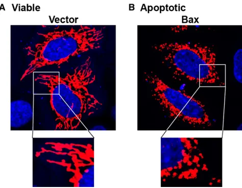
Whichever model is correct—noting that both mechanisms could well operate simultaneously—Bax/Bak activation results in the formation of a pore or channel in the mitochondrial outer membrane that permits release of multiple mitochondrial proteins into the cytosol (Figure 1). Aside from cytochromec, MOMP is also associated with efflux of Smac/DIABLO, a poten-tiator of caspase activation, as well as Omi/HtrA2, adenylate kinase 2, and many other mitochondrial intermembrane space proteins. As noted above, this event has catastrophic conse-quences for the cell, due to the swift activation of the Apaf-1/ caspase-9 apoptosome upon binding of cytochrome c. Because of the importance of this event for commitment to apoptosis, Bax/Bak-dependent cytochrome c release has been studied intensively over the past 10 years. During the course of such studies, it was also noticed that mitochondria, which form extensive interconnected networks in many cells, undergo extensive fragmentation practically simultaneous with apoptosis-associated mitochondrial cytochrome c release (Frank et al., 2001;Figure 2). Before we discuss the potential implications of this observation, we will first briefly discuss mitochondrial network dynamics in healthy cells.
Mitochondrial Fission and Fusion
[image:2.603.58.381.99.285.2]Contrary to the classical textbook view that mitochondria are small bean-shaped organelles scattered throughout the cytosol, many cells contain long tubular mitochondria that are exten-sively interconnected to form web-like networks that encom-pass the whole cell (Figure 2A). Mitochondrial networks are highly dynamic and undergo remodeling through continuous cycles of fission and fusion to produce shorter or longer mito-chondria (Chen and Chan, 2005; Suen et al., 2008). Indeed, experiments where cells with differentially labeled populations of mitochondria have been induced to undergo fusion indicate that intermixing of mitochondrial constituents is essentially complete within 10–12 hr (Legros et al., 2002). Furthermore, mitochondrial networks can undergo abrupt remodeling in response to cell stress (Tondera et al., 2009), changes in energy demands, and fluctuations in intracellular calcium levels
Figure 1. Bcl-2 Family Proteins Regulate Mitochondrial Outer Membrane Permeabilization
(A) In viable cells, members of the antiapoptotic subset of the Bcl-2 family (Bcl-2, Bcl-xL, A1, Mcl-1, Bcl-w, Bcl-b) inhibit the activity of the BH3-only proteins (Bad, Bid, Bim, Bik, Bmf, Noxa, Puma, Hrk) and prevent the activation of Bax and Bak.
(B) Upon activation of one or more members of the BH3-only subset of the Bcl-2 family, these proteins promote MOMP and consequent cyto-chromecrelease through neutralizing the antia-poptotic Bcl-2 proteins and/or triggering Bax/ Bak oligomerization within the mitochondrial outer membrane. Upon release of cytochromecinto the cytosol, this promotes assembly of the Apaf-1/ caspase-9 apoptosome and triggers a cascade of caspase activation events that culminate in apoptosis.
Figure 2. Mitochondrial Networks Undergo Fragmentation during Apoptosis
[image:2.603.313.550.488.672.2](Szabadkai et al., 2004) and in preparation for mitosis (Mitra et al., 2009).
Unsurprisingly, mitochondrial fission and fusion are highly regulated and serve several purposes, including ensuring equal partitioning of mitochondria to daughter cells during mitosis, mitochondrial replication, repair of defective mitochondria, and propagation of calcium waves (Chan, 2006). Furthermore, the state of the mitochondrial network may also influence the propensity of cells to enter apoptosis, as we shall discuss in more detail below.
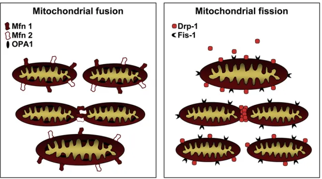
Mitochondrial fusion is controlled by a cohort of proteins that regulate the tethering of adjacent mitochondria, followed by fusion of their inner and outer membranes (Figure 3). Although the detailed mechanics of mitochondrial fusion are still not understood, it is well established that mitofusins (Mfn) 1 and 2, dynamin family GTPases that engage in homo- as well as hetero-typic interactions on adjacent mitochondria, play prominent roles in mitochondrial fusion (Chen and Chan, 2005; Chen et al., 2003). Mfn1 and Mfn2 are localized to mitochondrial outer membranes where regions within their C-termini are thought to interact, intrans, between neighboring mitochondria to promote tethering and fusion (Koshiba et al., 2004). Another dynamin family GTPase, Opa1, which is localized to the mitochondrial intermembrane space and the mitochondrial inner membrane, cooperates with the mitofusions in promoting mitochondrial fusion. Loss of Opa1, Mfn1, or Mfn2 leads to defective drial fusion and hence provokes fragmentation of the mitochon-drial network to varying degrees (Chen et al., 2003; Frezza et al., 2006; Griparic et al., 2004).
Mitochondrial fission is also regulated by a GTPase, dynamin-related protein 1 (Drp1), that can form ring-like oligomers at specific points along mitochondria (Figure 3). Once again, the details remain to be worked out, but Drp1 oligomers may be capable of physically constricting and pinching mitochondria apart in a drawstring-like mechanism. Interestingly, much of the cellular Drp1 content is not constitutively associated with mitochondrial membranes but translocates to discrete foci on mitochondria that often become future sites of fission. Fis1, a protein that is uniformly dispersed on mitochondrial outer membranes, is also involved in mitochondrial fission and might
act as a receptor for Drp1 in yeast (Mozdy et al., 2000), although that has yet to be confirmed in higher eukaryotes. Indeed, Youle and colleagues have found that Fis1 knockdown in mammalian cells does not interfere with the translocation of Drp1 to mito-chondria (Lee et al., 2004), suggesting either that Fis1 is redun-dant with other factors in mammals or that mammalian Fis1 is not a receptor for Drp1.
The importance of mitochondrial fission/fusion for cellular homeostasis is underscored by the phenotypes of mice mutant in many of the genes implicated as central players in mitochon-drial fission/fusion. Inactivation of the genes encoding either Mfn1, Mfn2, Opa1, or Drp1 in the mouse are all lethal (Chen et al., 2003; Frezza et al., 2006; Ishihara et al., 2009), and muta-tions in several of these genes in humans are associated with neurodegenerative conditions, most likely as a direct result of perturbing mitochondrial morphogenesis in neurons (Chan, 2006). Neurons, as well as other highly polarized cells, appear to be particularly sensitive to disturbances in mitochondrial network dynamics, as such cells, due to their extreme size and polarity, often require localization of mitochondria at specific sites, such as axon terminals where synapses are formed. This may be due to the high ATP demands at such sites and/or the role of mitochondria in cellular calcium buffering. It has also been found that the branching processes of such cells, the dendritic spines and axonal terminals, form in close association with mitochondrial network outgrowth, most likely due to the local high energy demands within these cellular regions (Li et al., 2004). Irrespective of the precise role of mitochondria at such sites, defects in mitochondrial fission/fusion often manifest in neurodegenerative diseases such as optic atrophy and Char-cot-Marie-Tooth disease type 2A (reviewed inChan, 2006).
Mitochondrial Fission Is Associated with Apoptosis
[image:3.603.54.378.100.281.2]Mitochondrial networks were first reported to undergo fragmen-tation during apoptosis almost 10 years ago (Desagher and Martinou, 2000; Frank et al., 2001). However, the fact that mito-chondrial fission is often associated with cell injury has been known for over 50 years, well before the term ‘‘apoptosis’’ was even coined (Claude and Fullam, 1945). Before delving into the possible causes of apoptosis-associated mitochondrial
Figure 3. Molecular Control of Mitochondrial Fission and Fusion
fragmentation, it should be said at the outset that opinion is divided about whether this phenomenon is simply a conse-quence of apoptosis or plays a more active role in the process. As we shall discuss below, some investigators have suggested that mitochondrial fission may promote cytochromecrelease and therefore act to drive caspase activation during apoptosis (Frank et al., 2001; Youle and Karbowski, 2005). However, other data suggest that apoptosis-associated mitochondrial fission is a consequence rather than a cause of apoptosis and reflects events that impinge upon some hitherto unrecognized connec-tion between members of the Bcl-2 family and the mitochondrial morphogenesis machinery (Arnoult et al., 2005; Delivani et al., 2006; Karbowski et al., 2006; Sheridan et al., 2008).
Irrespective of the functional role of apoptosis-associated mitochondrial fission, what is widely agreed upon is that this is a general feature of apoptosis in essentially all cell types and in response to most, if not all, proapoptotic stimuli (Frank et al., 2001; Karbowski et al., 2002; Arnoult et al., 2005; Sheridan et al., 2008). Not only do mitochondrial networks undergo frag-mentation during apoptosis, but these fragmented mitochondria also become clustered around the nucleus, and all mitochondrial movement appears to cease (Sheridan et al., 2008;Figure 2B). It is not clear exactly how this happens, but the end result is that mitochondria within apoptotic cells appear to become uncoupled from their normal attachment points to the cytoskel-eton, an observation that awaits further investigation.
Bax and Bak Can Promote Mitochondrial Fission
So, how does mitochondrial fission/fragmentation occur during apoptosis? Youle and colleagues have published a series of seminal studies demonstrating conclusively that activation of Bax and/or Bak leads rapidly to mitochondrial fragmentation (Frank et al., 2001; Karbowski et al., 2002, 2004). Furthermore, this event occurs very close in time to MOMP and associated mitochondrial cytochromecrelease (Arnoult et al., 2005). What is also highly compelling is that activated Bax—which can be distinguished from the inactive form of this protein using confor-mational-sensitive Bax antibodies—frequently decorates mito-chondrial scission sites (Karbowski et al., 2004). Apoptosis-asso-ciated mitochondrial fragmentation occurs upstream of caspase activation, as inhibition of caspase activity downstream of Bax/Bak activation fails to block this event (Frank et al., 2001; Sheridan et al., 2008). These data have led to the hypothesis that Bax/Bak-dependent mitochondrial fragmentation contrib-utes to the process of MOMP and promotes the release of cyto-chromecand other intermembrane space constituents that is seen during apoptosis (Desagher and Martinou, 2000; Youle and Karbowski, 2005).
Some other observations support the idea that mitochondrial fragmentation can facilitate apoptosis-associated cytochrome c release. A dominant-negative mutant of Drp1 (Drp1K38A) that
can impair mitochondrial fission has been observed to impair cytochrome c release and delay cell death in response to some proapoptotic agents (Frank et al., 2001; Brooks et al., 2007). However, it should be noted that the delay in cytochrome
crelease observed is relatively modest and has not been shown to translate into long-term survival. Indeed,Drp1 / null mice exhibit elevated levels of apoptosis, arguing that this protein is
dispensible for MOMP (Ishihara et al., 2009). The delayed kinetics of cytochromecrelease seen when Drp1 expression is ablated or functionally impaired may be a nonphysiological by-product of altered mitochondrial ultrastructure in such cells, because the release kinetics of Smac, Omi, adenylate kinase 2, and other intermembrane space proteins was unaltered under the same conditions (Estaquier and Arnoult, 2007; Parone et al., 2006; Ishihara et al., 2009). Because Smac and cytochromecare coreleased in a Bax/Bak-dependent manner (Munoz-Pinedo et al., 2006), this suggests that cells defective in Drp1 may exhibit artificially delayed cytochromecrelease kinetics. One possible reason for this is that remodeling of the cristae junctions in such cells—known to be required for efficient cytochrome c
release (Scorrano et al., 2002)—may be affected. However, because the release kinetics of Smac, Omi, adenylate kinase 2, and other intermembrane space proteins is overtly normal, this suggests that MOMP is unaffected through loss of Drp1.
Observations that, at first sight, appear to be at odds with the latter view have been made by Nunnari and colleagues using a pharmacological inhibitor of Drp-1, mdivi-1 (Cassidy-Stone et al., 2008). This inhibitor, which interferes with mitochondrial fission in yeast and mammalian cells, has been found to attenuate cytochromecrelease and apoptosis in response to staurosporine. However, mdivi-1 was also found to inhibit bid-dependent cytochrome c release from isolated mitochondria (Cassidy-Stone et al., 2008), which is puzzling, as purified mito-chondria do not undergo fission in vitro. This suggests either that mdivi-1 is also capable of inhibiting other (unknown) molecules that regulate MOMP, or that Drp1 plays a role in MOMP that is unrelated to its role in mitochondrial fission. Either way, this study does not provide support for a simple connection between mitochondrial fission and MOMP.
Overexpression of Mfn2, which produces highly fused mito-chondria, has been reported to delay cytochrome c release (Jahani-Asl et al., 2007). However, others find little impairment of apoptosis upon overexpression of Mfn1, Mfn2, or Drp1K38A (Sheridan et al., 2008), leading to the view that mitochondrial fission is unlikely to be essential for MOMP and apoptosis but acts as a contributory factor in the release of mitochondrial factors rather than a key driver of this process. So why does mitochondrial fragmentation invariably occur during apoptosis? A plausible interpretation of the available data is that mito-chondrial networks fragment during apoptosis because the events that open the Bax/Bak channel (i.e., activation of one or more BH3-only proteins) simultaneously perturb the function of members of this family involved in mitochondrial morphogen-esis, as well as MOMP. Put another way, we propose that one or more members of the Bcl-2 family play roles in mitochondrial morphogenesis that are functionally distinct from their role as regulators of MOMP. During apoptosis, the events that promote MOMP also coincidentally affect the role that these Bcl-2 family members play in mitochondrial network dynamics, resulting in mitochondrial fission.
and Shio, 2008; Sheridan et al., 2008; Tan et al., 2008; Berman et al., 2009). Another prediction is that it should be possible to provoke Bax/Bak-dependent mitochondrial fragmentation in healthy cells in the absence of MOMP. Once again, this predic-tion has also been fulfilled, as we shall discuss below. Further-more, it is also expected that members of the Bcl-2 family may interact with core constituents of the mitochondrial fission/fusion machinery such as Drp1, Mfn1, or Mfn2. Such evidence has also recently emerged (Delivani et al., 2006; Brooks et al., 2007; Li et al., 2008).
Bax/Bak-Induced Fission Can Be Uncoupled from
CytochromecRelease
The observation that Bax/Bak-induced mitochondrial fragmen-tation occurs almost simultaneously with the release of mito-chondrial cytochromecsuggested that these events could be related. However, several independent observations suggest that these events are merely coincident and can be functionally uncoupled.
Karbowski and Youle have shown that Bax/Bak double knockout cells have constitutively fragmented mitochondrial networks, despite being highly resistant to MOMP and apoptosis (Karbowski et al., 2006). Re-expression of Bak in these cells, under conditions where apoptosis was not provoked, led to increases in mitochondrial length, arguing that Bax and/or Bak can promote mitochondrial fusion/elongation in the absence of apoptosis. These authors also presented evidence that Bax/ Bak-induced mitochondrial fusion may occur via regulating Mfn2 function, although a direct interaction between these two proteins was not demonstrated in this study. These intriguing observations suggest that Bax and/or Bak contribute to mito-chondrial fusion quite apart from their role as positive regulators of MOMP. In this light, mitochondrial fragmentation during apoptosis could be interpreted as a direct consequence of neutralization of the Bax/Bak-dependent mitochondrial fusion activity due to the conformational changes that the latter proteins undergo upon activation (Dewson et al., 2008).
Additional evidence for an apoptosis-independent role for Bax/Bak in mitochondrial fusion comes from two independent studies on the CMV viral protein, vMIA, which binds Bax and Bak and inhibits apoptosis (Karbowski et al., 2006; Roumier et al., 2006). Both studies reported that vMIA provokes mito-chondrial fragmentation without either MOMP or apoptosis, once again suggesting that neutralization of Bax/Bak function disturbs the mitochondrial network in healthy cells, leading to mitochondrial fission in the absence of MOMP. However, it should be noted that there is also evidence to suggest that the effects of vMIA on mitochondrial fission-fusion dynamics may be Bax/Bak independent (Poncet et al., 2006).
Further evidence for an uncoupling of Bax/Bak-associated mitochondrial fission and MOMP comes from studies in which Bax or Bak was transiently expressed along with antiapoptotic members of the Bcl-2 family, such as Bcl-xL or Bcl-2 (Delivani et al., 2006; Sheridan et al., 2008). In all cases, Bcl-xL or Bcl-2 failed to suppress Bax-induced mitochondrial fission but readily suppressed MOMP and apoptosis downstream.
Collectively, these studies argue that perturbing the balance of interactions within the Bcl-2 family, even in situations in which
this does not lead to apoptosis, also impacts upon the connec-tivity of the mitochondrial network. These data argue that the Bcl-2 family may play a previously unsuspected role as regula-tors of mitochondrial morphogenesis, quite apart from their role in apoptosis. We will now take a look at further evidence for this.
Bcl-2 Family Members Can Remodel Mitochondrial Networks in Healthy Cells
Evidence from a number of laboratories is accumulating to suggest that members of the Bcl-2 family may influence mito-chondrial network dynamics in healthy cells. Perhaps the most persuasive evidence comes from Bcl-wand Bcl-xL knockout mice, where defects in mitochondrial networks have recently been reported (Liu and Shio, 2008; Berman et al., 2009).
UsingBcl-w / animals, Liu and Shio have made the unex-pected observation that mitochondria in cerebellar purkinje neurons are approximately 2-fold longer than normal in these animals (Liu and Shio, 2008). This suggests that Bcl-w may be required for normal rates of mitochondrial fission, although this was not directly measured in this study. The longer mitochondria seen in these animals may be due to normal rates of ongoing fusion, without counterbalancing fission, in the absence of Bcl-w. Somewhat surprisingly, the authors found little evidence that Bcl-w controls cell survival in the mouse, as cell numbers were overtly normal, but these mice did exhibit defective learning responses, which is very plausibly related to defective mitochon-drial fission. This is because mitochonmitochon-drial fission has been found to be required for normal synaptogenesis, as discussed earlier (Li et al., 2004).
Using conditional Bcl-xL / knockout mice, Hardwick and colleagues provide evidence that Bcl-xL can promote increased fission as well as fusion in healthy neurons. Mitochondria within healthy cortical and hippocampal neurons from Bcl-xL /
animals were shorter and more punctiform than their wild-type counterparts (Berman et al., 2009). Furthermore, transient expression of Bcl-xL in wild-type rat cortical neurons increased mitochondrial lengths (Berman et al., 2009). The simple interpre-tation of these data is that Bcl-xL is required for mitochondrial fusion. However, through careful analysis of rates of both fission and fusion in the presence of overexpressed Bcl-xL, Hardwick and colleagues found that this protein increased rates of mito-chondrial fusion as well as fission, and an effect on mitomito-chondrial biomass was also noted (Berman et al., 2009).
An intriguing connection between Bcl-xL and neuronal synap-togenesis, reported by Jonas and colleagues (Li et al., 2008), may also reflect a role for Bcl-xL in the regulation of mitochon-drial fission, rather than apoptosis. Li et al. found that Bcl-xL expression enhanced synapse formation and synaptic activity in neurons, an effect that could be abrogated by blocking mito-chondrial fission with dominant-negative Drp1K38A. As noted
turn may influence the availability of mitochondria for the forma-tion of new branches at axon terminals and dendritic spines.
Previous studies have also implicated other members of the Bcl-2 family as regulators of mitochondrial morphogenesis. Perhaps the first report to explicitly draw attention to the possi-bility that Bcl-2 may affect normal mitochondrial morphology in healthy cells was a study by Fiskum and colleagues ( Kowaltow-ski et al., 2002). Using flow cytometry to measure the size and granularity of isolated mitochondria, this report noted that Bcl-2-overexpressing cells contained mitochondria that were larger and more structurally complex than normal, as indicated by their greatly increased laser scattering properties.
Further evidence for a role for members of the Bcl-2 family in steady-state mitochondrial network dynamics has also been provided by studies in which Bcl-xL and Bcl-2 have been tran-siently expressed in a variety of mammalian cell types (Delivani et al., 2006; Sheridan et al., 2008). These studies reported that both Bcl-2 family proteins could promote increased rates of mitochondrial fusion or mitochondrial fission, depending on the concentration of expressed protein (Figure 4). In all cases, perturbation of mitochondrial network dynamics, through overexpression of Bcl-2 or Bcl-xL, led neither to MOMP nor to apoptosis. Furthermore, similar observations have been made with theC. elegansBcl-2 homolog, CED-9, that has been found to promote increased mitochondrial fusion upon ectopic expres-sion in either mammalian cells (Delivani et al., 2006),C. elegans
embryos (Rolland et al., 2009), or mature worm muscle tissue (Li et al., 2008; Rolland et al., 2009).
Mitochondrial Network Connectivity May Exert Long-Range Effects on the Threshold for MOMP
It may seem odd, at first sight, that members of the Bcl-2 family could impinge on the spatial organization of mitochondrial networks, given the overwhelming body of evidence that places this protein family firmly within the heart of the cell death machinery. However, these seemingly disparate functions may not be entirely unconnected.
[image:6.603.60.381.101.281.2]Accumulating evidence indicates that the efficiency of oxida-tive phosphorylation within the mitochondrial electron transport chain is affected by the degree of connectivity of mitochondria,
Figure 4. Bcl-2 Family Proteins Can Perturb Mitochondrial Networks in Healthy Cells
Mitochondrial networks in cells transfected with a mito-RFP plasmid along with empty vector plasmid (left panel) or Bcl-xL expression plasmid (middle and right panels). Bcl-xL expression results in cells with mitochondrial networks that are either fragmented (middle panel) or hyperag-gregated and/or fused (right panel). SeeDelivani et al. (2006)andSheridan et al. (2008)for further details.
with highly connected mitochondria correlating with increased efficiency of ATP production (Chen et al., 2005; Mitra et al., 2009; Tondera et al., 2009). For example, fragmentation of mitochondrial networks, through genetic inactivation of
Mfn1andMfn2or RNAi-mediated ablation of Opa1, led to mark-edly decreased rates of respiration (Chen et al., 2005). These defects could be reversed through restoration of mitochondrial fusion, suggesting that these defects were directly related to fusion incompetence and not due to a loss of mitochondrial DNA or other long-range effects (Chen et al., 2005).
Conversely, cells under starvation stress have been observed to increase mitochondrial connectivity and ATP production, which required Mfn-1 (Tondera et al., 2009), presumably as an adaptive response to the change in nutrient availability. Where stress-induced mitochondrial fusion was prevented, in Mfn1
null cells, for example, ATP production declined and cells died more rapidly (Tondera et al., 2009). On the other hand, where mitochondrial connectivity was restored through overexpression of Mfn-1, cell survival was enhanced.
However, the relationship between mitochondrial network status and respiratory function is clearly not a simple one, as knockdown of Drp1, which leads to mitochondrial elongation, can also lead to defective respiration (Estaquier and Arnoult, 2007; Benard et al., 2007; Parone et al., 2008), althoughDrp1 /
MEFs do not appear to suffer from such defects (Ishihara et al. 2009). However, these studies do provide further support for the view that mitochondrial interconnectivity can influence ATP production in either a positive or a negative way.
Evidence for Direct Interactions between Bcl-2 Proteins and the Fission/Fusion Machinery
So, if members of the Bcl-2 family contribute to the regulation of mitochondrial dynamics, how does this operate at a molecular level? This is where confusion currently reigns, because interac-tions have been reported between several Bcl-2 family proteins, both pro- and antiapoptotic, and several core constituents of the mitochondrial fission/fusion machinery. Bak has been reported to interact with Mfn1 and Mfn2 (Brooks et al., 2007). Bcl-xL and Bcl-2 have been reported to interact with Mfn2, but not Mfn1 (Delivani et al., 2006), and Drp1 has been found to interact with and to exhibit enhanced activity in response to Bcl-xL ( Ber-man et al., 2009). Furthermore, worm CED-9 has recently been found to interact with Fzo-1, the worm homolog of Mfn1/Mfn2 (Rolland et al., 2009). As yet, no consensus has emerged as to how Bcl-2 family members regulate mitochondrial fission and fusion dynamics, but it is plausible that members of this family could influence mitochondrial fission through stimulating Drp1 activity, as has been proposed (Berman et al., 2009), or through inhibiting interactions between Mfn1 and/or Mfn2. Simi-larly, because some members of the family have also been reported to promote mitochondrial fusion, this could occur as a result of enhancing Mfn1/Mfn2 interactions (Delivani et al., 2006; Rolland et al., 2009) or inhibiting Drp-1 function. Further studies are required to explore the range of interactions that members of the Bcl-2 family and mitochondrial morphogenesis proteins engage in, and to clarify the functional impact of these interactions.
Conclusion and Perspectives
As outlined above, evidence now suggests that members of the Bcl-2 family can influence the dynamics of mitochondrial fission and fusion. Precisely how this occurs remains unclear, but is likely to be mediated via direct interactions between one or more members of the Bcl-2 family and core components of the mitochondrial fission/fusion machinery, such as Drp1 and Mfn2. At present, it seems highly unlikely that Bcl-2 family proteins act as key constituents of the mitochondrial fission/ fusion machinery but rather as regulatory elements of this machinery, in a manner analogous to the way in which antiapop-totic Bcl-2 family proteins can regulate the autophagy machinery by interacting with Beclin-1 (Pattingre et al., 2005). Because Bcl-2 family proteins play such prominent roles as regulators of cell survival, it is perhaps unsurprising that members of this family may also survey the integrity of the mitochondrial network, given the importance of mitochondrial networks in cellular homeostasis.
It is worth noting that, apart from their central role in regulating cell death, Bcl-2 family proteins have also been implicated in seemingly diverse processes such as cell-cycle progression (Zinkel et al., 2006), inflammatory cytokine production (Bruey et al., 2007), and glucose metabolism (Danial et al., 2008) in recent years. Moreover, inDrosophila, where two Bcl-2 family members have been found, neither appears to be centrally involved in regulating MOMP or apoptosis in this organism. This begs the question as to what role, if any, these proteins subserve in the regulation of apoptosis in the fly. One possibility is that the Bcl-2 family has become an evolutionary remnant in
Drosophila. Another is that these proteins have been retained for the purpose of regulating mitochondrial morphogenesis in response to stress. Indeed, apoptosis-associated mitochondrial fragmentation has been observed in the fly (Goyal et al., 2007), despite the absence of a clear role for mitochondrial constituents for apoptosis in this organism. Thus, it remains unclear why mito-chondrial fragmentation occurs during apoptosis inDrosophila, but this may again reflect apoptosis-associated perturbation of Bcl-2 family proteins (and, in turn, mitochondrial networks) unre-lated to MOMP.
In a similar way, although CED-9 has also been found to be capable of influencing mitochondrial fission-fusion dynamics in mammalian cells and in the worm (Delivani et al., 2006; Li et al., 2008; Rolland et al., 2009), mitochondrial-derived factors do not appear to play any role in programmed cell death in this organism. Again, this leads us to ponder why CED-9 can impact upon mitochondrial network dynamics in the apparent absence of a role for mitochondria in worm cell death.
In conclusion, in addition to their role as regulators of MOMP within the cell death machinery, members of the Bcl-2 family may extend their sphere of influence by impacting upon processes—such as mitochondrial fission/fusion dynamics— that have long-range effects on cell viability, well before a cell is challenged with a direct proapoptotic insult.
ACKNOWLEDGMENTS
We thank Science Foundation Ireland (SRCG20336 and 08/IN.1/B203) for support of work in the Martin laboratory.
REFERENCES
Arnoult, D., Grodet, A., Lee, Y.J., Estaquier, J., and Blackstone, C. (2005). Release of OPA1 during apoptosis participates in the rapid and complete release of cytochrome c and subsequent mitochondrial fragmentation. J. Biol. Chem.280, 35742–35750.
Benard, G., Bellance, N., James, D., Parrone, P., Fernandez, H., Letellier, T., and Rossignol, R. (2007). Mitochondrial bioenergetics and structural network organization. J. Cell Sci.120, 838–848.
Berman, S.B., Chen, Y.B., Qi, B., McCaffery, J.M., Rucker, E.B., 3rd, Goeb-bels, S., Nave, K.A., Arnold, B.A., Jonas, E.A., Pineda, F.J., and Hardwick, J.M. (2009). Bcl-xL increases mitochondrial fission, fusion, and biomass in neurons. J. Cell Biol.184, 707–719.
Brooks, C., Wei, Q., Feng, L., Dong, G., Tao, Y., Mei, L., Xie, Z.J., and Dong, Z. (2007). Bak regulates mitochondrial morphology and pathology during apoptosis by interacting with mitofusins. Proc. Natl. Acad. Sci. USA 104, 11649–11654.
Bruey, J.M., Bruey-Sedano, N., Luciano, F., Zhai, D., Balpai, R., Xu, C., Kress, C.L., Bailly-Maitre, B., Li, X., Osterman, A., et al. (2007). Bcl-2 and Bcl-XL regu-late proinflammatory caspase-1 activation by interaction with NALP1. Cell129, 45–56.
Cassidy-Stone, A., Chipuk, J.E., Ingerman, E., Song, C., Yoo, C., Kuwana, T., Kurth, M.J., Shaw, J.T., Hinshaw, J.E., Green, D.R., and Nunnari, J. (2008). Chemical inhibition of the mitochondrial division dynamin reveals its role in Bax/Bak-dependent mitochondrial outer membrane permeabilization. Dev. Cell14, 193–204.
Chan, D.C. (2006). Mitochondria: dynamic organelles in disease, aging, and development. Cell125, 1241–1252.
Chen, H., Detmer, S.A., Ewald, A.J., Griffin, E.E., Fraser, S.E., and Chan, D.C. (2003). Mitofusins Mfn1 and Mfn2 coordinately regulate mitochondrial fusion and are essential for embryonic development. J. Cell Biol.160, 189–200.
Chen, H., Chomyn, A., and Chan, D.C. (2005). Disruption of fusion results in mitochondrial heterogeneity and dysfunction. J. Biol. Chem. 280, 26185–26192.
Chen, C.J., Kono, H., Golenbock, D., Reed, G., Akira, S., and Rock, K.L. (2007). Identification of a key pathway required for the sterile inflammatory response triggered by dying cells. Nat. Med.13, 851–856.
Claude, A., and Fullam, E.F. (1945). An electron microscope study of isolated mitochondria. J. Exp. Med.81, 51–65.
Colell, A., Ricci, J.E., Tait, S., Milasta, S., Maurer, U., Bouchier-Hayes, L., Fitzgerald, P., Guio-Carrion, A., Waterhouse, N.J., Li, C.W., et al. (2007). GAPDH and autophagy preserve survival after apoptotic cytochromecrelease in the absence of caspase activation. Cell129, 983–997.
Danial, N.N., Walensky, L.D., Zhang, C.Y., Choi, C.S., Fisher, J.K., Molina, A.J., Datta, S.R., Pitter, K.L., Bird, G.H., Wikstrom, J.D., et al. (2008). Dual role of proapoptotic BAD in insulin secretion and beta cell survival. Nat. Med.14, 144–153.
Delivani, P., Adrain, C., Taylor, R.C., Duriez, P.J., and Martin, S.J. (2006). Role for CED-9 and Egl-1 as regulators of mitochondrial fission and fusion dynamics. Mol. Cell21, 761–773.
Desagher, S., and Martinou, J.C. (2000). Mitochondria as the central control point of apoptosis. Trends Cell Biol.10, 369–377.
Dewson, G., Kratina, T., Sim, H.W., Puthalakath, H., Adams, J.M., Colman, P.M., and Kluck, R.M. (2008). To trigger apoptosis, Bak exposes its BH3 domain and homodimerizes via BH3:groove interactions. Mol. Cell30, 369–380.
Ekert, P.G., Read, S.H., Silke, J., Marsden, V.S., Kaufmann, H., Hawkins, C.J., Gerl, R., Kumar, S., and Vaux, D.L. (2004). Apaf-1 and caspase-9 accelerate apoptosis, but do not determine whether factor-deprived or drug-treated cells die. J. Cell Biol.165, 835–842.
Estaquier, J., and Arnoult, D. (2007). Inhibiting Drp1-mediated mitochondrial fission selectively prevents the release of cytochrome c during apoptosis. Cell Death Differ.14, 1086–1094.
Frank, S., Gaume, B., Bergmann-Leitner, E.S., Leitner, W.W., Robert, E.G., Catez, F., Smith, C.L., and Youle, R.J. (2001). The role of dynamin-related protein 1, a mediator of mitochondrial fission, in apoptosis. Dev. Cell1, 515–525.
Frezza, C., Cipolat, S., Martins de Brito, O., Micaroni, M., Beznoussenko, G.V., Rudka, T., Bartoli, D., Polishuck, R.S., Danial, N.N., De Strooper, B., and Scorrano, L. (2006). OPA1 controls apoptotic cristae remodeling indepen-dently from mitochondrial fusion. Cell126, 177–189.
Goyal, G., Fell, B., Sarin, A., Youle, R.J., and Sriram, V. (2007). Role of mito-chondrial remodeling in programmed cell death inDrosophila melanogaster. Dev. Cell12, 807–816.
Griparic, L., van der Wel, N.N., Orozco, I.J., Peters, P.J., and van der Bliek, A.M. (2004). Loss of the intermembrane space protein Mgm1/OPA1 induces swelling and localized constrictions along the lengths of mitochondria. J. Biol. Chem.279, 18792–18798.
Ishihara, N., Nomura, M., Jofuku, A., Kato, H., Suzuki, S.O., Masuda, K., Otera, H., Nakanishi, Y., Nonaka, I., Goto, Y.I., et al. (2009). Mitochondrial fission factor Drp1 is essential for embryonic development and synapse formation in mice. Nat. Cell Biol.11, 958–966.
Jahani-Asl, A., Cheung, E.C., Neuspiel, M., MacLaurin, J.G., Fortin, A., Park, D.S., McBride, H.M., and Slack, R.S. (2007). Mitofusin 2 protects cerebellar granule neurons against injury-induced cell death. J. Biol. Chem. 282, 23788–23798.
Karbowski, M., Lee, Y.J., Gaume, B., Jeong, S.Y., Frank, S., Nechushtan, A., Santel, A., Fuller, M., Smith, C.L., and Youle, R.J. (2002). Spatial and temporal association of Bax with mitochondrial fission sites, Drp1, and Mfn2 during apoptosis. J. Cell Biol.159, 931–938.
Karbowski, M., Arnoult, D., Chen, H., Chan, D.C., Smith, C.L., and Youle, R.J. (2004). Quantitation of mitochondrial dynamics by photolabeling of individual organelles shows that mitochondrial fusion is blocked during the Bax activa-tion phase of apoptosis. J. Cell Biol.164, 493–499.
Karbowski, M., Norris, K.L., Cleland, M.M., Jeong, S.Y., and Youle, R.J. (2006). Role of Bax and Bak in mitochondrial morphogenesis. Nature443, 658–662.
Kono, H., and Rock, K.L. (2008). How dying cells alert the immune system to danger. Nat. Rev. Immunol.8, 279–289.
Koshiba, T., Detmer, S.A., Kaiser, J.T., Chen, H., McCaffery, J.M., and Chan, D.C. (2004). Structural basis of mitochondrial tethering by mitofusin complexes. Science305, 858–862.
Kowaltowski, A.J., Cosso, R.G., Campos, C.B., and Fiskum, G. (2002). Effect of Bcl-2 overexpression on mitochondrial structure and function. J. Biol. Chem.277, 42802–42807.
Kroemer, G., and Martin, S.J. (2005). Caspase-independent cell death. Nat. Med.11, 725–730.
Kuwana, T., Bouchier-Hayes, L., Chipuk, J.E., Bonzon, C., Sullivan, B.A., Green, D.R., and Newmeyer, D.D. (2005). BH3 domains of BH3-only proteins differentially regulate Bax-mediated mitochondrial membrane permeabiliza-tion both directly and indirectly. Mol. Cell17, 525–535.
Lee, Y.J., Jeong, S.Y., Karbowski, M., Smith, C.L., and Youle, R.J. (2004). Roles of the mammalian mitochondrial fission and fusion mediators Fis1, Drp1, and Opa1 in apoptosis. Mol. Biol. Cell15, 5001–5011.
Legros, F., Lombes, A., Frachon, P., and Rojo, M. (2002). Mitochondrial fusion in human cells is efficient, requires the inner membrane potential, and is medi-ated by mitofusins. Mol. Biol. Cell13, 4343–4354.
Li, Z., Okamoto, K., Hayashi, Y., and Sheng, M. (2004). The importance of dendritic mitochondria in the morphogenesis and plasticity of spines and synapses. Cell119, 873–887.
Li, H., Chen, Y., Jones, A.F., Sanger, R.H., Collis, L.P., Flannery, R., McNay, E.C., Yu, T., Schwarzenbacher, R., Bossy, B., et al. (2008). Bcl-xL induces Drp1-dependent synapse formation in cultured hippocampal neurons. Proc. Natl. Acad. Sci. USA105, 2169–2174.
Liu, Q.A., and Shio, H. (2008). Mitochondrial morphogenesis, dendrite devel-opment, and synapse formation in cerebellum require both Bcl-w and the glutamate receptor delta2. PLoS Genet.4, e1000097. 10.1371/journal.pgen. 1000097.
Luthi, A.U., and Martin, S.J. (2007). The CASBAH: a searchable database of caspase substrates. Cell Death Differ.14, 641–650.
Mitra, K., Wunder, C., Roysam, B., Lin, G., and Lippincott-Schwartz, J. (2009). A hyperfused mitochondrial state achieved at G1-S regulates cyclin E buildup and entry into S phase. Proc. Natl. Acad. Sci. USA106, 11960–11965.
Mootha, V.K., Wei, M.C., Buttle, K.F., Scorrano, L., Panoutsakopoulou, V., Mannella, C.A., and Korsmeyer, S.J. (2001). A reversible component of mito-chondrial respiratory dysfunction in apoptosis can be rescued by exogenous cytochrome c. EMBO J.20, 661–671.
Mozdy, A.D., McCaffery, J.M., and Shaw, J.M. (2000). Dnm1p GTPase-medi-ated mitochondrial fission is a multi-step process requiring the novel integral membrane component Fis1p. J. Cell Biol.151, 367–380.
Munoz-Pinedo, C., Guio-Carrion, A., Goldstein, J.C., Fitzgerald, P., New-meyer, D.D., and Green, D.R. (2006). Different mitochondrial intermem-brane space proteins are released during apoptosis in a manner that is coordinately initiated but can vary in duration. Proc. Natl. Acad. Sci. USA
103, 11573–11578.
Parone, P.A., James, D.I., Da Cruz, S., Mattenberger, Y., Donze, O., Barja, F., and Martinou, J.C. (2006). Inhibiting the mitochondrial fission machinery does not prevent Bax/Bak-dependent apoptosis. Mol. Cell. Biol.26, 7397–7408.
Pattingre, S., Tassa, A., Qu, X., Garuti, R., Liang, X.H., Mizushima, N., Packer, M., Schneider, M.D., and Levine, B. (2005). Bcl-2 antiapoptotic proteins inhibit Beclin 1-dependent autophagy. Cell122, 927–939.
Poncet, D., Pauleau, A.L., Szabadkai, G., Vozza, A., Scholz, S.R., Le Bras, M., Brie`re, J.J., Jalil, A., Le Moigne, R., Brenner, C., et al. (2006). Cytopathic effects of the cytomegalovirus-encoded apoptosis inhibitory protein vMIA. J. Cell Biol.
174, 985–996.
Puthalakath, H., and Strasser, A. (2002). Keeping killers on a tight leash: transcriptional and post-translational control of the pro-apoptotic activity of BH3-only proteins. Cell Death Differ.9, 505–512.
Rolland, S.G., Lu, Y., David, C.N., and Conradt, B. (2009). The BCL-2-like protein CED-9 of C. elegans promotes FZO-1/Mfn1,2- and EAT-3/Opa1-dependent mitochondrial fusion. J. Cell Biol.186, 525–540.
Roumier, T., Szabadkai, G., Simoni, A.M., Perfettini, J.L., Paulau, A.L., Castedo, M., Metivier, D., Badley, A., Rizzuto, R., and Kroemer, G. (2006). HIV-1 protease inhibitors and cytomegalovirus vMIA induce mitochondrial fragmentation without triggering apoptosis. Cell Death Differ.13, 348–351.
Scorrano, L., Ashiya, M., Buttle, K., Weiler, S., Oakes, S.A., Mannella, C.A., and Korsmeyer, S.J. (2002). A distinct pathway remodels mitochondrial cristae and mobilizes cytochromecduring apoptosis. Dev. Cell2, 55–67.
Sheridan, C., Delivani, P., Cullen, S.P., and Martin, S.J. (2008). Bax- or Bak-induced mitochondrial fission can be uncoupled from cytochrome c
release. Mol. Cell31, 570–585.
Suen, D.F., Norris, K.L., and Youle, R.J. (2008). Mitochondrial dynamics and apoptosis. Genes Dev.22, 1577–1590.
Szabadkai, G., Simoni, A.M., Chami, M., Wieckowski, M.R., Youle, R.J., and Rizzuto, R. (2004). Drp-1-dependent division of the mitochondrial network blocks intraorganellar Ca2+ waves and protects against Ca2+-mediated apoptosis. Mol. Cell16, 59–68.
Tan, F.J., Husain, M., Manlandro, C.M., Koppenol, M., Fire, A.Z., and Hill, R.B. (2008). CED-9 and mitochondrial homeostasis in C. elegans muscle. J. Cell Sci.121, 3373–3382.
Taylor, R.C., Cullen, S.P., and Martin, S.J. (2008). Apoptosis: controlled demo-lition at the cellular level. Nat. Rev. Mol. Cell Biol.9, 231–241.
Tondera, D., Grandemange, S., Jourdain, A., Karbowski, M., Mattenberger, Y., Herzig, S., Da Cruz, S., Clerc, P., Raschke, I., Merkwirth, C., et al. (2009). SLP-2 is required for stress-induced mitochondrial hyperfusion. EMBO J.
28, 1589–1600.
Wei, M.C., Zong, W.X., Cheng, E.H., Lindsten, T., Panoutsakopoulou, V., Ross, A.J., Roth, K.A., MacGregor, G.R., Thompson, C.B., and Korsmeyer, S.J. (2001). Proapoptotic BAX and BAK: a requisite gateway to mitochondrial dysfunction and death. Science292, 727–730.
Willis, S.N., Fletcher, J.I., Kaufmann, T., van Delft, M.F., Chen, L., Czabotar, P.E., Ierino, H., Lee, E.F., Fairlie, W.D., Bouillet, P., et al. (2007). Apoptosis initi-ated when BH3 ligands engage multiple Bcl-2 homologs, not Bax or Bak. Science315, 856–859.
Wolter, K.G., Hsu, Y.T., Smith, C.L., Nechushtan, A., Xi, X.G., and Youle, R.J. (1997). Movement of Bax from the cytosol to mitochondria during apoptosis. J. Cell Biol.139, 1281–1292.
Youle, R.J., and Karbowski, M. (2005). Mitochondrial fission in apoptosis. Nat. Rev. Mol. Cell Biol.6, 657–663.
Youle, R.J., and Strasser, A. (2008). The BCL-2 protein family: opposing activ-ities that mediate cell death. Nat. Rev. Mol. Cell Biol.9, 47–59.