Original Article
MicroRNA-3646 promotes cell proliferation, migration
and invasion by targeting RhoA in breast cancer
Lei Pei, Meirong Zhou, Zhonghua Tang, Enxiang Zhou, Pingfang Xie, Wenjun Yi
Department of Breast and Thyroid Surgery, The Second Xiangya Hospital, Central South University, Changsha, Hunan, P. R. China
Received October 17, 2016; Accepted October 26, 2016;Epub January 1, 2017; Published January 15, 2017
Abstract: MicroRNAs (miRNAs) are small single-stranded non-coding RNA molecules that are often located in ge-nomic breakpoint regions and play an important role in regulating gene expression and other biological processes in human cancer, including tumor cell invasion and migration. MiR-3646 has been recorded to participate in tumori-genic progression in breast and bladder cancer, but in breast cancer its potential functions and exact mechanistic roles are still indistinct. In this study, we found that the expression level of miR-3646 in breast cancer cells and tis-sues was markedly increased when compared with normal breast cells and no tumor tistis-sues. In addition, we found
that miR-3646 promotes breast cells proliferation, migration and invasion. RhoA was identified as a direct target of
miR-3646 by using luciferase reporter and western blot assays. The expression of RhoA and miR-3646 in the breast cancer cells had a positive correlation. Upregulation of miR-3646 in breast cancer cells increased RhoA protein level and downregulation of miR-3646 decreased the protein level of RhoA. Moreover we found that inhibit RhoA activity by C3 can block the miR-3646 to regulate RhoA.
Keywords: MiR-3646, breast cancer, proliferation, migration, invasion, RhoA
Introduction
Breast cancer is the most common cause of cancer related mortality among females with more than 508,000 deaths and it is the second most common cancer with nearly 1.67 million new cases diagnosed in 2012 [1, 2]. Breast cancer is a heterogeneous disease character-ized by many kinds of pathological features [3]. Despite many great advances has made in the early diagnosis and treatment strategi- es of breast cancer, the long-term survival in patients, especially patients with metastatic breast cancer, remains ungratified [4]. The mor -tality of breast cancer increased derives from tumor metastasis [5]. It has been illustrated that tumor metastasis is a complicated, poly-stage and multi-factor process [6], and thus, it is very important to understand the molecular and cellular mechanisms that cause primary tumors to metastasize.
RhoA is a representative member of the Rho GTPase family, and plays a key role in the regu-lation of cytoskeleton reorganization. RhoA play
an important role in many variety of biological process, such as cell motility [7], cytokinesis [8], morphogenesis and tumor development [9, 10]. Previous researchers have discovered that RhoA is over-expressed in several kinds of human cancers, including colorectal [11] and breast cancer [12], gastric cancer [13], and changing the expression level of intrace- llular RhoA can directly influence the process of metastasis and invasion in a variety of tumors [10, 14, 15].
posttranscriptional gene expression [20-22]. MiRNA-3646 has been found to be frequently upregulated and acted as a tumor promoter in many cellular functions [23]. The expression of miR-3646 in breast cancer and the relation-ship between the progression and develop-ment of it has not yet been elucidated.
In the present study, we aimed to detect the expression of miR-3646 in non-tumoral and tumoral breast tissue and cell lines and con-firm the role of miR-3646 in breast cancer cell lines proliferation, colony formation and mi- gration. In addition, we also want to identify RhoA as a target gene of miR-3646.
Materials and methods
Cell lines and patient samples
Human breast cancer cell lines MDA-MB-231, MCF-7 and a normal epithelial breast cell line MCF10A were purchased from the Institute of Biochemistry and Cell Biology of the Chinese Academy of Sciences (Shanghai, China). MDA-MB-231 and MCF-7 cells were cultured in RP- MI 1640 medium supplemented with heat-in- activated 10% FBS and penicillin/streptomy- cin (100 U/ml and 100 mg/ml, respectively). MCF10A cells were maintained in DMEM/F12 (1:1) medium supplemented with 5% horse se- rum, 100 ng/mL cholera enterotoxin, 0.5 mg/ mL hydrocortisone, 10 µg/mL insulin and 20 ng/mL epidermal growth factor. All cells were cultured at 37°C in a humidified atmosphere of 5% CO2. The tissues were acquired from the cancer center of our hospital.
RNA extraction and real-time quantitative PCR (RT-qPCR)
Total RNA was extracted by using TRIzol Re- agent (Invitrogen, Carlsbad, CA, USA) according to the operating instructions. RNA was quan- tified using UV absorbencies at 260 and 280 nm (A260/280). Subsequently the RNA was reverse-transcribed into cDNA by using rever- se transcription system (Thermo Scientific, CA, USA). The mRNA expression level of miR-3646 was detected by the ABI PRISM 7500 Sequen- ce Detection System (ABI) using the TaqMan MicroRNA assay kits (Applied Biosystems, Ca- lifornia, USA). U6 small nuclear RNA (snRNA) was used as the control normalize. The gene expression of RhoA also analyzed by SYBR
Green and normalized with β-actin. The judg -ment of primer sequences’ specificity was based on dissociation curve, 2-ΔΔCt (cycle
thresh-old) was used to calculate the relative gene expression levels.
Lentivirus production and infection
The miR-3646 NC (an oligonucleotide not on behalf of any known miRNA), miR-3646 mimic and miR-3646 inhibitor were purchased from Gene-Chem, Shanghai, China. Cells were trans-fected using Lipofectamine 2000 reagent (In- vitrogen, Carlsbad, California, USA) according to the manufacturer’s protocols.
Plasmid construction and dual luciferase activ-ity assay
The eukaryotic expression vector pcDNA3.1 (+) was subcloned with full-length RhoA cDNA which lacking the 3’-UTR (Invitrogen, Carlsbad, California, USA). The RhoA 3’UTR target site for miR-3646 was amplified by PCR and cloned into the XhoI site of pGL3 control (Promega, Madison, USA). This vector was called WT RhoA 3’UTR. The Quick-change mutagenesis kit (Strata-gene, Heidelberg, Germany) was used to carry out the site-directed mutagenesis of the miR-3646 target-site in the RhoA 3’UTR and known as Mut RhoA 3’UTR. For the lucifer-ase activity assay, Wt or Mut RhoA 3’UTR vector and the control vector pRL-CMV (cytomegalovi -rus) coding for Renilla luciferase, Promega) were cotransfected. Dual-Luciferase Reporter Assay System (Promega, Madison, USA) was used to detect the luciferase activity 36 hours after transfection.
Western blot and immunohistochemical analy-sis
chemistry assays were conducted as previously mentioned [24].
Cell invasion assay
Cells transfected with the miR-3646 mimics, miR-3646 inhibitor and control oligo for 24 h.
[image:3.612.94.524.78.281.2]Then, the cells were plated onto the 24-well upper chamber with a membrane that was pre-treated with Matrigel (100 μg per well; BD Biosciences, San Jose, CA, USA). In the lower portion of the chamber, fresh medium con-tained 10% FBS was added. After the cells were cultured for 24 h at 37°C, we carefully Figure 1. MiR-3646 is over-expressed in breast cancer cell lines and tissues. The expression level of miR-3646 in breast cancer cell lines and tissues was detected by using qRT-PCR. A. miR-3646 was markedly higher in breast cancer tissues (Tumor) when compared with adjacent non-tumor tissues (Normal), *P<0.05. B. miR-3646 was sig-nificantly increased in breast cancer cell lines (MCF-7 and MDA-MB-231) than that in the normal human breast cell
line (MCF10A), *P<0.05, **P<0.01.
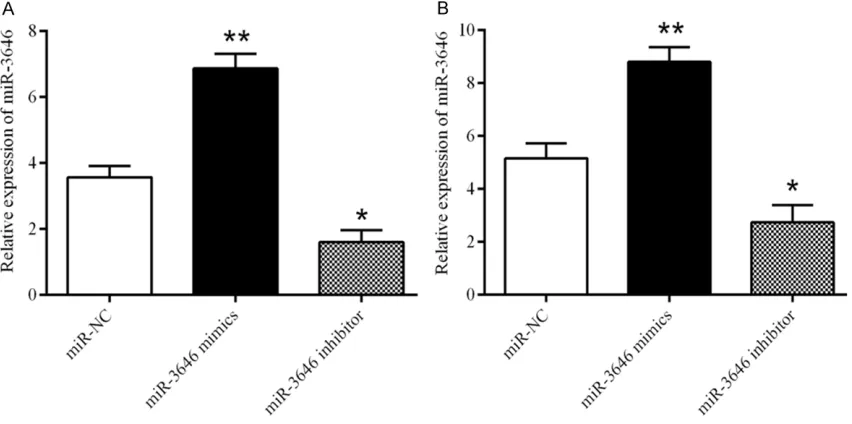
Figure 2. The expression level of miR-3646 in breast cancer cell lines transfected with miR-NC, miR-3646 mimic or miR-3646 inhibitor. A. The expression level of miR-3646 in MCF-7 cells, *P<0.01, **P<0.05. B. The expression level
[image:3.612.94.520.370.582.2]removed the cells in the upper chamber. In- vaded cells were fixed with 4% formaldehyde, stained with 0.5% crystal violet, and counted under a microscope.
Cell proliferation assay
For the colony formation test, MDA-MB-231 and MCF-7 cells treated with miR-3646 NC, miR-3646 mimics, miR-3646 inhibitor were se- eded in 6-well plates. Cultured after 14 days, the cells were stained with methylene blue to the colonies and then photographed.
Wound healing assay
Human breast cancer cells were seeded into 6-well plate. Wounds were created in the con-fluent cell monolayer using a 200 μl pipette tip, and any other free-floating cells and debris were removed by washing with PBS. Medium was then added, and the culture plates were incubated at 37°C. Wound healing within the scraped wound line was measured at a 48 h time point.
In vivo tumor growth assay
In this study all involved in animals proced- ures and experiments were performed in con-formity to the National Institutes of Health Guide for Care and Use of Laboratory Animals. The 6-week-old nude mice [BALB/cA-nu (nu/ nu)] were purchased from Shanghai SLAC La- boratory Animal Co., Ltd. (Shanghai, China) and housed in the Animal Resource Facility at La- boratory Animal Centre. 5×10⁶ human breast cancer cells stably expressing either miR-3646 or miRNA control was injected subcutaneous- ly in the both flanks of nude mice. Every two days to observe the palpable tumor formation. Thirty days after the implantation, removed the xenografts from mice and weighed the xeno-grafts. Tumor volume was calculated using the following formula: 4π/3×(width/2)2×(length/2).
Statistical analysis
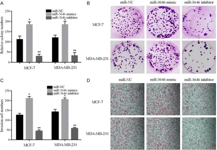
[image:4.612.94.524.75.373.2]All data were presented as mean ± SD. Two-tailed student’s t-test was used to analyze sig-nificant differences between groups. P<0.05 Figure 3. Colony formation and invasion assays indicated that miR-3646 promotes breast cancer cells proliferation and invasion. A, B. Colony formation assay showed that miR-3646 could increase the colony ability of breast cancer cells and downregulated miR-3646 suppressed the colony ability, *P<0.05, **P<0.01. C, D. Invasion assay showed
was accepted as significant. SPSS 18.0 and Graph Pad Prism 6.0 software were used for data analysis.
Results
MiR-3646 expression level is upregulated in both breast cancer cells and tissues
In order to declare the role of miR-3646 in tumor metastasis and tumor growth, we sub- sequently investigated the expression level of
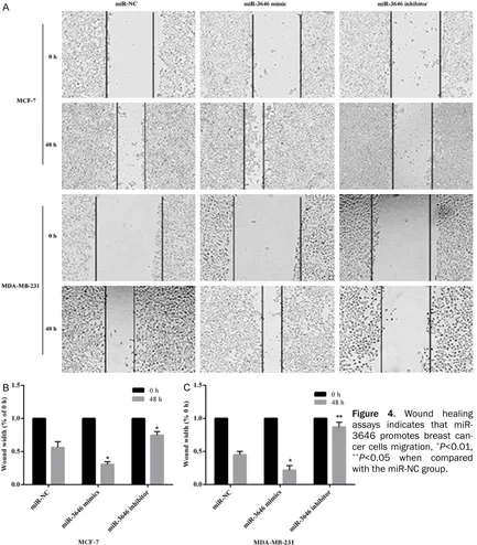
[image:5.612.93.526.75.569.2]miR-3646 in breast cancer tissues and whe- ther miR-3646 influenced the growth, inva-sion and migration of breast cancer cells. We first surveyed the level of miR-3646 in human breast cancer tissues. Results showed that the expression of miR-3646 in human breast cancer tissues was higher than that in nor- mal breast tissues (Figure 1A). Furthermore, the expression level of miR-3646 in breast cancer cell lines MDA-MB-231 and MCF-7 was higher than in the normal epithelial breast cell Figure 4. Wound healing assays indicates that miR-3646 promotes breast can-cer cells migration, *P<0.01, **P<0.05 when compared
line MCF10A (Figure 1B). To sum up, the results showed that in human breast cancer tissues and cells the miR-3646 overexpressed.
MiR-3646 promotes the proliferation, migra-tion and invasion of breast cancer cells
After we noted the high level expression of miR-3646 in breast cancer tissue and cells, we next examined the role of miR-3646 in the pro-liferation and invasion of breast cancer cells. MiR-NC, miR-3646 mimic, or miR-3646 inhibi-tor were transfected into MCF-7 and MDA-MB- 231 cells and the expression of miR-3646 was
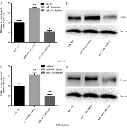
detected by qRT-PCR. The expression level of miR-3646 in the mimic group was markedly increase when compared with the control, while in miR-3646 inhibitor group its expression re- markably suppressed in both cell lines (Figure 2A and 2B). The results illustrated that the level of miR-3646 can be regulated. In our colony formation assay, results showed that transfection of miR-3646 mimic obviously en- hanced the proliferation of MCF-7 and MDA-MB-231 cells; one the other hand, inhibited the level of miR-3646 significantly suppressed the proliferation of MCF-7 and MDAMB-231 cells (Figure 3A and 3B). Furthermore, the inva-Figure 5. Expression of RhoA varies with the level of miR-3646 expression in breast cancer cells. A, B. Expression level of RhoA changes with the level of miR-3646 expression in MCF-7 cells, *P<0.01, **P<0.05 when compared with
[image:6.612.94.519.70.499.2]sion experiment results revealed that enhanced expression of miR-3646 resulted in a signifi -cant increase in invasive ability of breast can-cer cells, while decreased the level of miR-3646 led to a remarkable reduction of cell in- vasive ability of both breast cancer cell lines (Figure 3C and 3D). A similar tendency was dis-covered in the wound healing assays (Figure 4A-C). Taken together, all the results showed that miR-3646 play an important role in pro-moting breast cancer cells growth.
Expression of RhoA changes with the expres-sion of miR-3646 in breast cancer cell lines
To verify that whether promote migration, prolif-eration and invasion by miR-3646 is related with changes in RhoA expression, we transfect-ed the breast cancer cells with miR-3646 mim-ics or the inhibitor then detected the protein and gene levels of RhoA by western blot and RT-PCR. Results showed that, as the miR-3646 upregulated the levels of RhoA increased, when miR-3646 was downregulated the expression of RhoA decreased (Figure 5A-D). These results
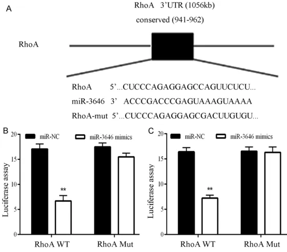
breast cancer cells. The results demonstrated that in MCF-7 and MDA-MB-231 cell lines miR-3646 significantly inhibited the luciferase ac-tivity of the RhoA WT 3’-UTR but had no influ -ence on the mutant (Figure 6B and 6C). These findings illustrated that RhoA is a direct down -stream target for miR-3646 in breast cancer cells.
Inhibition of miR-3646 suppressed the growth of breast cancer cells in vivo
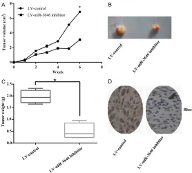
[image:7.612.92.372.74.320.2]Based on the effect of miR-3646 promoting cell growth in both MCF-7 and MDA-MB-231 cells, we further measured the effect of miR-3646 tumor angiogenesis and growth in vivo. Transfection of MDA-MB-231 cells with miR-3646 inhibitor caused a significant decreased in the tumor volume and tumor weight of sub- cutaneous xenograft tumors on nude mice when compared with those cells who stably transfected with LV-control (Figure 7A-C). What is more, the expression level of RhoA was also declined by stable transfection of miR-3646 inhibitor (Figure 7D). In conclusion, these re- Figure 6. RhoA 3’UTR is a potential miR-3646 target gene. A. Sequence
alignment of miR-3646 and the RhoA 3’-UTR, which contains one predicted miR-3646-biding site. B. Luciferases say in MCF-7 cell lines co-transfected with miR-3646 a luciferase reporter containing the RhoA 3’-UTR (WT) or a mutant (Mut). C. Luciferas assay in MDA-MB-231 cell lines co-transfected with miR-3646 a luciferase reporter containing the RhoA 3’-UTR (WT) or a mutant (Mut).
confirmed that miR-3646 reg -ulated RhoA expression.
RhoA 3’UTR is a targeting gene of miR-3646
sults indicated that inhibition of miR-3646 could suppress angiogenesis and growth of breast cancer cells in vivo.
After treated MDA-MB-231 cells with RhoA in-hibitor C3 the migratory ability had no change with the levels of miR-3646 expression
To further make sure that whether miR-36- 46 suppresses migration in a Rho-dependent manner, we using Rho inhibitor C3 at a final concentration of 2.0 μg/ml to blocked the ac-tivity of RhoA. Results showed that the RhoA mRNA and protein levels decreased after tre- atment with C3 when compared with the un- treated cells (Figure 8A-C). Furthermore, after treatment with C3, the migratory ability of the MDA-MB-231 cells transfected with the miR-3646 mimics or the miR-miR-3646 inhibitor was neither increased nor decreased when com-pared with the control cells (Figure 9). These results illustrated that miR-3646 does not reg-ulate RhoA when the RhoA pathway is blocked.
Discussion
Breast cancer is the most common diagnosed cancer, which is caused by a lot of factors and
meaning of miRNAs in breast cancer develop-ment and metastasis [34, 35]. Previous studies indicated that miR-3646 was over expressed in patients with bladder cancer, and breast can-cer and it play a critical role in cell proliferation, invasion and metabolism [23, 36].
[image:8.612.92.370.73.324.2]In this study, the results provided the evidence that miR-3646 accelerated cell proliferation and cell migration in breast cancer cell lines. First we researched the expression of miR-3646 in tumoral and non-tumoral breast tis-sues and cell lines. Results indicated that the expression level of miR-3646 was markedly in- creased in breast cancer cells and tumor sues than that in normal breast cells and tis-sues. Up-regulation of miR-3646 significantly promoted cell proliferation, while knockdown of miR-3646 significantly suppressed cell growth. In order to further explore the effect of miR-3646 in breast cancer cells, we examined the role of miR-3646 in the growth of subcutane-ous xenograft tumors in nude mice. In confor-mity to the in vitro researches, inhibit the ex- pression of miR-3646 suppressed the grow- th of xenograft tumors. These results suggest Figure 7. Inhibition of miR-3646 suppressed the growth of breast cancer
cells in vivo. A, B. Stable transfection of U87 cells with miR-3646-inhibitor
reduced the tumor size. C. Downregulation miR-3646 significantly dimin -ished the mean tumor weight. D. Immuno-histochemical staining showed that stable transfection of miR-3646-inhibitor leaded the expression of RhoA to reduce.
that in the progression of breast cancer miR-3646 might act as a carcinogen.
Rho/ROCK signaling channel has been verified to participate in the molecular mechanism of tumor metastasis [37]. Rho-subfamily protein is one of the members of the Ras superfamily with GTP enzyme activity, and mediates a wide variety of cellular effects, such as mem-brane trafficking, apoptosis/survival [38], cyto -skeletal reorganization [39], proliferation, cell polarity, cell cycle, cell adhesion [40] and gene transcription. As a major member of the Rho GTPase family, RhoA has been found to be over-expressed in several types of human tumors, including breast [12, 41] and colorectal cancer [11, 42], hepatocellular carcinoma [43, 44] and gastric cancer [13]. Previous researchers have found that change the intracellular RhoA ex- pression level can directly influence the pro
-cess of invasion and metastasis in many can-cers [10, 15]. The results of our study suggest that miR-3646 directly targets RhoA, and that the expression of RhoA change as the expres-sion of miR-3646, and may be involved in the migration of breast cancer cells.
[image:9.612.98.518.75.196.2]According to our results, we showed that in the breast cancer cell lines, miR-3646 directly mo- dulated the target gene RhoA with the 3’UTR, finally inhibiting the RhoA expression, and thus controlling the migration and invasion of cells. In order to confirm that miR-3646 targeting of RhoA 3’UTR we used luciferase reporter and western blot demonstrated in breast cancer cells. In addition, we used Rho inhibitor C3 to block RhoA activity, results showed that miR-3646 was unable to regulate RhoA. These re- sults further verified the possibility that miR-3646 inhibited migration via a RhoA-dependent pathway.
Figure 8. The expression level of RhoA in MDA-MB-231 cells treated with C3. A, B. The relative protein expression
levels of RhoA were measured by western blot assay, and normalized to β-actin. C. Expression of RhoA mRNA in
MDA-MB-231 cells treated with or without C3 was measured by real-time RT-PCR. **P<0.01 vs. the control group.
[image:9.612.93.521.260.428.2]Taken together, our results suggested that miR-3646 is involved in breast cancer cell lines and tissue specimens. In addition, we demonstrat-ed that miR-3646 functions as an important role in the metastasis of breast cancer, at least, through upregulation of RhoA. This is the first study combine miR-3646 with RhoA in breast cancers, and miR-3646 could be a potential target to treatment breast cancers in the future.
Acknowledgements
This study was supported by the national sci-ence foundation of China (No. 89487593).
Disclosure of conflict of interest
None.
Address correspondence to: Wenjun Yi, Depart- ment of Breast and Thyroid Surgery, The Second Xiangya Hospital, Central South University, 139 Middle Renmin Road, Changsha 410011, Hunan, P. R. China. Tel: +86-18608403318; Fax: +86-731-85295666; E-mail: xywenjunyi@126.com
References
[1] Bray F, Jemal A, Grey N, Ferlay J and Forman D. Global cancer transitions according to the human development index (2008-2030): a population-based study. Lancet Oncol 2012; 13: 790-801.
[2] Ferlay J, Soerjomataram I, Dikshit R, Eser S, Mathers C, Rebelo M, Parkin DM, Forman D and Bray F. Cancer incidence and mortality worldwide: sources, methods and major pat-terns in GLOBOCAN 2012. Int J Cancer 2015; 136: E359-386.
[3] Polyak K. Heterogeneity in breast cancer. J Clin Invest 2011; 121: 3786-3788.
[4] Jones SE. Metastatic breast cancer: the treat-ment challenge. Clin Breast Cancer 2008; 8: 224-233.
[5] Steeg PS. Tumor metastasis: mechanistic in-sights and clinical challenges. Nat Med 2006; 12: 895-904.
[6] Vanharanta S and Massague J. Origins of met -astatic traits. Cancer Cell 2013; 24: 410-421. [7] Zegers MM and Friedl P. Rho GTPases in
col-lective cell migration. Small GTPases 2014; 5: e28997.
[8] Chircop M. Rho GTPases as regulators of mito-sis and cytokinemito-sis in mammalian cells. Small GTPases 2014; 5.
[9] Gonzalez-Billault C, Munoz-Llancao P, Henri- quez DR, Wojnacki J, Conde C and Caceres A. The role of small GTPases in neuronal
morpho-genesis and polarity. Cytoskeleton (Hoboken) 2012; 69: 464-485.
[10] Orgaz JL, Herraiz C and Sanz-Moreno V.
Rho GTPases modulate malignant transforma-tion of tumor cells. Small GTPases 2014; 5: e29019.
[11] Cai K, Mulatz K, Ard R, Nguyen T and Gee SH. Increased diacylglycerol kinase zeta expres-sion in human metastatic colon cancer cells augments Rho GTPase activity and contributes to enhanced invasion. BMC Cancer 2014; 14: 208.
[12] Dulong C, Fang YJ, Gest C, Zhou MH,
Patte-Mensah C, Patte-Mensah-Nyagan AG, Vannier JP, Lu H, Soria C, Cazin L, Mei YA, Varin R and Li
H. The small GTPase RhoA regulates the ex-pression and function of the sodium chan- nel Nav1.5 in breast cancer cells. Int J Oncol 2014; 44: 539-547.
[13] Duan JT, Wang XM, Zhang SQ and Zhao GJ. Effect of RhoA gene silencing on proliferation and migration of gastric MGC-803 cells. Int J Clin Exp Med 2015; 8: 14410-14415.
[14] Guan R, Xu X, Chen M, Hu H, Ge H, Wen S, Zhou S and Pi R. Advances in the studies of roles of Rho/Rho-kinase in diseases and the development of its inhibitors. Eur J Med Chem 2013; 70: 613-622.
[15] Yu OM and Brown JH. G protein-coupled re- ceptor and RhoA-stimulated transcriptional
re-sponses: links to inflammation, differentiation,
and cell proliferation. Mol Pharmacol 2015; 88: 171-180.
[16] Liu SX, Zhang YJ, Guo HF, Hao J, Liu QJ, Liu JR, Guo JW, Liu JH and Zuo LF. The regulatory effect of the p38 signaling pathway on valde-coxib-induced apoptosis of the Eca109 cell line. Oncol Rep 2009; 22: 313-319.
[17] Steinfeld I, Navon R, Ach R and Yakhini Z. miR-NA target enrichment analysis reveals directly active miRNAs in health and disease. Nucleic Acids Res 2013; 41: e45.
[18] Tang J, Ahmad A and Sarkar FH. The role of microRNAs in breast cancer migration, inva-sion and metastasis. Int J Mol Sci 2012; 13: 13414-13437.
[19] Guo L, Zhao Y, Yang S, Cai M, Wu Q and Chen F. Genome-wide screen for aberrantly expressed
miRNAs reveals miRNA profile signature in
breast cancer. Mol Biol Rep 2013; 40: 2175-2186.
[20] van Kouwenhove M, Kedde M and Agami R. MicroRNA regulation by RNA-binding pro-teins and its implications for cancer. Nat Rev Cancer 2011; 11: 644-656.
[22] Dalmay T. Mechanism of miRNA-mediated re-pression of mRNA translation. Essays Biochem 2013; 54: 29-38.
[23] Zhang K, Zhao S, Wang Q, Yang HS, Zhu J and
Ma R. Identification of microRNAs in nipple
discharge as potential diagnostic biomarkers for breast cancer. Ann Surg Oncol 2015; 22 Suppl 3: S536-544.
[24] Zhang H, Qi M, Li S, Qi T, Mei H, Huang K, Zheng L and Tong Q. microRNA-9 targets ma-trix metalloproteinase 14 to inhibit invasion, metastasis, and angiogenesis of neuroblasto-ma cells. Mol Cancer Ther 2012; 11: 1454-1466.
[25] Citi S, Spadaro D, Schneider Y, Stutz J and Pulimeno P. Regulation of small GTPases at epithelial cell-cell junctions. Mol Membr Biol 2011; 28: 427-444.
[26] Wojnacki J, Quassollo G, Marzolo MP and Caceres A. Rho GTPases at the crossroad of signaling networks in mammals: impact of Rho-GTPases on microtubule organization and dynamics. Small GTPases 2014; 5: e28430. [27] Wheler JJ, Atkins JT, Janku F, Moulder SL,
Yelensky R, Stephens PJ and Kurzrock R. Multiple gene aberrations and breast cancer: lessons from super-responders. BMC Cancer 2015; 15: 442.
[28] Yi T, Zhai B, Yu Y, Kiyotsugu Y, Raschle T, Etzkorn M, Seo HC, Nagiec M, Luna RE, Reinherz EL, Blenis J, Gygi SP and Wagner G. Quantitative phosphoproteomic analysis re-veals system-wide signaling pathways down-stream of SDF-1/CXCR4 in breast cancer stem cells. Proc Natl Acad Sci U S A 2014; 111: E2182-2190.
[29] Wang D, Xu J, Shi G and Yin G. Molecular
mark-ers’ progress of breast cancer treatment effi -cacy. J Cancer Res Ther 2015; 11 Suppl 1: C11-15.
[30] Matsumoto A, Jinno H, Ando T, Fujii T, Nakamura T, Saito J, Takahashi M, Hayashida T and Kitagawa Y. Biological markers of inva-sive breast cancer. Jpn J Clin Oncol 2016; 46: 99-105.
[31] Prisack HB, Karreman C, Modlich O, Audretsch W, Danae M, Rezai M and Bojar H. Predictive biological markers for response of invasive breast cancer to anthracycline/cyclophospha-mide-based primary (radio-)chemotherapy. An- ticancer Res 2005; 25: 4615-4621.
[32] Utikal J, Abba M, Novak D, Moniuszko M and
Allgayer H. Function and significance of
Micro-RNAs in benign and malignant human stem cells. Semin Cancer Biol 2015; 35: 200-211.
[33] Seven M, Karatas OF, Duz MB and Ozen M. The role of miRNAs in cancer: from pathogen-esis to therapeutic implications. Future Oncol 2014; 10: 1027-1048.
[34] Chan SH and Wang LH. Regulation of cancer metastasis by microRNAs. J Biomed Sci 2015; 22: 9.
[35] Bouyssou JM, Manier S, Huynh D, Issa S, Roccaro AM and Ghobrial IM. Regulation of microRNAs in cancer metastasis. Biochim Biophys Acta 2014; 1845: 255-265.
[36] Rogler A, Hoja S, Socher E, Nolte E, Wach S, Wieland W, Hofstadter F, Goebell PJ, Wullich B, Hartmann A and Stoehr R. Role of two single nucleotide polymorphisms in secreted frizzl- ed related protein 1 and bladder cancer risk. Int J Clin Exp Pathol 2013; 6: 1984-1998. [37] Matsuoka T and Yashiro M. Rho/ROCK
signal-ing in motility and metastasis of gastric can- cer. World J Gastroenterol 2014; 20: 13756-13766.
[38] Street CA and Bryan BA. Rho kinase proteins--pleiotropic modulators of cell survival and apoptosis. Anticancer Res 2011; 31: 3645-3657.
[39] Duluc L and Wojciak-Stothard B. Rho GTPases in the regulation of pulmonary vascular barrier function. Cell Tissue Res 2014; 355: 675-685. [40] Baranwal S and Alahari SK. Rho GTPase effec-tor functions in tumor cell invasion and metas-tasis. Curr Drug Targets 2011; 12: 1194-1201. [41] Alho I, Costa L, Bicho M and Coelho C. Low mo-lecular weight protein tyrosine phosphatase isoforms regulate breast cancer cells migra-tion through a RhoA dependent mechanism. PLoS One 2013; 8: e76307.
[42] Ariake K, Ohtsuka H, Motoi F, Douchi D, Oikawa M, Rikiyama T, Fukase K, Katayose Y, Egawa S and Unno M. GCF2/LRRFIP1 promotes color- ectal cancer metastasis and liver invasion through integrin-dependent RhoA activation. Cancer Lett 2012; 325: 99-107.
[43] Wang SC, Lin XL, Li J, Zhang TT, Wang HY, Shi JW, Yang S, Zhao WT, Xie RY, Wei F, Qin YJ, Chen L, Yang J, Yao KT and Xiao D. MicroRNA- 122 triggers mesenchymal-epithelial transi-tion and suppresses hepatocellular carcinoma cell motility and invasion by targeting RhoA. PLoS One 2014; 9: e101330.