Original Article
Plasma cell myeloma with dual expression of kappa
and lambda light chains
Andrew S Jiang1, Zhao Wu2, Eric X Wei3, Hongyu Ni4, Bei You1, Tao Yang5, Jie-Gen Jiang1,2
1Department of Pathology and Laboratory Medicine, Rutgers New Jersey Medicine School, 185 South Orange
Avenue, Newark, NJ 07103, USA; 2Department of Pathology, Genoptix Medical Laboratory, 2110 Rutherford Road,
Carlsbad, California 92008, USA; 3Department of Pathology, LSU Health, 1501 Kings Highway, Shreveport, LA
71130, USA; 4Department of Pathology, University of Illinois at Chicago, 840 S. Wood St., Suite 130 CSN, Chicago,
IL 60612, USA; 5Department of Pathology, Los Alamitos Medical Center, 3751 Katella Avenue, Los Alamitos, CA
90720, USA
Received July 21, 2018; Accepted August 24, 2018; Epub September 1, 2018; Published September 15, 2018
Abstract: Plasma cell myeloma is a clonal proliferation of neoplastic plasma cells and typically expresses a monoclo- nal heavy and/or light chain immunoglobulin. Plasma cell myeloma with dual expression of kappa and lambda light chains in a single clone is extremely rare. Here we report three cases of plasma cell myeloma with a co-expression
of both kappa and lambda light chains. All three cases were confirmed by comprehensive workup including IHC, ISH and flow cytometry analysis to detect light chain expression patterns at the mRNA and protein levels. We also
reviewed three cases so far published in the literature. Our study suggests that plasma cell myeloma with dual light chain expression may be more likely to be light chain only myeloma. It may have a high frequency of complex cyto-genetic and/or FISH abnormalities, associated with a high-risk disease.
Keywords:Dual expression of kappa and lambda, plasma cell myeloma, high risk disease
Introduction
Plasma cell myeloma is characterized by a mul-tifocal clonal proliferation of plasma cells based in bone marrow and the presence of paraproteins in the blood and/or urine with associated end organ damage. It is the second most common hematological malignancy accounting for approximately 10-15% of hema-topoietic cancers [1]. The diagnosis of plasma cell myeloma requires ≥10% clonal plasma cells in the bone marrow. Plasma cell myeloma is further classified into asymptomatic (smol-dering) myeloma, symptomatic myeloma, non-secretory myeloma, and plasma cell leukemia based on a combination of clinicopathological and radiological features [1, 2].
Normal plasma cells are terminally differentiat-ed effector B cells developdifferentiat-ed from naïve mar-ginal zone B cells and follicular B cells after an antigen encounter [3]. The immunoglobulins produced by normal plasma cells are central to the body’s adaptive immune response to
for-eign antigens. Immunoglobulins are either secretory or cell surface bound proteins which are composed of two heavy chains (α, γ, δ, ε or μ) and two light chains (κ or λ). Neoplastic plas-ma cells in the plas-majority of plasplas-ma cell myeloplas-ma cases retain the ability to produce either com-plete immunoglobulins or at least a fragment of paraproteins.
Table 1. Characteristics of plasma cell myeloma with dual expression of kappa and lambda light chains
CASE# Age (year)/sex Pathology diagnosis Serum immunoglobulin analysis Urine protein analysis Flow cytometry IHC test of plasma cells ISH test of plasma cells Karyotype and/or FISH
CASE 1 65/M Plasma cell myeloma Increased IgA kappa and free kappa
chain
Increased IgA kappa and free kappa chain
Kappa+, Lambda+ Kappa+, lambda+, IgA+ Kappa+, Lambda+ Normal karyotype
FISH: -13
CASE 2 58/M Plasma cell myeloma Increased free lambda light chain,
no increased Ig Increased free lambda chain Kappa+, Lambda+ Kappa+, Lambda+, No heavy chains Kappa+, Lambda+ Complex karyotype
CASE 3 76/F Plasma cell myeloma Increased free kappa chain, no
increased Ig
NA Kappa+, Lambda+ Kappa+, Lambda+, No
heavy chains
Kappa+, Lambda+ Normal karyotype FISH: 1q+, +5 and
13q-CASE 4 [6] 68/F Plasma cell myeloma
and plasmacell leu-kemia
Increased IgG kappa and free lambda
Increased both free kappa and lambda chains
Kappa+, Lambda+ Kappa-, Lambda+ Kappa+, Lambda+ Complex karyotype
CASE 5 [7] 52/M Plasma cell myeloma Increased free lambda chain, no
increased Ig
NA Kappa+, Lambda+ Kappa-, Lambda+, Heavy
chains: not done
Kappa+, Lambda+ Complex karyotype
CASE 6 [7] 58/F Plasma cell myeloma Increased IgG lambda and free
lambda
Increased free lambda chain
been studied in detail [6, 7]. Here we report three cases of plasma cell myeloma with dual expression of both kappa and lambda light chains. We summarize their clinical, pathologi-cal, flow cytometric, cytogenetic, FISH, and molecular characteristics along with previously reported three cases.
Case studies
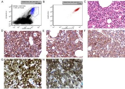
The first case is a 65-year-old male patient presenting with anemia and lytic bone lesions in 2011 (Case 1 in Table 1). Serum protein electrophoresis revealed monoclonal kappa light chain gammopathy. A quantitative serum immunoglobulin analysis showed decreased IgM (<5 mg/dL; reference range 48-271 mg/dl) and IgG (211 mg/dl; reference range 694-1618 mg/dl) levels, and an increased IgA (4481 mg/ dl; reference range 81-463 mg/dl) level. A serum immunoglobulin free light chain study revealed increased kappa free light chain (1370 mg/dl; reference range 158-502 mg/dl) and reduced lambda free light chain (23 mg/dl; ref-erence range 100-317 mg/dl) levels. The kappa/lambda ratio was increased to 59.57 (reference range: 1.35-2.95). The urine immu-noelectrophoresis revealed clonal IgA kappa and a free kappa light chain. Flow cytometric analysis of the marrow aspirate detected a dis-tinct population of abnormal plasma cells expressing CD38, CD138 (Figure 1A), IgA, and both cytoplasmic kappa and lambda light chains (Figure 1B) with negativity for CD56. A morphological evaluation revealed markedly increased plasma cells (~60-70% of total mar-row cellularity, Figure 1C and 1D) in a hypercel-lular marrow (80-90%). Nodules of plasma cells were seen. Plasma cells demonstrated atypical features including large sizes, a high nuclear to cytoplasmic ratio, centrally located nuclei, and visible nucleoli. Immunohistochemistry (IHC) stains and mRNA in-situ hybridization (ISH) tests confirmed that these plasma cells were positive for both kappa and lambda light chains (Figure 1E-H). Plasma cells were positive for IgA and negative for IgG, IgM, IgD. This patient was diagnosed with plasma cell myeloma with a dual expression of kappa and lambda light chains. No significant pleomorphic or plasma-blastic features were evident. Karyotype was normal. FISH studies (including probes for t(4;14), +5, +7, +11q, t(11;14), -13, 13q-, t(14;16) and TP53(17p-) detected monosomy
13. A molecular test by IgH PCR detected a clonal immunoglobulin heavy chain gene rear-rangement with a single high peak, indicating the presence of a single clonal plasma cell population.
The second case is a 58-year-old male patient with a history of stage IIIB IgG lambda multiple myeloma diagnosed in October 2003 (case 2 in Table 1). At that time serum protein electro-phoresis and immunofixation revealed a mono-clonal lambda light chain. Quantitative serum immunoglobulin analysis showed decreased IgM (6 mg/dL; reference range 40-230 mg/dl) and IgG (491 mg/dl; reference range 700-1600 mg/dl) levels and normal IgA (111 mg/dl; refer-ence range 70-400 mg/dl) level. The urine immunoelectrophoresis revealed a monoclonal free lambda light chain. The monoclonal free lambda chain was at 20.6% of the total urine protein. This patient was in remission after treatment with Revlimid and Dexamethasone followed by an autologous stem cell transplant. The patient then presented with lytic bone lesions and anemia in November 2011. Flow cytometry detected clonal plasma cells ex- pressing CD56 and both cytoplasmic kappa and lambda light chains in bone marrow aspi-rate. A core biopsy revealed large aggregates of atypical plasma cells, representing around 20-40% of total marrow cellularity. Plasma cells strongly expressed both kappa and lambda light chains by IHC stains and ISH studies. Immunostains for IgA, IgD, IgM, and IgG heavy chains were negative. The findings are consis-tent with recurrent plasma cell myeloma with dual expression of kappa and lambda light chains. Cytogenetic analysis showed a complex karyotype with several bizarre structural and numerical changes: 44~46, del(X)(q24), -Y, del(1)(p13p22), t(2;8)(p12;q11.2), der(2)(2;8) (p12;q11.2), add(3)(p14), add(5)(p15.1), del(7) (q22), -8, i(12)(q10;q10), -14, -15, -18, add(22) (q13), +1-4mar[cp4]/46, XY [16].
light chain study revealed increased kappa free light chain (294.9 mg/l; reference range 3.3-19.4 mg/l) and normal lambda free light chain (20.8 mg/l; reference range 5.7-26.3 mg/l) lev-els. The Kappa/lambda ratio was increased at 14.18 (reference range 0.26-1.65). Bone lytic lesions were not detected. A bone marrow biop-sy was performed. Flow cytometry detected abnormal plasma cells expressing CD56 and both kappa and lambda light chains. The core biopsy revealed ~15% atypical plasma cells with no detectable IgA, IgD, IgM, and IgG heavy chains. The plasma cells strongly expressed both kappa and lambda by IHC and ISH studies. The findings are consistent with a plasma cell myeloma with a dual expression of kappa and lambda light chains. The IgH PCR test for clon-ality was negative and compatible with nega-tive IHC stains for heavy chains (neganega-tive for IgA, IgG, IgM, IgD). Her karyotype was normal.
However, the FISH analysis with probes for 1p/1q, 5p/5q, 13q14/13q34, IGH rearrange-ment, and TP53 detected 1q+, +5 and 13q-.
Discussion
There are few reported cases of plasma cell myeloma with a dual expression of kappa and lambda light chains in the literature. Table 1
[image:4.612.89.518.74.381.2]summarizes the clinical, pathological, flow cyto-metric, cytogenetic, and FISH characteristics of these three cases published in the literature [6, 7] and three new cases in our current report. The final diagnoses as plasma cell myeloma with a dual expression of kappa and lambda light chains in all six cases were confirmed by comprehensive workups including ISH, IHC, and flow cytometric analysis to detect light chain expression patterns at the mRNA and protein levels. A single clonal population of
Figure 1. Plasma cell myeloma with dual expression of kappa and lambda light chains (case 1 in Table 1). A and B: Flow cytometric analysis revealing CD138 positive plasma cells with dual expression of kappa and lambda light
chains. C: Hematoxylin and eosin staining showing diffuse infiltration of bone marrow by myeloma cells in a core
plasma cells was confirmed by IgH PCR test for heavy chain rearrangements when heavy chain was expressed (Table 1, case 1).
Myeloma usually occurs around the age of 60th and is more common in men than women. IgG and IgA are more frequent [1]. Similarly, the median age is 61.5 years old (range from 52 to 76) and IgG and IgA myeloma are common in these six plasma cell myeloma cases. However, no male predominance is noted. 3 cases are men and 3 cases are women (Table 1). Interestingly all six plasma cell myeloma cases with dual expression of kappa and lambda light chains harbor either complex karyotype(s) or FISH evidence of chromosome abnormalities including monosomy 13 and +1q (Table 1), which have been reported to be associated with a high risk disease [8-11].
Approximately 0.5-2% of normal B cells express dual kappa and lambda light chains [12, 13]. These normal B cells may be the neoplastic counterparts leading to the formation of B cell lymphoma with dual surface immunoglobulin expression [14-16]. It is not clear whether myeloma with dual expression of kappa and lambda light chains is the result of neoplastic transformation of the dual light chain express-ing B-cells.
The mechanism by which B or plasma cells prevent the production of multiple heavy chain classes or light chain classes is defined gener-ally as allelic exclusion, but the actual molecu-lar mechanisms are still poorly understood. The locus encoding for the heavy chain portion undergoes rearrangement first, and if success-ful, it is followed by a rearrangement of the kappa light chain locus. Only if the kappa light chain rearrangement for both alleles is ineffec-tive does lambda light chain rearrangement occur. Interestingly, half of the cases (3 out 6) described in our study express dual light chains with no detectable heavy chains (Table 1), which is significantly higher than it is in the gen-eral myeloma population (~11%) [1]. The lack of heavy chains in myeloma can be due to a vari-ety of reasons, including illegitimate switch recombination and/or deletion of the rear-ranged IGH locus. The phenomenon raises a possibility that clonal plasma cells with unsuc-cessful heavy light chain rearrangements may more likely have failed allelic exclusion machin-ery, leading to further kappa and lambda light chain gene rearrangements.
Our above observation reveals some interest-ing features of plasma cell myeloma with dual expression of kappa and lambda light chains. A high frequency of complex cytogenetic and/or FISH abnormalities associated with an unfavor-able prognosis may exist in this group of patients. However, due to the limited number of cases reported, further investigation is war-ranted to shed more light on this condition.
Disclosure of conflict of interest
None.
Address correspondence to: Dr. Jie-Gen Jiang, Department of Pathology and Laboratory Medicine, Rutgers New Jersey Medical School, 185 South Orange Avenue, Newark, NJ 07103, USA. Tel: 973-972-2236; E-mail: [email protected]
References
[1] McKenna RW, Kyle RA, Kuehl WM, Harris NL, Coupland RW, Fend F. Plasma cell neoplasms. In: Swerdlow SH, Campo E, Harris NL, et al. editors. Revised 4th ed. WHO classification of tumours of haematopoietic and lymphoid tis-sues. Lyon (France): IARC; 2017. pp. 241-258. [2] Palumbo A, Anderson K. Multiple myeloma. N
Engl J Med 2011; 364: 1046-1060.
[3] Shapiro-Shelef M, Calame K. Regulation of plasma-cell development. Nat Rev Immunol 2005; 5: 230-242.
[4] Greipp PR, San Miguel J, Durie BG, Crowley JJ, Barlogie B, Bladé J, Boccadoro M, Child JA, Avet-Loiseau H, Kyle RA, Lahuerta JJ, Ludwig H, Morgan G, Powles R, Shimizu K, Shustik C, Sonneveld P, Tosi P, Turesson I, Westin J. International staging system for multiple my-eloma. J Clin Oncol 2005; 23: 3412-3420. [5] García-García P, Enciso-Alvarez K, Diaz-Espada
F, Vargas-Nuñez JA, Moraru M, Yebra-Bango M. Biclonal gammopathies: retro.spective study of 47 patients. Rev Clin Esp 2015; 215: 18-24. [6] Gentry M, Pettenati M, Pang CS. Biclonal light
chain gammopathy with aberrant CD33 ex-pression in secondary plasma cell leukemia. Int J Clin Exp Pathol 2013; 6: 2224-2229. [7] Jiwani S, Bornhost J, Alapat D. Biphenotypic
plasma cell myeloma: two cases of plasma cell neoplasm with a coexpression of kappa and lambda light chains. Int J Clin Exp Pathol 2015; 8: 8536-8544.
Pineda-Roman M, Tricot G, van Rhee F, Sawyer J, Alsayed Y, Walker R, Zangari M, Crowley J, Barlogie B. A validated gene expression model
of high-risk multiple myeloma is defined by de -regulated expression of genes mapping to chromosome 1. Blood 2007; 109: 2276-2284. [9] Nemec P, Zemanova Z, Kuglik P, Michalova K,
Tajtlova J, Kaisarova P, Oltova A, Filkova H, Holzerova M, Balcarkova J, Jarosova M, Rabasova J, Hruba M, Spicka I, Gregora E, Adam Z, Scudla V, Maisnar V, Schutzova M, Hajek R; Czech Myeloma Group. Complex
karyotype and translocation t(4;14) define pa -tients with high-risk newly diagnosed multiple myeloma: results of CMG2002 trial. Leuk Lymphoma 2012; 53: 920-927.
[10] Binder M, Rajkumar SV, Ketterling RP, Greipp PT, Dispenzieri A, Lacy MQ, Gertz MA, Buadi FK, Hayman SR, Hwa YL, Zeldenrust SR, Lust JA, Russell SJ, Leung N, Kapoor P, Go RS, Gonsalves WI, Kyle RA, Kumar SK. Prognostic implications of abnormalities of chromosome 13 and the presence of multiple cytogenetic high-risk abnormalities in newly diagnosed multiple myeloma. Blood Cancer J 2017; 7: e600.
[11] Chesi M, Bergsagel PL. Molecular pathogene-sis of multiple myeloma: basic and clinical up-dates. Int J Hematol 2013; 97: 313-323. [12] Diaw L, Siwarski D, DuBois W, Jones G, Huppi
K. Double producers of kappa and lambda
de-fine a subset of B cells in mouse plasmacyto -mas. Mol Immunol 2000; 37: 775-781. [13] Burtin P, Buffe D. Synthesis of human
immuno-globulins in germinal centers of lymphoid or-gans. J Immunol 1967; 98: 536.
[14] Xu D. Dual surface immunoglobulin light-chain expression in B-cell lymphoproliferative disor-ders. Arch Pathol Lab Med 2006; 130: 853-856.
[15] González D, van der Burg M, García-Sanz R, Fenton JA, Langerak AW, González M, van Dongen JJ, San Miguel JF, Morgan GJ. Immu-noglobulin gene rearrangements and the pathogenesis of multiple myeloma. Blood 2007; 110: 3112-312.