Original Article
GF109203X attenuates RANKL-induced
osteoclastogenesis and suppresses
osteolysis in a mouse model
Dawang Xie1*, Jun Yao1*, Wenyu Feng1, Yan Wei2, Huiping Lu2, Jia Li2, Qingjun Wei1
Departments of 1Orthopaedic Surgery, 2Pathology, The First Affiliated Hospital of Guangxi Medical University, Nan
-ning, Guangxi, P. R. China. *Equal contributors.
Received October 25, 2016; Accepted November 25, 2016; Epub February 1, 2017; Published February 15, 2017
Abstract: Aseptic loosening after joint replacement is primarily caused by wear particle-induced osteolysis, which shortens the life of the prosthesis. Research shows that there are many osteolytic cytokines around prostheses that are loosened due to wear particles. Among these, receptor activator of nuclear factor κB ligand (RANKL) is the only factor that can directly stimulate the formation and functional activation of osteoclasts. This study aimed to inhibit the signaling path of RANKL-induced osteoclastogenesis is a promising prevention and treatment strategy for wear particle-induced osteolysis. Based on our previous research, it was confirmed that GF109203X attenuates osteo -clastogenesis by inhibiting RANKL-induced osteoclast differentiation. We established a mouse model of polyethyl -ene (PE) particle-induced osteolysis to study the effect of a protein kinase C (PKC) inhibitor on osteoclastog-enesis in vivo. We found that by inhibiting the combination of RANKL and receptor activator of nuclear factor κB (RANK), which mediates the signaling paths of osteoclastogenesis, GF109203X acted in a dose-dependent manner to suppress the differentiation and functional activity of osteoclasts, reduce osteoclast formation, decrease the expression of osteoclast-associated receptor (OSCAR) and cross linked C-telopeptide of type I collagen (CTX-1), and promote the expression of osteoprotegerin (OPG). In addition, the degree of bone destruction was inhibited, bone resorption pitting and area were reduced, and the symptoms of osteolytic disease were lessened. This research helped clarify that GF109203X may provide a new option for the treatment of osteolytic disease.
Keywords: PKC inhibitor, GF109203X, RANKL, osteoclast, osteolysis
Introduction
At present, joint replacement therapy is the primary treatment option for osteoarthritis. However, the most important complication after joint replacement surgery is loosening of the prosthesis, which has no ideal treatment [1, 2]. Although the most common method of
treating aseptic loosening of artificial joints is
joint revision surgery, this method cannot guar-antee the stability of the prosthesis and even affect the patient’s daily activity and quality of life. Researchers have increasingly studied the wear particle-induced aseptic loosening of the
prostheses, since finding and developing a specific drug to overcome bone dissolution by
inhibiting or blocking the progress of wear particle-induced osteolysis [3], would reduce patients’ pain and economic burden greatly.
Aseptic loosening of the prosthesis is mainly due to mechanical and biological factors and
results from a biological inflammatory
media-tor reaction [4-6]. Recent studies consider the release of wear particles into the surrounding prosthesis and the resultant bone dissolution to be the key factor in triggering prosthetic
aseptic loosening [7, 8]. Crotti et al [9] and Kim
et al [10]. found that loosening of the prosthe-sis is not only associated with a large number of
inflammatory cells and cytokines produced by
surrounding tissue, such as interleukelin-6
(IL-6), interleukelin-1 (IL-1), tumor necrosis factor-α (TNF-α), prostaglandin E2 (PGE-2), etc., but also is associated with the expression of RANKL, RANK and OPG. Moreover, studies have sug -gested that these cells, especially macrophage, produce a variety of cytokines which are the
RANKL and RANK promotes osteoclast differ -entiation and maturation and prevents apopto-sis. It is the key factor leading to osteolysis around the prosthesis. The osteoblast and
mar-row stromal cells produce OPG, which com
-bined with RANK and RANKL, blocks
RANKL-induced osteoclast differentiation and matu-
ration and benefits bone reconstruction [12,
13]. Therefore, inhibiting the signaling
path-ways that lead to RANKL-induced osteoclasto -genesis and reducing the biological effects induced by wear particles are crucial steps for aseptic loosening of the prosthesis.
RANKL is the key cytokine in osteoclast
diff-erentiation. It is a member of the tumor necro-sis factor (TNF) super family, and is primarily
secreted by the ossification cells, T lympho
-cytes, and B lymphocytes [14]. The preliminary research identified that in the RANKL-mediated signaling pathways, RANKL links to RANK and causesinhibitor kappa B kinase β (IKKβ) phos -phorylation. It then induces the degradation
of IκB, eventually leading to the release of nuclear factor kappa B (NF-κB) into the nucle -us, which prevents osteoclast apoptosis and maintains normal differentiation [15, 16]. In the osteoclast precursors (OCPs), Nuclear factor of activated T-cells, cytoplasmic 1 (NFATc1) is activated by calcineurin dephosphorylation. It participates in all aspects of osteoclast forma-tion and activaforma-tion; extracellular regulated
pro-tein kinases (ERK) signal pathways are involved
in regulating the expression of NFATc1 gene transcription factors, participate in OCPs prolif-eration, differentiation, and survival together with NFATc1, and enhance the expression of
RANK [17-19]. Because the NF-κB, NFAT, and ERK signaling pathways are involved in the
formation and differentiation of osteoclasts, whose activation and phosphorylation is
medi-ated by RANKL and RANK in combination, tar
-geting the RANKL-mediated signal pathway by
attenuating the signal transduction of osteo-clastogenesis should available to bone resorp-tion disease caused by osteoclasts.
PKC regulates the proliferation, differentiation,
and survival of several cellular components.
The PKC family includes more than 10 kinds of isomers. Among these, PKC-β has two isomers: PKC-βI and PKC-βII. The expression of these two PKC-β isomers increases during the pro -cess of osteoclast differentiation. Studies have
shown that PKC-β is a key factor in regulating
osteoclast differentiation by participating in
the RANKL-mediated downstream signaling pathway of NF-κB and ERK [20, 21]. Previous studies found that GF109203X acts as a PKC inhibitor by intervening in the activity of NF-κB,
the NFAT signaling pathway, and then inhibits
RANKL-induced osteoclastogenesis functional activation, suggesting that GF109203X has
therapeutic potential in osteolytic disease [22].
However, these studies on GF109203X were
only at the cellular level, and have not yet been applied to animal models. Therefore, we set up a mouse model of particle-induced surface bone erosion to observe the degree of damage to the calvarium and detect the expression of
osteoclasts and related specific cytokines of osteoblasts. We inferred that GF109203X can
inhibit osteoclast formation, explored the po-
tential protective effect of GF109203X against
osteolysis, and addressed clinical applications for the prevention and treatment of bone dis-solution disease.
Materials and methods
Polyethylene particle preparation
Pure PE particles were purchased from Clariant
(Gersthofer, Germany) [23]. To remove adher -ent endotoxins, the particles were soaked in 100% ethanol for 48 h, prepared for washing [24], and then resuspended in a mixture of phosphate-buffer solution and 1% normal mouse serum at a concentration of 1000 mg/ mL. The mean particle size was 1.84 ± 1.50
μm (range 0.14-12.1); more than 32% of the particles were smaller than 1 μm [25].
Reagent preparation and groups of mice
GF109203X was purchased from Sigma (Castle
Hill, New South Wales, Australia). Eight-week
old LPS-resistant C57BL/6 mice were pur -chased from the Laboratory Animal Center,
Academy of Military Medical Sciences, PLA,
China (mean weight 18.3 ± 0.6 g, range 17.5-19.2 g) and randomly divided into four groups of six: Control group (no PE particle-induced
and injected with PBS); Vehicle group (PE parti
-cle-induced and injected with PBS);
GF109-203X-low group (PE particle-induced and
treat-ment with 100 μl GF109203X); and
GF1092-03X-high group (PE particle-induced and
and Pathology Laboratory. Ethical approval
was ratified in accordance with the principles
and procedures by the Animal Care Committ-
ee of Guangxi Medical University, China
[No.201509105].
Model of polyethylene particle-induced cal
-varial osteolysis
The mouse model of PE particle-induced cal-varial osteolysis was based on a model
estab-lished by Wedemeyer et al [23]. Mice were raised for two weeks before being sacrificed.
Two percent pentobarbital (0.01 mL/g) was used to anesthetize the mice. Two mm of cal-varial skin was exposed and the cranial perios-teum was dissected. A control group with no PE particle implantation was also injected with
PBS (100 μl). In the other three groups, PE par
-ticles (30 μl, 1000 mg/mL) were implanted
under the periosteum at the middle incision of the calvarium, and the skin was stapled. Two days after implantation of PE particles, the
pos-itive group was injected with 100 μl PBS, and
the other two groups were treated with
GF109203X; these were separately injected with a 100 μl or 200 μl dosage into the perios
-teum. PBS or GF109203X was injected every other day for 14 days. Blood was collected from
the femoral artery after the mice were ana-
esthetized and then sacrificed by cervical
dis-location for ELISA detection. The calvaria were stored in 10% formaldehyde and prepared for micro-CT analysis and histological evalua-tion (tartrate-resistant acid phosphatase [TR- AP] staining). No adverse effects or mortality occurred.
Micro-computed tomography (micro-CT) scan
-ning
PE particles were removed by mixing in 10% formaldehyde for 24 h before scanning to exclude interference. The mice calvaria were constructed by a high-resolution micro-CT
sys-tem (Skyscan 1076; Skyscan, Kontich, Belgium)
using the following settings: X-ray voltage, 75
kV; electric current, 120 µA; rotation step, 0.5°.
Representative three-dimensional images of the mouse calvaria in each group were export-ed to the computer for scanning. In order to analyze the reconstructed micro-CT images quantitatively, a square region of interest (ROI) 6 mm long × 6 mm wide around the area of PE particle implantation was selected for further
qualitative and quantitative analysis. Bone vol
-ume to tissue vol-ume (BV/TV), number of pits,
and percentage of porosity of each sample were measured.
Histomorphometric analysis of osteocytes The mouse calvaria were recycled and deca-
lcified in 10% EDTA (pH 7.4) at 4°C for one month, and then embedded in paraffin. The
eroded section of each calvarium was prepared for TRAP staining. A high-powered microscope was used to examine and photograph the spec-imens for quantitative analysis; the number of TRAP+ multinucleated osteoclasts was count-ed, and the bone erosion area was measured. Pathological analysis of the liver and the small
intestine
In addition, the liver and the small intestines of the mice were collected and stored in 10%
formaldehyde after sacrifice, and then hema
-toxylin and eosin- (H&E-) stained and TUNEL
staining. Each section was evaluated micros- copically.
Serum analysis
Blood collected in the Eppendorf tubes was
centrifuged for extraction of serum to analyze
the levels of RANKL, OSCAR, CTX-1, and OPG, as per ELISA kit protocols (Wuhan Hi-tech Med,
Hubei, China).
Statistical analysis
Values are presented as the mean ± SD of
results obtained from three or more experi-ments. Statistical analyses were performed
using one-way analysis of variance (ANOVA) followed by the Student-Newman-Keuls test (GraphPad Prism 6 for Windows). P < 0.05 was considered significant.
Results
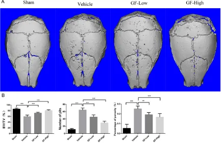
GF109203X attenuated the bone destruction of PE particle-induced osteolysis in C57BL/6
mice
In order to analyze the therapeutic effect of
GF109203X on particle-induced bone dissolu
-tion and confirm the func-tion of GF109203X in
suppressing calvarial osteolysis in an animal model, we set up a murine mouse calvarial osteolysis model of PE particle-induced
osteol-ysis to evaluate the effect of GF109203X on
of PE particle implantation showed severe os- teolysis, as evidenced by the extensive surface erosion on the calvaria in the vehicle group as
compared to the sham group. By contrast, treatment with GF109203X significantly
redu-ced the extent of PE particle-induredu-ced bone destruction, as the scan images showed that
bone loss of mice in the high GF109203X
gro-up was markedly less than that of mice in the
low GF109203X group (Figure 1A). The quanti-tative analysis of bone mass parameters was
further identified, and confirmed these results. The significant increase in BV/TV and marked
reduction in the number of resorptive pits and percentage of porosity of the calvaria (Figure 1B and Supplementary Table 1) led to the
con-clusion that GF109203X effectively induced
and ameliorated particle-induced calvarial dissolution.
GF109203XGF reduced the number of osteo
-clasts and suppressed PE particle-induced
calvarial inflammatory response
To investigate the inhibitory function of
GF109-203X on particle-induced osteolysis and
con-firm that GF109203X can reduce the number of
osteoclasts and the inflammatory reaction,
we then analyzed the pathological changes in the mouse skulls by TRAP staining. In the area where PE particle injection resulted in bone
destruction, inflammatory cells, such as lym -phocytes, macrophages, and particularly os- teoclasts, were aggregated along the bone surface erosion, as revealed by TRAP staining (10 ×) (Figure 2A). In addition, consistent with micro-CT quantitation, histological assess- ment and histomorphometric analysis demon-strated that both low-dose and high-dose
GF109203X markedly ameliorated PE
particle-induced osteoclastogenesis in a dose-depen-dent manner, as revealed by the reduced num-ber of TRAP (+) osteoclasts and bone erosion area in the treatment groups (Figure 2B and Supplementary Table 2). Thus, our results
con-firmed that GF109203X can attenuate the for -mation of osteoclast.
GF109203X suppresses the expression of RANKL and OSCAR
Previous studies have shown that GF109203X can inhibit the signaling pathways of
[image:4.612.90.525.74.353.2]tion of bone dissolution after treatment with
GF109203X. We detected the expression of
CTX-1 using ELISA. The data showed that the concentration of CTX-1 was higher in the ve- hicle group than in the sham, low-dose, and
high-dose groups. Treatment with GF109203X
markedly reduced CTX-1 expression in a dose-dependent manner, the higher the medication dosage, the lower the expression of CTX-1 (Figure 3C and Supplementary Table 3C). These
results indicated that GF109203X may amelio -rate osteoclast-induced bone resorption.
GF109203X promotes the expression of osteo
-blast cytokine OPG
The OPG expressed in the osteoblasts is a pro -tective factor in bone reconstruction. We
mea-sured OPG quantitatively using ELISA for study -ing whether it has an effect on the expression of osteoblasts. The results showed that the
concentration of OPG was lower in the vehicle
group than in the sham group. Collectively,
treatment with GF109203X significantly incre-ased OPG expression as compared to the vehi -cle group, and the higher the dosage, the
high-induced osteoclastogenesis. To confirm that the primary mechanism of GF109203X inhibi -tion of osteoclast forma-tion is interference with
the RANKL-mediated downstream signaling pathways and attenuation of RANKL expres -sion feedback, we used ELISA kits to detect the
expression of RANKL and OSCAR for quantita -tive analysis. The results indicated that
expres-sion of RANKL and OSCAR was higher in the vehicle group than in the sham group. More-over, the levels of serum RANKL and OSCAR
conspicuously decreased under treatment with
GF109203X, and their concentration in the GF109203X-high group was significantly lower than in the GF109203X-low group (Figure 3A, 3B and Supplementary Table 3A, 3B).
Expression of RANKL and OSCAR was reduced after treatment with GF109203X, which is
associated with osteoclast differentiation. GF109203X inhibits bone resorption and
decreases bone dissolution
To further explore whether GF109203X is also
effective for particle-induced bone destruction in animal models and to evaluate the
[image:5.612.91.519.76.347.2]prosthesis and increasing mechanical wear and tear between the implant and bone can cause prosthetic loosening [26, 27]. Since the generation of wear particles is inevitable, researchers have studied the signaling path-ways that result in osteolysis and used drugs to inhibit or block the cytokines and signaling pathways that induce osteoclast formation. Such as, Jimi et al suppressed the signaling
pathways of NF-κB by inhibiting NEMO
(IKKγ)-binding domain peptide, and found that this could reduce osteoclast formation and bone
erosion in arthritis, confirming that the signal
-ing system of NF-κB plays a crucial role in the
process of osteoclast formation [28, 29].
Similarly, we do research into GF109203X sup
-presses the signaling pathways that RANKL
mediates osteoclast formation.
Ultra-high molecular weight polyethylene (UH-MWPE) particle-induced bone dissolution is er the concentration of OPG (Figure 3D and
Supplementary Table 3D). Taken together,
these data imply that GF109203X regulates
the balance of osteoclast and osteoblast differ-entiation in bone destruction, and the expres-sion of osteoblasts get promotion is mainly related to the inhibition of osteoclast.
Assessment of GF109203X damage to the liver and the small intestine
PKC participates in a variety of cell processes. GF109203X, as a PKC inhibitor, has been wide -ly used in research on anti-tumor drugs. In order to further understand the metabolism of
GF109203X in C57 mice, and whether it causes
damage to the visceral organs, we extracted the liver and the small intestine for pathological study. H&E pathological staining demonstrated that the pathology of the liver and the small
intestine did not significantly differ between the
[image:6.612.91.371.73.375.2]groups (Figure 4A). The TUNEL staining (Figure
Figure 3. Biochemical analyses of the murine serum. A. RANKL concentra -tion in particle-induced mouse model of osteolysis (n = 6). B. OSCAR con -centration in particle-induced mouse model of osteolysis (n = 6). C. CTX-1 concentration in particle-induced mouse model of osteolysis (n = 6). D. OPG concentration in particle-induced mouse model of osteolysis (n = 6). The val-ues are presented as mean ± SD. Significance was considered as P < 0.05 (*p-value < 0.05, **p-value < 0.01, ***p-value < 0.001).
4B) and the apoptosis cell count (Figure 4C and Supple- mentary Table 4) were furth- er examined in the liver and the small intestine, and
con-firmed that there was no dif -ference in pathology between each groups. These results
show that GF109203X causes
no damage to the body’s metabolism.
Overall, our results suggest
that GF109203X attenuat-es RANKL-induced osteoclast
formation and suppresses wear particle-induced osteol-ysis in vivo. This is mainly re-
flected in the reduced expre-ssion of RANKL, OSCAR, and
CTX-1 and increased
expres-sion of OPG, which is benefi -cial to the reconstruction of bone. Finally, the number of osteoclasts was reduced and bone destruction was ameli- orated.
Discussion
the downstream signal pathway of NF-κB, NFAT, and ERK phosphorylation in the process of
osteoclastogenesis [29, 32, 33]. In the present study, in accordance with previous signaling
pathway studies on inhibition of RANKL-induced NF-κB and NFAT transcription factor activity, GF109203X was shown to attenuate
RANKL-induced osteoclast differentiation, suppress osteolysis in C57 mice, and promote the
pro-tective effect of osteoblast formation, finally
appeared as decreasing the expression of
RANKL, OSCAR, and CTX-1 and increasing the expression of OPG. Collectively, the severity of
bone dissolution got improvement. Further- more, pathological staining of the liver and the
small intestine showed that GF109203X did
not damage the metabolism of the mice. In
view of GF109203X’s effective reduction and
amelioration of particle-induced calvarial oste-olysis, it will provide a therapeutic option for commonly used in osteolysis models, and plays
a large role in the process of bone erosion sur-rounding prosthetics compared to metal parti-cles and bone cement partiparti-cles [30]. Similar to other researches on Ti particle-induced osteol-ysis [31], we established a mouse model of PE particle-induced calvarial osteolysis and simu-lated the organizational environment of
pros-thetic aseptic loosening to study how
GF10-9203X inhibits osteoclast formation induced
by RANKL and observe and analyze the cura
-tive effect of GF109203X on osteolysis. The
results of micro-CT and TRAP staining showed
that GF109203X inhibited osteoclast formation
in a dose-dependent manner and reduced the number of osteoclasts and bone resorption pits, so in the area of bone resorption, the level of the mice calvarial damage decreased.
[image:7.612.90.519.72.392.2]During osteoclast differentiation, RANKL binds to osteoclast receptor RANK and can mediate
components retrieved post mortem. J Bone Joint Surg Am 2012; 94: 1877-1885.
[3] Shin DK, Kim MH, Lee SH, Kim TH, Kim SY. In -hibitory effects of luteolin on titanium particle-induced osteolysis in a mouse model. Acta Biomater 2012; 8: 3524-3531.
[4] Harris WH. Osteolysis and particle disease in hip replacement. A review. Acta Orthop Scand 1994; 65: 113-123.
[5] Konttinen YT, Zhao D, Beklen A, Ma G, Takagi M, Kivela-Rajamaki M, Ashammakhi N, Santa -virta S. The microenvironment around total hip replacement prostheses. Clin Orthop Relat Res 2005; 28-38.
[6] Goodman SB, Gibon E, Yao Z. The basic sci -ence of periprosthetic osteolysis. Instr Course Lect 2013; 62: 201-206.
[7] Jiang Y, Jia T, Wooley PH, Yang SY. Current re-search in the pathogenesis of aseptic implant loosening associated with particulate wear de-bris. Acta Orthop Belg 2013; 79: 1-9.
[8] Purdue PE, Koulouvaris P, Nestor BJ, Sculco TP. The central role of wear debris in periprosthet-ic osteolysis. HSS J 2006; 2: 102-113. [9] Crotti TN, Smith MD, Findlay DM, Zreiqat H,
Ahern MJ, Weedon H, Hatzinikolous G, Capone M, Holding C, Haynes DR. Factors regulating osteoclast formation in human tissues adja-cent to peri-implant bone loss: expression of receptor activator NFkappaB, RANK ligand and osteoprotegerin. Biomaterials 2004; 25: 565-573.
[10] Kim KJ, Kotake S, Udagawa N, Ida H, Ishii M, Takei I, Kubo T, Takagi M. Osteoprotegerin in -hibits in vitro mouse osteoclast formation in-duced by joint fluid from failed total hip arthro -plasty. J Biomed Mater Res 2001; 58: 393-400.
[11] Holding CA, Findlay DM, Stamenkov R, Neale SD, Lucas H, Dharmapatni AS, Callary SA, Shrestha KR, Atkins GJ, Howie DW, Haynes DR. The correlation of RANK, RANKL and TNFalpha expression with bone loss volume and polyeth-ylene wear debris around hip implants. Bioma -terials 2006; 27: 5212-5219.
[12] Simonet WS, Lacey DL, Dunstan CR, Kelley M, Chang MS, Luthy R, Nguyen HQ, Wooden S, Bennett L, Boone T, Shimamoto G, DeRose M, Elliott R, Colombero A, Tan HL, Trail G, Sullivan J, Davy E, Bucay N, Renshaw-Gegg L, Hughes TM, Hill D, Pattison W, Campbell P, Sander S, Van G, Tarpley J, Derby P, Lee R, Boyle WJ. Os -teoprotegerin: a novel secreted protein in-volved in the regulation of bone density. Cell 1997; 89: 309-319.
[13] Hofbauer LC. Osteoprotegerin ligand and os-teoprotegerin: novel implications for osteoclast biology and bone metabolism. Eur J Endocrinol 1999; 141: 195-210.
osteolytic disease. However, the key to bone reconstruction lies in the balance between bone resorption of osteoclasts and bone
for-mation of osteoblasts. Whether GF109203X has influence on the conduction of signaling
pathways in osteoblast formation and how it
affects, the effect of other subtypes of PKC
inhibitor on bone dissolution disease still require further study.
In conclusion, the results showed that polye- thylene particles can induce osteolysis, while
GF109203X can suppress bone dissolution
in a dose-dependent manner by attenuating
RANKL-induced osteoclastogenesis and func
-tional activation of osteoclasts. Therefore,
GF-109203X may have clinical applications for the prevention and treatment of wear particle-induced aseptic prosthetic loosening, and is likely to provide a new treatment option for osteolytic diseases in the future.
Acknowledgements
This study was supported by Guangxi scientific
research and technological development pro-gram [1598012-41] and the National Natural Science Foundation of China [81560371].
Disclosure of conflict of interest
None.
Address correspondence to: Qingjun Wei, Depart- ment of Orthopedic Surgery, The First Affiliated Ho-spital of Guangxi Medical University, 6 Shuangyong Rd., Nanning 530021, Guangxi, P. R. China. Tel: +86-771-5359339; E-mail: weiqingjungxnn@163.com; Jia Li, Department of Pathology, The First Affiliated Hospital of Guangxi Medical University, 6 Shuang-yong Rd., Nanning 530021, Guangxi, P. R. China. Tel: +86-18677149533; E-mail: jialee2005@126.com
References
[1] Tsutsumi R, Hock C, Bechtold CD, Proulx ST, Bukata SV, Ito H, Awad HA, Nakamura T, O’Keefe RJ, Schwarz EM. Differential effects of biologic versus bisphosphonate inhibition of wear debris-induced osteolysis assessed by longitudinal micro-CT. J Orthop Res 2008; 26: 1340-1346.
[24] Li Y, Zhang C, Zhou X, Wang H, Mao Y, Wang X. Parthenolide inhibits polyethylene particle-in-duced mouse calvarial osteolysis in vivo. J Surg Res 2014; 187: 176-181.
[25] von Knoch M, Sprecher C, Barden B, Saxler G, Loer F, Wimmer M. [Size and shape of com -mercially available polyethylene particles for in-vitro and in-vivo-experiments]. Z Orthop Ihre Grenzgeb 2004; 142: 366-370.
[26] Holt G, Murnaghan C, Reilly J, Meek RM. The biology of aseptic osteolysis. Clin Orthop Relat Res 2007; 460: 240-252.
[27] von Knoch F, Heckelei A, Wedemeyer C, Saxler G, Hilken G, Henschke F, Loer F, von Knoch M. The effect of simvastatin on polyethylene parti-cle-induced osteolysis. Biomaterials 2005; 26: 3549-3555.
[28] Jimi E, Aoki K, Saito H, D’Acquisto F, May MJ, Nakamura I, Sudo T, Kojima T, Okamoto F, Fu -kushima H, Okabe K, Ohya K, Ghosh S. Selec -tive inhibition of NF-kappa B blocks osteoclas -togenesis and prevents inflammatory bone destruction in vivo. Nat Med 2004; 10: 617-624.
[29] Boyle WJ, Simonet WS, Lacey DL. Osteoclast differentiation and activation. Nature 2003; 423: 337-342.
[30] Kadoya Y, Revell PA, Kobayashi A, Al-Saffar N, Scott G, Freeman MA. Wear particulate spe -cies and bone loss in failed total joint arthro-plasties. Clin Orthop Relat Res 1997; 118-129.
[31] Qiu S, Zhao F, Tang X, Pei F, Dong H, Zhu L, Guo K. Type-2 cannabinoid receptor regulates pro -liferation, apoptosis, differentiation, and OPG/ RANKL ratio of MC3T3-E1 cells exposed to Ti -tanium particles. Mol Cell Biochem 2015; 399: 131-141.
[32] Asagiri M, Takayanagi H. The molecular under -standing of osteoclast differentiation. Bone 2007; 40: 251-264.
[33] Teitelbaum SL. Bone resorption by osteoclasts. Science 2000; 289: 1504-1508.
[14] Pacifici R. Role of T cells in ovariectomy in -duced bone loss-revisited. J Bone Miner Res 2012; 27: 231-239.
[15] Vaira S, Alhawagri M, Anwisye I, Kitaura H, Fac -cio R, Novack DV. RelA/p65 promotes osteo -clast differentiation by blocking a RANKL-in -duced apoptotic JNK pathway in mice. J Clin Invest 2008; 118: 2088-2097.
[16] Novack DV. Role of NF-kappaB in the skeleton. Cell Res 2011; 21: 169-182.
[17] Takayanagi H, Kim S, Koga T, Nishina H, Isshiki M, Yoshida H, Saiura A, Isobe M, Yokochi T, In -oue J, Wagner EF, Mak TW, Kodama T, Tanigu -chi T. Induction and activation of the transcrip-tion factor NFATc1 (NFAT2) integrate RANKL signaling in terminal differentiation of osteo-clasts. Dev Cell 2002; 3: 889-901.
[18] Takayanagi H. Osteoimmunology: shared me- chanisms and crosstalk between the immune and bone systems. Nat Rev Immunol 2007; 7: 292-304.
[19] Ross FP, Teitelbaum SL. alphavbeta3 and mac-rophage colony-stimulating factor: partners in osteoclast biology. Immunol Rev 2005; 208: 88-105.
[20] Lee SW, Kwak HB, Chung WJ, Cheong H, Kim HH, Lee ZH. Participation of protein kinase C beta in osteoclast differentiation and function. Bone 2003; 32: 217-227.
[21] Tiedemann K, Hussein O, Sadvakassova G, Guo Y, Siegel PM, Komarova SV. Breast can-cer-derived factors stimulate osteoclastogene-sis through the Ca2+/protein kinase C and transforming growth factor-beta/MAPK signal -ing pathways. J Biol Chem 2009; 284: 33662-33670.
[22] Yao J, Li J, Zhou L, Cheng J, Chim SM, Zhang G, Quinn JM, Tickner J, Zhao J, Xu J. Protein ki -nase C inhibitor, GF109203X attenuates os -teoclastogenesis, bone resorption and RANKL-induced NF-kappaB and NFAT activity. J Cell Physiol 2015; 230: 1235-1242.