Original Article
Long noncoding RNA expression profile of endothelial
progenitor cells from deep vein thrombosis patients
identified by microarray analysis
Ai-Min Qian*, Wen-Dong Li*, Ling-Shang Kong, Jian-Jie Rong, Feng-Rui Lei, Xiao-Long Du, Cheng-Long Li, Xiao-Qiang Li
Department of Vascular Surgery, The Second Affiliated Hospital of Soochow University, Suzhou, China. *Equal
con-tributors.
Received September 20, 2016; Accepted September 27, 2016; Epub December 1, 2016; Published December 15, 2016
Abstract:The aim of this study was to identify differentially expressed lncRNAs in endothelial progenitor cells (EPCs) derived from the peripheral blood of deep vein thrombosis (DVT) patients compared with healthy controls. First, EPCs were obtained from DVT patients and healthy controls. Then, LncRNA gene expression profile analysis was per -formed by microarray. Bioinformatic analyses (gene ontology, pathway and network analysis) were applied. Finally, qRT-PCR analysis was performed to validate the results of microarray in EPCs derived from 21 DVT patients and nine healthy subjects. We found 275 lncRNA and 363 mRNAs that were abnormally expressed in EPCs derived from DVT patients (fold change ≥2.0, P<0.05 and FDR<0.05) with the genome-wide lncRNAs and mRNAs expression profile analysis. We also found that TCONS_00013536, ENST00000577218, ENST00000511042 and TCONS_00013917 were up-regulated, whereas ENST00000544704 was down-regulated. Expression of these five lncRNAs was signifi -cantly correlated to their nearby coding genes. Together, our results indicated that the lncRNA expression profile in EPCs from DVT patients was dramatically changed compared with healthy controls, and we identified a series of novel DVT-related lncRNAs which will provide important insights about the lncRNAs in DVT pathogenesis and may provide a new diagnosis way for DVT.
Keywords:lncRNAs, microarray, expression profiles, endothelial progenitor cell, deep vein thrombosis
Introduction
Deep venous thrombosis (DVT) is a common peripheral vascular disease with an incidence of about 1.0 person per 1,000 population per year [1], and a major cause of morbidity and mortality in various medical conditions. DVT may also lead to abnormal swelling and ulcer-ation of lower limbs, post-thrombotic syndrome or even pulmonary embolism, which could
result in an over 15% death rate in the first 3
months after diagnosis [2]. Anticoagulation therapy is currently the standard method for treatment of DVT. However, failure to remove existing thrombus and risk of pulmonary embo-lism hinder the use of the therapy [3]. Because
of the lack of efficient therapy in DVT treat -ment, research to improve our understanding of the biology of DVT is urgently needed and novel strategies are required to identify targets
for therapy and improve early detection of the disease.
previ-ous studies, we also found that EPCs, as a promising therapeutic choice for DVT, played an important role in the process of venous thrombosis resolution [4, 10, 11]. This may open a new way for possible clinical translation and targeted cellular therapy for DVT [4]. Long noncoding RNAs (lncRNAs), a class of RNA molecules longer than 200 nucleotides, play critical roles in a series of biological processes, including genetic imprinting, immune response, tumorigenesis, cellular development and metabolism through comprehensive mecha-nisms [12-14]. Besides, LncRNAs also play criti-cal roles in a variety of human diseases, such as cancer, neurodegeneration disease and car-diovascular disease [15-18]. Deregulation of lncRNAs has been demonstrated to be associ-ated with angiogenesis, such as Tie1 antisense lncRNA, SENCR, and lncRNA MALAT1 [19-21]. Tie1 antisense lncRNA controls endothelial cell homeostasis and junction contacts in vivo, and was increased in patients with vascular anoma-lies, indicating therapeutic potential [19]. Des-
Suzhou, China. The DVT patients were
con-firmed by Color Doppler Ultrasound and lower
extremity angiography and did not have a his-tory of hypertension, diabetes mellitus and other chronic diseases. Patients and healthy controls were matched by age, gender and other risk factors (Table 1). The protocols were approved by the Institutional Review Board of
the Second Affiliated Hospital of Soochow University and written informed consent was
obtained from each participant. Isolation and identification of EPCs
EPCs were isolated and characterized accord-ing to previous methods [22-24]. Peripheral blood mononuclear cells (PBMCs) were isolated using Ficoll-Isopaque Plus (Histopaque-1077;
Sigma, MO, USA) gradient centrifugation meth
[image:2.612.91.396.85.190.2]-od. PBMCs were seeded onto a fibronectin- coated cell culture flask, cultured in endothelial basal medium-2 (EBM-2; Lonza, MD, USA) sup -plemented with 20% fetal bovine serum (FBS), vascular endothelial growth factor (VEGF; R&D
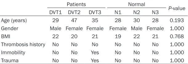
Table 1. Clinical characteristics of the patients
Patients Normal P-value
DVT1 DVT2 DVT3 N1 N2 N3
Age (years) 29 47 35 28 30 28 0.193
Gender Male Female Female Female Male Female 1.000
BMI 22 20 21 19 22 21 0.768
Thrombosis history No No No No No No 1.000
Immobility No No Yes No No No 1.000
Trauma No No Yes No No No 1.000
BMI: Body Mass Index.
Table 2. Primers used in qRT-PCR
Gene Name Primer sequence (°C)Tm length (bp)Product ENST00000544704 F: 5’CAAATCTGTGACAATGCCCC3’
R: 5’GTTATGACTCAAGCGAAAATGG3’ 60 72
TCONS_00013536 F: 5’TGAGCACACCATTGGAGAACCT3’
R: 5’TTTGGGATTGGAGGGAGAGG3’ 60 87
ENST00000577218 F: 5’CCGCCTCTAGTCCTCACAC3’
R: 5’CGGAATGAATGGATGGTCT3’ 60 235
ENST00000511042 F: 5’GCTGGATGTATGAACCCTGCT3’
R: 5’TCCTGCTGGACTCTCGTGT3’ 60 64
TCONS_00013917 F: 5’ATCCACCGAAGGTTTGAGG3’
R: 5’GGGAGACGGGACTATATCCAG3’ 60 98
GAPDH F: 5’ACGGTGGTGGAGGAGCTCTT3’
R: 5’GCCGGTTCAGGTACTCAGTCAT3’ 60 157
F: Forward primer; R: Reverse primer.
pite these exciting deve- lopments, many more lncRNAs that play crucial roles in EPCs of DVT patients remain to be
clarified.
LncRNAs expression
pro-files may help provide
important insights into pathogenesis and a pos-sible diagnostic strategy for DVT. Thus, in this study we examined lnc- RNAs and mRNAs that were differentially expre- ssed in EPCs from DVT patients and healthy con- trols.
Materials and methods
Subjects
From January 2014 to January 2015, eighty mil-liliter of peripheral blood were collected from DVT patients (n = 3) and con-trol subjects (n = 3) at the
[image:2.612.92.396.240.408.2]Systems, MN, USA), human recombinant long
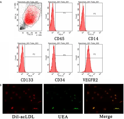
insulin-like growth factor-1 (R3-IGF), ascorbic acid, and hydrocortisone and maintained at 37°C, 5% CO2. EPCs were characterized by
con-focal microscopy and flow cytometry. The cells were incubated with agglutinin 1 (FITC-UEA-1;
Sigma Deisenhofen, Germany) and 1, 19- dioctadecyl-3, 3,3939-tetramethylindocar-bo- cyanine perchlorate (DiI)-labeled acetylated low density lipoprotein (Dil-Ac-LDL) as described previously [25]. Incorporation of DiI-Ac-LDL and
binding of FITCUEA-1 were detected under a
confocal microscope (Leica Microsystems GmbH, Germany). Cells with double positive
staining of DiI-Ac-LDL and UEA-1 were identi
-fied as EPCs. EPCs were analyzed for surface
expression of CD14, CD34, CD45, CD133 and 2. Antibodies CD34, CD133 and VEGFR-2 were purchased from Miltenyi Biotec, Bergisch, Germany; CD14 and CD45 were pur-chased from BD Biosciences Pharmingen, CA,
USA. The second passage of EPCs was used.
RNA extraction
Total RNA exaction was performed using TRIzol
reagent (Life Technology, USA) according to the
manufacturer’s instructions. The quantity and
quality of RNA were verified by a Nano Drop
ND-1000 spectrophotometer. RNA integrity and genomic DNA contamination was exam-ined by denatured agarose gel electropho- resis.
Microarray analysis and computational analy-sis
Sample preparation and microarray hybridiza-tion were performed by Kangchen Bio-tech, Shanghai P. R. China. The methods were similar
to previous reports [14]. Briefly, RNA was puri
-fied from 1 μg total RNA after removal of rRNA
(mRNA-ONLY Eukaryotic mRNA Isolation Kit,
Epicentre). Each sample was amplified and transcribed into fluorescent cRNA along the
entire length of the transcripts without 3’ bias using a random priming method. The labeled cRNAs were hybridized onto the Human LncRNA Array v3.0 (Arraystar). After the slides were washed, the arrays were scanned by the Agilent Scanner G2505C. Agilent Feature Extraction software (version 11.0.1.1) was used to ana-lyze acquired array images. Quantile normaliza-tion and subsequent data processing were car-ried out using the GeneSpring GX v12.1
soft-ware package (Agilent Technologies). Differ- entially expressed lncRNAs and mRNAs were
identified through fold change filtering (Fold Change ≥2.0 or ≤0.5), paired t-test (P<0.05) and multiple hypothesis testing (FDR<0.05). P values and FDR were calculated by Microsoft Excel and MATLAB respectively. Pathway analy-sis and GO analyanaly-sis were used to determine the roles of these differentially expressed mRNAs in these biological pathways or GO terms. R programme with Top GO package [26] was used to analyze the enrichment of differentially regu-lated mRNAs. We also used the KEGG (Kyoto Encyclopedia of Genes and Genomes) data-base (http://www.genome.ad.jp/kegg/) to ana-lyze the potential functions of these target genes in the pathways [27, 28].
Quantitative real-time polymerase chain reac-tion (qRT-PCR) validareac-tion and statistical analy-sis
To validate the results of microarray analysis, other EPCs samples from 21 DVT patients and nine control subjects were obtained. Total RNA was reverse-transcribed using I SuperScriptTM III Reverse Transcriptase according to the man-ufacturer’s protocol. Real-time PCR was per-formed with SYBR Premix Ex Taq (TaKaRa) on ABI7500 Real-time PCR System according to the manufacturer’s protocol. PCR primers are listed in Table 2. The relative fold change was calculated using the 2-ΔΔCt method normalized
to GAPDH. Differences of lncRNAs between patients and normal were analyzed using paired t-test. A P value <0.05 was considered
as statistically significant.
Results
EPCs characterization
First, EPCs were identified by morphology, fluo
-rescence double-staining and flow cytometry. The flow cytometry analysis results (Figure 1A) matched with the previously described EPC phenotype [23, 29]. Most adherent cells were
double stained by DiI-AcLDL and FITC-UEA-I
(Figure 1B). These results were consistent with the characterization of late-outgrowth EPCs. Expression profile of lncRNAs and mRNAs in EPCs from DVT patients
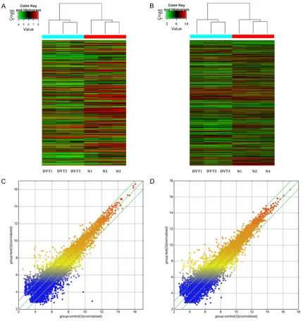
com-pared with controls, we performed microarray
analysis and identified 30,586 differentially
expressed lncRNAs and 26,109 mRNAs. After expression level normalization, we calculated the fold-change (DVT vs. control) and p value. A total of 275 lncRNAs and 363 mRNAs we-
re confirmed as significantly differentially
expressed in EPCs from DVT patients com-pared with EPCs from control subjects (fold
change ≥2.0 or ≤0.5, P<0.05 and FDR<0.05). Among these, 83 lncRNAs and 127 mRNAs were consistently up-regulated in all DVT groups, and 192 lncRNAs and 236 mRNAs were consistently down-regulated. The number of deregulated lncRNAs and mRNAs varied in different patients. The differentially
expres-sion of these lncRNAs suggest that they may play important roles in the regulation of the function of EPCs during the development of DVT.
We used hierarchical clustering analysis to arrange samples into groups based on their expression levels, which allowed us to hypoth-esize on the relationships among samples (Figure 2). The resulting dendrogram shows the relationships between the lncRNA and mRNA expression patterns between samples.
LncRNA classification and subgroup analysis
[image:4.612.93.522.73.485.2]LncRNAs are classified into different sub -groups, such as antisense lncRNAs, lncRNAs
with enhancer-like functions and large intergen-ic noncoding RNAs [14]. To further investigate potential functions of these DVT-associated
lncRNAs, lncRNA classification and subgroup
analysis was performed.
Natural antisense lncRNA transcripts are RNA molecules that are transcribed from the opposite DNA strand and overlap in part with
sense RNA, and can exert regulatory effects on the sense RNA through epigenetic
mecha-nisms. The profiling data indicated that five
such lncRNAs were dysregulated in DVT samples.
Using the GENCODE annotation [30] of the human genome, Orom et al. defined a set of
[image:5.612.92.522.71.527.2]lncRNAs with enhancer-like functions in human
cell lines [31]. The profiling data showed that
four such lncRNAs were differentially expressed in our DVT samples.
According to previous report [32], we also selected lncRNAs and nearby mRNAs to
per-form the analyses. The profiling data indicated
[image:6.612.92.373.72.571.2]enriched among the downregulated transcri- pts. Among them, leukocyte transendothelial migration and cytosolic DNA-sensing pathway were the most enriched networks (Figure 4). Of note, pathways associated with migration may play important roles in EPCs homing to thrombi.
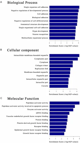
Figure 3. Gene ontology (GO) enrichment analysis for differentially expressed mRNAs. A: GO analysis of mRNAs according to biological process. B: GO anal-ysis of mRNAs according to cellular component. C: GO analanal-ysis of mRNAs according to molecular function.
that five such lncRNAs were
dysregulated in EPCs from DVT samples, and these were located in nearby aberrantly expressed coding genes. Bioinformatic analysis of mRNA expression in DVT
Our results identified a total
of 363 mRNAs that were dif-ferentially expressed in DVT
patients. Using R program -ming and top GO package [26], we analyzed the enrich-ment of these 363 differen-tially regulated mRNAs. The
most significant functional
groups consisted of cell adhe-sion, cell differentiation and the regulation of developmen-tal processes (Figure 3). The
most significant molecular
function included enzyme ac- tivator activity, VEGF recep- tor binding, platelet-derived growth factor binding and other growth factor receptor binding. These changes may contribute to EPCs homing and migrating into thrombi and differentiating into ma- ture endothelial cells.
Pathway analysis
Significant pathways of differ -ential genes were compared with the KEGG database to further identify target mRNAs
among the 3,278 identified
genes. Through the pathway
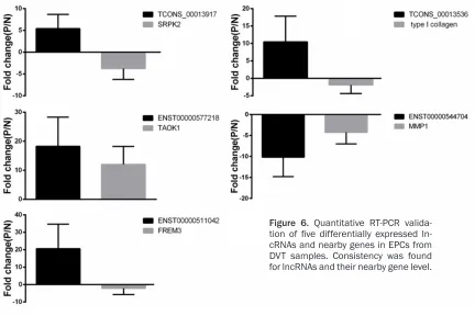
Table 3. The 14 pathways that corresponded to up-regulated transcripts
Pathway ID Definition Fisher-P value FDR Enrichment score
hsa04670 Leukocyte transendothelial migration-Homo sapiens (human) 0.004721208 0.4491231 2.325947
hsa00512 Mucin type O-Glycan biosynthesis-Homo sapiens (human) 0.007848341 0.4491231 2.105222
hsa05160 Hepatitis C-Homo sapiens (human) 0.00838558 0.4491231 2.076467
hsa05162 Measles-Homo sapiens (human) 0.008686624 0.4491231 2.061149
hsa05164 Influenza A-Homo sapiens (human) 0.009043315 0.4491231 2.043672
hsa05202 Transcriptional misregulation in cancer-Homo sapiens (human) 0.009589817 0.4491231 2.01819
hsa05168 Herpes simplex infection-Homo sapiens (human) 0.01169454 0.4694521 1.932017
hsa05412 Arrhythmogenic right ventricular cardiomyopathy (ARVC)-Homo sapiens (human) 0.01668933 0.5862126 1.777561
hsa00983 Drug metabolism-other enzymes-Homo sapiens (human) 0.02297849 0.7174396 1.638678
hsa04640 Hematopoietic cell lineage-Homo sapiens (human) 0.02941917 0.7687683 1.53137
hsa04380 Osteoclast differentiation-Homo sapiens (human) 0.03009413 0.7687683 1.521518
hsa04514 Cell adhesion molecules (CAMs)-Homo sapiens (human) 0.04366333 0.9110974 1.359883
hsa05161 Hepatitis B-Homo sapiens (human) 0.04475318 0.9110974 1.349176
hsa05416 Viral myocarditis-Homo sapiens (human) 0.04539275 0.9110974 1.343013
Table 4. The 30 pathways that corresponded to down-regulated transcripts Pathway
ID Definition PFisher value FDR Enrichmentscore
hsa04623 Cytosolic DNA-sensing pathway-Homo sapiens (human) 0.00263103 0.3459388 2.579874
hsa03440 Homologous recombination-Homo sapiens (human) 0.003006624 0.3459388 2.521921
hsa04961 Endocrine and other factor-regulated calcium reabsorption-Homo sapiens (human) 0.003782637 0.3459388 2.422205
hsa04978 Mineral absorption-Homo sapiens (human) 0.004924395 0.3459388 2.307647
hsa00240 Pyrimidine metabolism-Homo sapiens (human) 0.007990745 0.3916327 2.097413
hsa04640 Hematopoietic cell lineage-Homo sapiens (human) 0.01240606 0.3916327 1.906366
hsa05215 Prostate cancer-Homo sapiens (human) 0.01306934 0.3916327 1.883746
hsa03430 Mismatch repair-Homo sapiens (human) 0.01323153 0.3916327 1.87839
hsa04510 Focal adhesion-Homo sapiens (human) 0.01472141 0.3916327 1.832051
hsa04919 Thyroid hormone signaling pathway-Homo sapiens (human) 0.01528542 0.3916327 1.815723
hsa04720 Long-term potentiation-Homo sapiens (human) 0.01533082 0.3916327 1.814435
hsa05034 Alcoholism-Homo sapiens (human) 0.0170253 0.3986758 1.768905
hsa05218 Melanoma-Homo sapiens (human) 0.01928739 0.4169044 1.714726
hsa04916 Melanogenesis-Homo sapiens (human) 0.02305891 0.4552372 1.637161
hsa00230 Purine metabolism-Homo sapiens (human) 0.03373287 0.4552372 1.471947
hsa04621 NOD-like receptor signaling pathway-Homo sapiens (human) 0.03566406 0.4552372 1.447769
hsa05221 Acute myeloid leukemia-Homo sapiens (human) 0.03566406 0.4552372 1.447769
hsa04725 Cholinergic synapse-Homo sapiens (human) 0.03723153 0.4552372 1.429089
hsa04726 Serotonergic synapse-Homo sapiens (human) 0.03861753 0.4552372 1.413216
hsa04151 PI3K-Akt signaling pathway-Homo sapiens (human) 0.0394896 0.4552372 1.403517
hsa04512 ECM-receptor interaction-Homo sapiens (human) 0.03986393 0.4552372 1.39942
hsa00533 Glycosaminoglycan biosynthesis-keratan sulfate-Homo sapiens (human) 0.04152981 0.4552372 1.38164
hsa04012 ErbB signaling pathway-Homo sapiens (human) 0.04157717 0.4552372 1.381145
hsa04730 Long-term depression-Homo sapiens (human) 0.04189233 0.4552372 1.377865
hsa04974 Protein digestion and absorption -Homo sapiens (human) 0.04333446 0.4552372 1.363167
hsa03030 DNA replication-Homo sapiens (human) 0.04344184 0.4552372 1.362092
hsa04540 Gap junction-Homo sapiens (human) 0.04513596 0.4552372 1.345477
hsa04810 Regulation of actin cytoskeleton-Homo sapiens (human) 0.04601429 0.4552372 1.337107
hsa05414 Dilated cardiomyopathy-Homo sapiens (human) 0.04698177 0.4552372 1.328071
hsa05219 Bladder cancer-Homo sapiens (human) 0.04974438 0.4605383 1.303256
Genomic location of differentially expressed lncRNAs
Several studies have suggested that lncRNAs can regulate high order chromosomal
[image:7.612.90.527.297.665.2]expression of adjacent protein-coding genes [34, 35]. The relationships of lncRNAs and
with nine control subjects. The expression
pat-tern results of the five lncRNAs were consistent
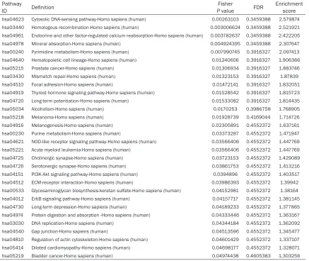
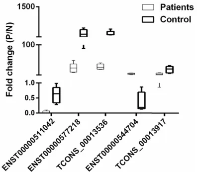
with the microarray data. TCONS_00013536, ENST00000577218, ENST00000511042 and TCONS_00013917 were upregulated, where- as ENST00000544704 was downregulated in EPCs from DVT samples compared with con-trols (Figure 5). Additionally, microarray data also provided the nearby coding genes of these differentially expressed lncRNAs. We found that the changes of lncRNAs and their nearby coding genes are basically identical to the results of qRT-PCR analysis (Figure 6). These results, which were consistent with the previ-ous reports [14], provided strong evidence that lncRNAs had intrinsic cis-regulatory capacity to their own locus.
Discussion
[image:8.612.92.373.72.368.2]During the past decades, the roles of EPCs in the prognosis of thrombosis have been exten-sively studied [4, 8-11]. However, the molecular mechanisms of EPCs in the resolution of throm-bosis have not yet been fully elucidated.
Figure 4. Pathway analysis. The top 10 pathways of the coding genes associ-ated with upregulassoci-ated lncRNAs (A) and downregulassoci-ated lncRNAs (B) are listed.
Figure 5. Quantitative RT-PCR validation of five differentially expressed lncRNAs in DVT sam-ples. TCONS_00013536, ENST00000577218, ENST00000511042 and TCONS_00013917 were upregulated, whereas ENST00000544704 was downregulated in five pairs of DVT samples com -pared with control samples.
nearby coding genes
identi-fied here include bidirection -al, exon sense overlapping, intergenic, intro sense over-lapping, intronic antisense and natural antisense. The 275 differentially expressed lncRNAs in DVT samples in- cluded 24 directional sequ- ences, 29 exon sense-over-lapping sequences, 123 inter-genic sequences, 12 intro sense-overlapping sequenc-es, 42 intronic antisense and 37 natural antisense.
Five dysregulated lncRNAs in DVT samples
To verify the microarray data,
[image:8.612.91.289.421.594.2]MicroRNAs (miRNAs) are a class of noncoding small RNAs that exert a critical role in the func-tion regulafunc-tion of EPCs. Altered expression of miRNAs in EPCs has been shown to be involved in thrombi resolution [10, 11]. However, the roles of lncRNAs in EPCs and in thrombi forma-tion and resoluforma-tion have not yet been deter-mined. Although the majority of lncRNA could be functional, only few have been demonstrat-ed to be involvdemonstrat-ed in biological processes, such as regulation of basal transcription machinery functions, RNA splicing and translation, and epigenetic regulation [36-38]. Moreover, the exact roles of lncRNA in cells are less clear compared with those of miRNAs. Therefore, we conducted the present study to obtain insight into the roles of lncRNA in EPCs and a role in pathogenesis of DVT.
Recent evidences have shown that lncRNAs play an important role in gene expression regu-lation through cis (i.e., on neighboring genes) or trans (distant genes) manners [39]. However,
prediction of the specifics is relative difficulty
based on lncRNA sequence [40, 41]. LncRNAs have been implicated in multiple functions, par-ticularly in vascular diseases such as in athero-sclerosis [42] and varicose vein disease [43]. Previous studies have shown expression of the
hypoxia-inducible factor 1α (HIF1α) gene, which
is commonly involved in angiogenic events [44, 45], is regulated through lncRNA-p21 [46]. Thus, the aberrant expression of at least one lncRNA has been linked to vascular events. To date, there have been no reports of differen-tially expressed lncRNAs in DVT samples. Our
data is the first to show a total of 275 differen -tially expressed lncRNAs and 363 mRNAs in DVT samples, with a fold change of 2 or more. GO and pathway analyses predicted that down-regulated and updown-regulated transcripts of mRNAs were associated with cellular process (ontology: biological process), cell (ontology: cellular component), and binding (ontology: molecular function). We found that these genes were mainly involved in cell adhesion and cell differentiation, which may suggest that the dif-ferently expressed lncRNAs could regulate
migration and differentiation via influencing the
expression of these genes. To further under-stand the functions of lncRNAs, we used path-way analysis to associate these differentially expressed lncRNAs with their target genes and found 44 pathways that corresponded to tran-scripts; the most enriched network was the cytosolic DNA-sensing pathway, which was composed of 64 targeted genes. One of these
[image:9.612.93.525.71.359.2]pathways, the gene category ‘leukocyte tran-sendothelial migration and focal adhesion pathways’, has been reported to be involved in the homing of EPCs [4].
Here we demonstrated that TCONS_000135- 36, ENST00000577218, ENST00000511042 and TCONS_00013917 were upregulated and ENST00000544704 was downregulated in EPCs from DVT samples compared with
con-trols. Furthermore, expressions of five lncRNAs were significantly correlated with their nearby
coding genes, which have not been described before. For example, the downregulated lncRNA ENST000000544704 in DVT was found to be located near the gene encoding MMP1. MMP-1 functions to promote proliferation and migra-tion of vascular cells (vascular smooth muscle cells and endothelial cells) [47, 48], vessel remodeling, angiogenesis and tumor progres-sion [49]. A natural antisense association between the downregulated lncRNA ENST0- 00000544704 and MMP1 gene may help illu-minate how lncRNAs regulate gene expression in vessel components. The upregulated lncRNA ENST00000511042 FREM3 is important in the regulation of in the structure and function of basement membrane [50] and therefore may be an important regulator in angiogenesis. The current study of lncRNAs in EPCs from DVT samples is a proof-of-principle that lncRNAs have a probable role in vein thrombosis patho-genesis. Further studies should be made to fully understand this disease with the aims of developing effective treatment strategies. Our present study on the potential relationships between lncRNAs and EPCs in DVT presents a novel area for further investigations into the targeted lncRNAs and the potential therapeutic strategies for this disease.
Acknowledgements
This work was supported by grants from the
Scientific and Technological Research Projects
of Jiangsu Province (No. BL2014043), Graduate Research and Innovation Program in Colleges
and Universities of Jiangsu Province (No.
KYLX15_1202).
Disclosure of conflict of interest
None.
Address correspondence to: Xiao-Qiang Li, De- partment of Vascular Surgery, The Second Affiliated Hospital of Soochow University, 1055 Sanxiang Rd, Suzhou 215000, Jiangsu, China. E-mail: [email protected]; [email protected]
References
[1] Saez-Gimenez B, Berastegui C, Loor K, Lopez-Meseguer M, Monforte V, Bravo C, Santamaría A, Roman A. Deep vein thrombosis and pulmo-nary embolism after solid organ transplanta-tion: an unresolved problem. Transplant Rev (Orlando) 2015; 29: 85-92.
[2] Piazza G, Goldhaber SZ. Acute pulmonary em-bolism: part I: epidemiology and diagnosis. Circulation 2006; 114: e28-32.
[3] Weitz JI, Eikelboom JW, Samama MM. New an-tithrombotic drugs: Anan-tithrombotic Therapy and Prevention of Thrombosis, 9th ed: Ame- rican College of Chest Physicians Evidence-Based Clinical Practice Guidelines. Chest 2012; 141: e120S-151S.
[4] Li WD, Li XQ. Endothelial progenitor cells ac-celerate the resolution of deep vein thrombo-sis. Vascul Pharmacol 2016; 83: 10-16. [5] Grisar JC, Haddad F, Gomari FA, Wu JC.
Endothelial progenitor cells in cardiovascular disease and chronic inflammation: from bio -marker to therapeutic agent. Biomark Med 2011; 5: 731-744.
[6] Bakogiannis C, Tousoulis D, Androulakis E, Briasoulis A, Papageorgiou N, Vogiatzi G, Kampoli AM, Charakida M, Siasos G, Latsios G, Antoniades C, Stefanadis C. Circulating endo-thelial progenitor cells as biomarkers for pre-diction of cardiovascular outcomes. Curr Med Chem 2012; 19: 2597-2604.
[7] Smadja DM, Duong-van-Huyen JP, Dal Cortivo L, Blanchard A, Bruneval P, Emmerich J, Gaussem P. Early endothelial progenitor cells in bone marrow are a biomarker of cell therapy success in patients with critical limb ischemia. Cytotherapy 2012; 14: 232-239.
[8] Alessio AM, Beltrame MP, Nascimento MC, Vicente CP, de Godoy JA, Silva JC, Bittar LF, Lorand-Metze I, de Paula EV, Annichino-Bizzacchi JM. Circulating progenitor and ma-ture endothelial cells in deep vein thrombosis. Int J Med Sci 2013; 10: 1746-1754.
[9] Modarai B, Burnand KG, Sawyer B, Smith A. Endothelial progenitor cells are recruited into resolving venous thrombi. Circulation 2005; 111: 2645-2653.
Target PIK3R2. J Cell Biochem 2015; 116: 1613-1623.
[11] Wang W, Li C, Li W, Kong L, Qian A, Hu N, Meng Q, Li X. MiR-150 enhances the motility of EPCs in vitro and promotes EPCs homing and throm-bus resolving in vivo. Thromb Res 2014; 133: 590-598.
[12] Ponting CP, Oliver PL, Reik W. Evolution and functions of long noncoding RNAs. Cell 2009; 136: 629-641.
[13] Wang KC, Chang HY. Molecular mechanisms of long noncoding RNAs. Mol Cell 2011; 43: 904-914.
[14] Zhu J, Liu S, Ye F, Shen Y, Tie Y, Zhu J, Jin Y, Zheng X, Wu Y, Fu H. The long noncoding RNA expression profile of hepatocellular carcinoma identified by microarray analysis. PLoS One 2014; 9: e101707.
[15] Gupta RA, Shah N, Wang KC, Kim J, Horlings HM, Wong DJ, Tsai MC, Hung T, Argani P, Rinn JL, Wang Y, Brzoska P, Kong B, Li R, West RB, van de Vijver MJ, Sukumar S, Chang HY. Long non-coding RNA HOTAIR reprograms chromatin state to promote cancer metastasis. Nature 2010; 464: 1071-1076.
[16] Johnson R. Long non-coding RNAs in Hunting- ton’s disease neurodegeneration. Neurobiol Dis 2012; 46: 245-254.
[17] Faghihi MA, Modarresi F, Khalil AM, Wood DE, Sahagan BG, Morgan TE, Finch CE, St Laurent G 3rd, Kenny PJ, Wahlestedt C. Expression of a noncoding RNA is elevated in Alzheimer’s dis-ease and drives rapid feed-forward regulation of beta-secretase. Nat Med 2008; 14: 723-730.
[18] Fiedler J, Breckwoldt K, Remmele CW, Hartmann D, Dittrich M, Pfanne A, Just A, Xiao K, Kunz M, Müller T, Hansen A, Geffers R, Dandekar T, Eschenhagen T, Thum T. De- velopment of Long Noncoding RNA-Based Strategies to Modulate Tissue Vascularization. J Am Coll Cardiol 2015; 66: 2005-2015. [19] Li K, Blum Y, Verma A, Liu Z, Pramanik K, Leigh
NR, Chun CZ, Samant GV, Zhao B, Garnaas MK, Horswill MA, Stanhope SA, North PE, Miao RQ, Wilkinson GA, Affolter M, Ramchandran R. A noncoding antisense RNA in tie-1 locus regu-lates tie-1 function in vivo. Blood 2010; 115: 133-139.
[20] Bell RD, Long X, Lin M, Bergmann JH, Nanda V, Cowan SL, Zhou Q, Han Y, Spector DL, Zheng D, Miano JM. Identification and initial function -al characterization of a human vascular cell-enriched long noncoding RNA. Arterioscler Thromb Vasc Biol 2014; 34: 1249-1259. [21] Yan B, Yao J, Liu JY, Li XM, Wang XQ, Li YJ, Tao
ZF, Song YC, Chen Q, Jiang Q. lncRNA-MIAT regulates microvascular dysfunction by func-tioning as a competing endogenous RNA. Circ Res 2015; 116: 1143-1156.
[22] Hill JM, Zalos G, Halcox JP, Schenke WH, Waclawiw MA, Quyyumi AA, Finkel T. Circulating endothelial progenitor cells, vascular function, and cardiovascular risk. N Engl J Med 2003; 348: 593-600.
[23] Kalka C, Masuda H, Takahashi T, Kalka-Moll WM, Silver M, Kearney M, Li T, Isner JM, Asahara T. Transplantation of ex vivo expanded endothelial progenitor cells for therapeutic neovascularization. Proc Natl Acad Sci U S A 2000; 97: 3422-3427.
[24] Kong L, Hu N, Du X, Wang W, Chen H, Li W, Wei S, Zhuang H, Li X, Li C. Upregulation of miR-483-3p contributes to endothelial progenitor cells dysfunction in deep vein thrombosis pa-tients via SRF. J Transl Med 2016; 14: 23. [25] Ma FX, Zhou B, Chen Z, Ren Q, Lu SH,
Sawamura T, Han ZC. Oxidized low density lipo-protein impairs endothelial progenitor cells by regulation of endothelial nitric oxide synthase. J Lipid Res 2006; 47: 1227-1237.
[26] Alexa A, Rahnenfuhrer J. topGO: Enrichment analysis for Gene Ontology. R Package Version 2010; 2230.
[27] Kanehisa M, Goto S, Kawashima S, Okuno Y, Hattori M. The KEGG resource for deciphering the genome. Nucleic Acids Res 2004; 32: D277-280.
[28] Zhang C, Han L, Zhang A, Yang W, Zhou X, Pu P, Du Y, Zeng H, Kang C. Global changes of mRNA expression reveals an increased activity of the interferon-induced signal transducer and acti-vator of transcription (STAT) pathway by repres-sion of miR-221/222 in glioblastoma U251 cells. Int J Oncol 2010; 36: 1503-1512. [29] Rehman J, Li J, Orschell CM, March KL.
Peripheral blood “endothelial progenitor cells” are derived from monocyte/macrophages and secrete angiogenic growth factors. Circulation 2003; 107: 1164-1169.
[30] Harrow J, Denoeud F, Frankish A, Reymond A, Chen CK, Chrast J, Lagarde J, Gilbert JG, Storey R, Swarbreck D, Rossier C, Ucla C, Hubbard T, Antonarakis SE, Guigo R. GENCODE: producing a reference annotation for ENCODE. Genome Biol 2006; 7 Suppl 1: S4 1-9.
[31] Orom UA, Derrien T, Beringer M, Gumireddy K, Gardini A, Bussotti G, Lai F, Zytnicki M, Notredame C, Huang Q, Guigo R, Shiekhattar R. Long noncoding RNAs with enhancer-like function in human cells. Cell 2010; 143: 46-58.
[34] Martianov I, Ramadass A, Serra Barros A, Chow N, Akoulitchev A. Repression of the hu-man dihydrofolate reductase gene by a non-coding interfering transcript. Nature 2007; 445: 666-670.
[35] Feng J, Bi C, Clark BS, Mady R, Shah P, Kohtz JD. The Evf-2 noncoding RNA is transcribed from the Dlx-5/6 ultraconserved region and functions as a Dlx-2 transcriptional coactiva-tor. Genes Dev 2006; 20: 1470-1484.
[36] Dinger ME, Amaral PP, Mercer TR, Mattick JS. Pervasive transcription of the eukaryotic ge-nome: functional indices and conceptual impli-cations. Brief Funct Genomic Proteomic 2009; 8: 407-423.
[37] Mercer TR, Dinger ME, Mattick JS. Long non-coding RNAs: insights into functions. Nat Rev Genet 2009; 10: 155-159.
[38] Dong R, Jia D, Xue P, Cui X, Li K, Zheng S, He X, Dong K. Genome-wide analysis of long noncod-ing RNA (lncRNA) expression in hepatoblasto-ma tissues. PLoS One 2014; 9: e85599. [39] Khachane AN, Harrison PM. Mining
mammali-an trmammali-anscript data for functional long non-cod-ing RNAs. PLoS One 2010; 5: e10316. [40] Hung T, Chang HY. Long noncoding RNA in
ge-nome regulation: prospects and mechanisms. RNA Biol 2010; 7: 582-585.
[41] Qureshi IA, Mattick JS, Mehler MF. Long non-coding RNAs in nervous system function and disease. Brain Res 2010; 1338: 20-35. [42] Hu YW, Zhao JY, Li SF, Huang JL, Qiu YR, Ma X,
Wu SG, Chen ZP, Hu YR, Yang JY, Wang YC, Gao JJ, Sha YH, Zheng L, Wang Q. RP5-833A20.1/ miR-382-5p/NFIA-dependent signal transduc-tion pathway contributes to the regulatransduc-tion of cholesterol homeostasis and inflammatory re -action. Arterioscler Thromb Vasc Biol 2015; 35: 87-101.
[43] Li X, Jiang XY, Ge J, Wang J, Chen GJ, Xu L, Xie DY, Yuan TY, Zhang DS, Zhang H, Chen YH. Aberrantly expressed lncRNAs in primary vari-cose great saphenous veins. PLoS One 2014; 9: e86156.
[44] Kumarswamy R, Bauters C, Volkmann I, Maury F, Fetisch J, Holzmann A, Lemesle G, de Groote P, Pinet F, Thum T. Circulating long noncoding RNA, LIPCAR, predicts survival in patients with heart failure. Circ Res 2014; 114: 1569-1575. [45] Vausort M, Wagner DR, Devaux Y. Long non-coding RNAs in patients with acute myocardial infarction. Circ Res 2014; 115: 668-677. [46] Yang F, Zhang H, Mei Y, Wu M. Reciprocal
regu-lation of HIF-1alpha and lincRNA-p21 modu-lates the Warburg effect. Mol Cell 2014; 53: 88-100.
[47] Shi ZD, Ji XY, Qazi H, Tarbell JM. Interstitial flow promotes vascular fibroblast, myofibroblast, and smooth muscle cell motility in 3-D colla-gen I via upregulation of MMP-1. Am J Physiol Heart Circ Physiol 2009; 297: H1225-1234. [48] Blackburn JS, Brinckerhoff CE. Matrix
metallo-proteinase-1 and thrombin differentially acti-vate gene expression in endothelial cells via PAR-1 and promote angiogenesis. Am J Pathol 2008; 173: 1736-1746.
[49] Mazor R, Alsaigh T, Shaked H, Altshuler AE, Pocock ES, Kistler EB, Karin M, Schmid-Schönbein GW. Matrix metalloproteinase-1-mediated up-regulation of vascular endothe-lial growth factor-2 in endotheendothe-lial cells. J Biol Chem 2013; 288: 598-607.