Original Article
Immunohistochemical differentiation between type B3
thymomas and thymic squamous cell carcinomas
Xue-Ying Su*, Wei-Ya Wang*, Jin-Nan Li, Dian-Ying Liao, Wei-Lu Wu, Gan-Di Li
Department of Pathology, West China Hospital of Sichuan University, Chengdu 610041, China. *Equal contribu-tors.
Received February 27, 2015; Accepted April 15, 2015; Epub May 1, 2015; Published May 15, 2015
Abstract: Type B3 thymomas and thymic squamous cell carcinomas have some overlapping histological features, so it is difficult to make the differential diagnosis between these two entities, especially when the biopsy specimen is small. Only a few markers such as CD5 and CD 117 were applied to the differential diagnosis, the purpose of this study is to identify other diagnostic markers to help making the differential diagnosis more accurate. GLUT-1, MUC-1, CD117, CD5, CEA, P63, CK19, CK5/6, CD1a and TdT were evaluated using 16 cases of type B3 thymoma and 20 cases of thymic squamous cell carcinoma. Staining scores were obtained by calculating the percentage of positive cells. The sensitivity of GLUT-1 or MUC-1 for thymic squamous cell carcinomas was highest (100%), followed by CK5/6 (95%), CD117 (90%), P63 (85%), CD5 (80%) and CEA (75%). The specificities of CD5, CD117 and CEA for thymic squamous cell carcinomas all were 100%, next was MUC-1 (56.3%), followed by GLUT-1 (50%), P63 (25%), CK5/6 (12.5%). The sensitivities of CK19, TdT, and CD1a for type B3 thymomas were 100%, 93.8% and 87.5%, respectively. The specificity of CD1a for type B3 thymomas was highest (100%), followed by TdT (95%), CK19 (10%). The reactivity of GLUT-1, MUC-1, CD117, CD5, CEA, CD1a and TdT in thymic squamous cell carcinomas and type B3 thymomas had significant difference. Usually a panel of markers is needed, if we combine GLUT-1 or MUC-1 which sensitivity for thymic squamous cell carcinomas is highest with CD5, CD117, CEA, CD1a or TdT which have high specificity, we can make the differential diagnosis effectively.
Keywords: Type B3 thymoma, thymic squamous cell carcinoma, immunohistochemistry, differentiation
Introduction
In 2004, the World Health Organization (WHO)
classified epithelial thymic tumors into type A,
AB, B1, B2, B3 thymoma and thymic carcinoma [1]. Type B3 thymoma is predominantly epithe-lial type, with a few immature T lymphocytes. The tumor cells usually palisade around the perivascular space, with mild to moderate cel-lular atypia. Foci of squamous metaplasia could be found in some cases. Thymic carcinomas usually have obvious cellular atypia, with a small number of mature lymphocytes and plas-ma cells. Thymic carcinoplas-mas include several histological types which are similar to the same histological types of extrathymic carcinomas without organotypical features of thymic differ-entiation. Squamous cell carcinoma is the most frequent type [1].Either type B3 thymoma or thymic carcinoma can present as invasive growth, the tumor cells of type B3 thymoma
may have obvious atypia. Sometimes it is diffi
-cult to make the differential diagnosis between
them histologically, especially between type B3 thymoma and squamous cell carcinoma, par-ticularly when the biopsy specimen is small [2].
Some markers such as CD5 and CD 117 were
used to the differential diagnosis, however, the positive rate of CD5 and CD117 in thymic
carci-noma had been reported to be 70%, 50%-90%, respectively [3-7]. Moreover, not all neo -plastic cells in thymic carcinomas were stained
positive for these markers. Meanwhile, the
tumor cells in a few number of type B3 thymo-mas could be positive for CD5 or CD 117 [8, 9]. So it is necessary to identify other diagnostic
markers to help the differential diagnosis. GLUT-1, CEA and MUC-1 were reported to be
Differentiation of type B3 thymoma and TSCC
were related to these markers and the cases
involved in these studies were very limited, so
the value of these markers needs to be further analyzed. As we know, CD1a and TdT are always
stained positive for the immature T cells in type
B3 thymomas, CK5/6 and P63 are usually posi -tive for squamous cell carcinomas, if these
markers could help to make the differential
diagnosis more reliable? In this study, we evalu-ated a panel of antibodies and try to choose the
markers which can help the differentiation of
thymic squamous cell carcinomas (TSCCs) and type B3 thymomas.
Materials and methods
Subjects
Two hundred and forty-nine surgically treated cases of thymic epithelial tumors were collect-ed from the Department of Pathology, West
China Hospital of Sichuan University since
1999 to 2009. According to the WHO 2004
classification schema, there were 18 cases of
type A, 97 of type AB, 22 of type B1, 63 of type B2, 16 of type B3 and 33 of thymic carcinoma. The thymic carcinomas consisted of 8 cases of
keratinizing squamous carcinomas, 13 cases of nonkeratinizing carcinomas, 6 cases of neu -roendocrine carcinomas, 4 cases of
lymphoep-ithelioma-like carcinomas, 1 sarcomatoid carci -noma and 1 adenocarci-noma. 16 cases of type B3 thymoma and 20 cases of thymic squamous cell carcinoma were included in this study.
The specimens were fixed with 10% neutral buffered formalin and embedded in paraffin. 4-6 μm sections were stained using hematoxy -lin and eosin (H&E). All the cases were reviewed
and reclassified according to the 2004 WHO classification system by two pathologists.
Some complicated cases were reviewed with the famous German pathologist Dr.
Müller-Hermlink using a multiheaded microscope. The
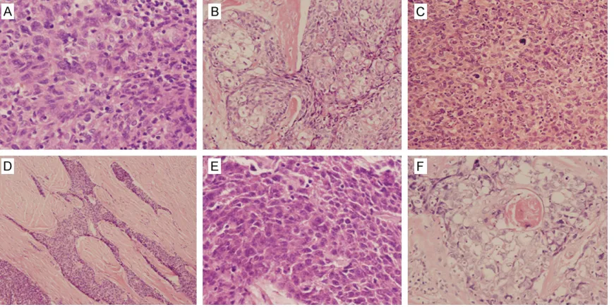
discrepancies were resolved by joint discus-sion. The diagnosis of TSCC was made after excluding metastases from other sites. Representative features of type B3 thymoma and TSCC were shown in Figure 1.
Immunohistochemistry
All the primary antibodies used in this study were listed in Table 1. The immunostaining was
performed on 4 μm paraffin sections for each case. The slides were deparaffinized in xylene,
then rehydrated through graded alcohol series.
The endogenous peroxidase was blocked using 3% hydrogen peroxide. Slides were immunos -tained with the primary antibodies shown in
Table 1. The sections were washed with PBS
and stained using DAKO EnVision + (Dako, Carpinteria, CA, USA) system. Finally the slides
were counterstained with hematoxylin.
Only membrane based reactivity was regarded as positive for CD1a, CD5 and CD117,
cyto-plasm for CEA, CK5/6 and CK19, both mem
-brane and cytoplasm for MUC-1, GLUT-1, nucle -ar based reactivity was reg-arded as positive for P63 and TdT.
For GLUT-1, MUC-1, CD117, CD5, CEA, P63, CK19 and CK5/6, the immunohistochemical stains were recorded as positivity when >10%
of tumor cells showed positive. For CD1a and TdT, the immunohistochemical stains were
recorded as positivity when >10% of lympho -cytes showed positive. Staining scores were made by calculating the percentage of positive
cells per slide: 0-10% (negative), 10%-25% (weak), 25%-50% (moderate) and >50%
(strong).
Statistical analysis
SPSS17.0 system was used for statistical anal-ysis. The results of immunohistochemical
stain-ing were evaluated by the χ2-test or Fisher’s
exact test. A significant level of 0.05 was used,
P-value <0.05 was considered as significant
difference.
Results
Clinical pathological features
The ages of 16 cases of type B3 thymoma ranged from 26 to 76 years old (median =43.5), male to female ratio was 1.67:1. All the tumors were located in the anterior mediastinum in which 7 cases were in the anterosuperior medi-astinum. The largest tumor dimension ranged from 3.5 cm to 13 cm (mean =8.3 cm). The Perivascular spaces with palisading of the tumor cells were found in 12 cases. Foci of squamous metaplasia presented in 8 cases in
I, 6 of Masaoka II, 6 of Masaoka III and 3 of Masaoka IV.
The ages of 20 cases of TSCC ranged from 20 to 70 years old (median =52.5), male to female ratio was 1:1. 19 cases were located in the anterior mediastinum in which 14 cases were in the anterosuperior mediastinum, 1 case was in middle mediastinum. The largest tumor dimension ranged from 4.3 cm to 12 cm (mean =8.4 cm). None presented organotypical fea-tures related to thymic differentiation. There
were 8 cases of keratinizing squamous cell car
-cinoma and 12 cases without t keratinization.
This group of thymomic carcinomas included
16 of Masaoka III and 4 of Masaoka IV, none was in Masaoka I or Masaoka II.
Immunohistochemical studies
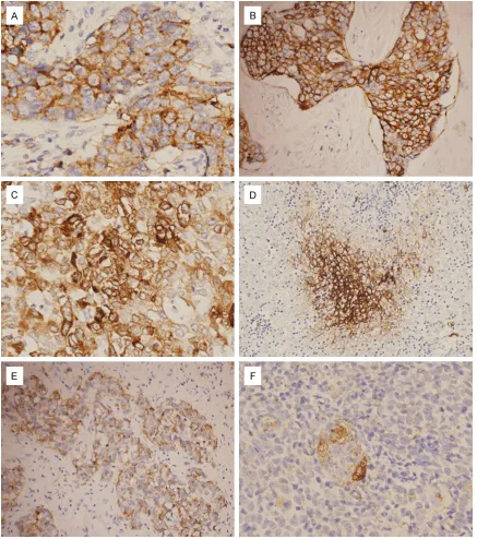
CD5: 16 cases of TSCC reacted with CD5 (Figure 2A), none of the type B3 thymomas was positive for CD5. The sensitivity of CD5 for
TSCCs was 80% while the specificity was 100%.
The reactivity of CD5 in TSCCs and type B3
thy-momas had significant difference (P=0.000).
CD117: 18 cases of TSCC were stained positive
for CD117 (Figure 2B), none of the type B3 thy-momas reacted with it. The sensitivity of CD117
for TSCCs was 90% while the specificity was 100%. The reactivity of CD117 in TSCCs and type B3 thymomas had significant difference
(P=0.000).
GLUT-1: 20 cases of TSCC were all strongly
pos-itive for GLUT-1 (Figure 2C) while only 8 cases of type B3 thymoma reacted with it. Only one type B3 thymoma presented as strong
positivi-ty, the others were recorded as weakly positive.
The positive signals tended to be more intense in the central area of the tumor nests than in the peripheral area (Figure 2D). The sensitivity
of GULT-1 for TSCCs was 100% while the speci
[image:3.612.92.527.72.290.2]-ficity was 50%. The reactivity of GLUT-1 in
Table 1. Antibody information
Antibody Clone Company Ag retrieval Dilution
CD5 SP19 Maxim EDTA 1:100
CD117 Polyclone Dako -- 1:100
CEA CEA-31 ZYMED PC 1:100
GLUT-1 Polyclone Maxim PC 1:100
MUC-1 MRQ-17 ZYMED PC Prediluted
CK5/6 D5/16B4 Maxim EDTA 1:100
P63 4A4 ZYMED EDTA 1:100
CK19 RCK108 Dako PC 1:100
CD1a 010 Dako EDTA 1:100
TdT SEN28 Maxim EDTA 1:100
[image:3.612.91.307.371.519.2]Ag: Antigen; PC: Pressure cooker.
Differentiation of type B3 thymoma and TSCC
TSCCs and type B3 thymomas had significant
difference (P=0.000).
MUC-1: 20 cases of TSCC were all stained
positive for MUC-1 (Figure 2E) and 15 cases were strongly positive. Only 7 cases of type B3
thymoma reacted with MUC-1 in which 5 cases showed weakly positive and 2 cases were mod
-erate positive (Figure 2F). The sensitivity of
MUC-1 for TSCCs was 100% while the specifici
-ty was 56.3%. The reactivi-ty of MUC-1 in TSCCs and type B3 thymomas had significant differ -ence (P=0.000).
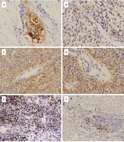
[image:4.612.89.526.71.565.2]negative for it. The sensitivity of CEA for TSCCs
was 75% while the specificity was 100%. The
reactivity of CEA in TSCCs and type B3
thymo-mas had significant difference (P=0.000).
P63: 17 cases of TSCC were positive for P63 (Figure 3B) and 12 cases of type B3 thymomas reacted with it. The sensitivity of P63 for TSCCs
was 85% while the specificity was only 25%. We
couldn’t conclude the reactivity of P63 in TSCCs
and type B3 thymomas had significant differ -ence (P=0.369).
CK5/6: 19 cases of TSCC were positive for
CK5/6 while 14 cases of type B3 thymomas
[image:5.612.91.522.71.565.2]Differentiation of type B3 thymoma and TSCC
CK5/6 for TSCCs was 95% while the specificity was only 12.5%. We couldn’t conclude the reac
-tivity of CK5/6 in TSCCs and type B3 thymomas had significant difference (P=0.415).
CK19:16 cases of TSCC were positive for CK19
while 18 cases of type B3 thymomas reacted with it (Figure 3D). The sensitivity of CK19 for TSCCs was 100% while the specificity was only 10%. We couldn’t conclude the reactivity of CK19 in TSCCs and type B3 thymomas had sig
-nificant difference (P=0.302).
CD1a: The lymphocytes in 16 cases of type B3
thymomas were positive for CD1a (Figure 3E) while the lymphocytes in all TSCCs were nega-tive for it. The sensitivity of CD1a for type B3
thymomas was 87.5% while the specificity was 100%. The reactivity of CD1a in TSCCs and type B3 thymomas had significant difference
(P=0.000).
TdT: The lymphocytes in 15 cases of type B3 thymomas were positive for TdT while the lym-phocytes in only one TSCC were positive for it (Figure 3F). The sensitivity of TdT for type B3
thymomas was 93.8% while the specificity was 95%. The reactivity of TdT in TSCCs and type B3 thymomas had significant difference (P=0.000). The reactivity of the above antibodies was sum-marized in Table 2.
Discussion
Thymic squamous cell carcinoma (TSCC) is the most common histological type of thymic carci-noma. Sometimes it is hardly to be distin-guished from type B3 thymoma only based on HE stained slides because of the overlapping morphological features. In this study, we select-ed 20 surgically removselect-ed TSCCs and 16 type B3 thymomas to evaluate the immunohisto-chemical differential diagnosis between them.
CD5 and CD117 are usually applied to the dif-ferential diagnosis between these two entities. In our study, the sensitivities of CD5 and CD117
for TSCC were 80% and 100%, respectively. The specificities of CD5 and CD117 for TSCCs both were 100%. We thought these two mark -ers were very useful. But there were four cases of TSCC stained negative for CD5, two cases of TSCC were negative for CD117. For one case,
the tumor cells didn’t react with both markers, so we thought some other markers were need
-ed to help making a correct diagnosis.
Glucose transporter 1 (GLUT-1) is one of 14
members of the mammalian facilitative glu-cose transporter family of passive carriers that function as an energy independent system for transport of glucose down a concentration gra-dient [11, 12]. It is thought to be a possible
intrinsic marker of hypoxia. GLUT-1 is expressed
in a variety of malignant tumors including renal cell carcinoma, breast carcinoma, ovarian car-cinoma, lung cancer and malignant pleural mesothelioma [13-15]. The study about the
expression of GLUT-1 in thymic tumors is very limited. Kojika et al. reported the sensitivity
and specificity of GLUT-1 for thymic carcinomas were 70% and 100%, respectively [2]. However
in a research of Hayashi’s,GLUT-1 was found to
be not very useful for the differential diagnosis
because of the low specificity [16]. In our study, all the TSCCs were strongly positive for GLUT-1, the sensitivity was 100%. Though 8 cases of
type B3 thymoma also reacted with it, the
sig-nals in 7 cases were weak. Moreover, the posi
-tive signals of GLUT-1 for type B3 thymomas
were more intense in the central area of the tumor nests than in the peripheral area.
Although the specificity was not so high, the
staining pattern in type B3 thymomas was
dif-ferent from TSCCs, so we thought GLUT-1 was
helpful to the differential diagnosis.
The transmembrane mucin MUC-1 is a heavily
[image:6.612.89.525.84.164.2]O-glycosylated protein expressed on most
Table 2. Summary of the antibodies reactivity
CD5 CD117 CEA CK5/6 P63 GLUT-1 MUC-1 CK19 CD1a TdT
+ - + - + - + - + - + - + - + - + - +
-TSCC
% 80 20 90 10 75 25 9516 4 18 2 15 5 19 15 17 3 20 085 15 100 0 10020 00 90 10 018 2 0 10020 51 1920 B3
% 0 16 0 16 0 160 100 0 100 0 100 87.5 12.5 72 25 50 50 43.7 56.3 100 0 87.5 12.5 93.8 6.214 2 12 4 8 8 7 9 16 0 14 2 15 1
secretory epithelium. MUC-1 consists of a heav -ily O-glycosylated excellilar domain, a trans-membrane domain and a cytoplasmic tail of 72 amino acids [17, 18]. It was reported that
MUC-1 oncoprotein aberrantly expressed at
high levels in most human neoplasms, and
MUC-1 played important roles in development
and progression of malignant tumors [19-21].
The precise function of MUC-1 overexpression in tumorigenesis is still unknown, although vari
-ous domains of MUC-1 had been implicated in
cell adhesion, cell signaling, and
immunoregu-lation. The overexpression of MUC-1 had been
also reported to be associated with bad prog-nosis in many malignant tumors [20, 22]. There are only a few studies related to the expression
of MUC-1 in thymic epithelial tumors. Kaira et al.reported MUC-1 was stained positive in 94%
(16/17) thymic carcinomas, all type B3
thymo-mas in his study were negative for MUC-1 [10].
In another research, the sensitivity and
speci-ficity of MUC-1 for thymic squamous cell carci
-nomas were 33% and 100%, respectively [2]. In our study, all the TSCCs were positive for MUC-1, the sensitivity was 100%. 7 cases of type B3 thymoma weakly reacted with it and the speci
-ficity was 56.3%. The sensitivity in our study
was higher than which was reported in the
lit-eratures, but the specificity was not so high. We found 75% (15/20) TSCCs was strong positive for MUC-1 while all the thymomas were weakly positive, so we thought MUC-1 was also useful
to the differential diagnosis.
CEA was found as a marker for colon adenocar -cinoma. The positive rate of CEA was higher in gastrointestinal adenocarcinoma, the adeno-carcinoma of lung; breast and ovary also could
express it in varied degree [23-25]. Kojika et al.
tried to use CEA as the marker for thymic carci
-noma. There were 6 cases of keratinizing carci
-noma and 6 cases non-keratinizing carci-noma
in his study, only 3 cases of thymic carcinoma stained positive for it, the sensitivity was only
25% while the specificity was 100% [2]. In our study, the sensitivity of CEA was 75%, none of
type B3 thymoma reacted with it. If CEA was combined with CD5 and CD117, the sensitivity
for TSCCs reached 100%.
CK5/6 or P63 was often used as the marker for
squamous cell carcinomas. But only a few
stud-ies were related to the expression of CK5/6 or P63 in TSCC. Kojika et al. revealed CK5/6 was
useless in the differential diagnosis of TSCC
and thymoma [2]. However, Khoury’s research showed that CK5/6 was one of the best mark -ers for the differential diagnosis. The sensitivity
and specificity of CK5/6 for TSCCs were 75% and 88.2%, respectively [8]. In our study, the sensitivity of CK5/6 for TSCCs was 95%.
Though the sensitivity was even higher than
which was reported, the specificity was only 12.5%, we didn’t think CK5/6 was very useful. Khoury showed 100% (17/17) TSCCs reacted with P63, the sensitivity was 100%, but the specificity was only 8.3% [8]. In our study, the sensitivity and specificity of P63 for TSCCs were 85% and 25%, respectively. The value of
P63 for the differential diagnosis was questionable.
Kuo et al. found all thymic epithelial cells were
stained positive for CK19 regardless of differ -ent epithelial compartm-ents [26]. However, four thymomas studied by Savino and Dardenne
were all negative for CK19 [27]. In a study of Fukai et al.,it was revealed that different types of thymoma could not be distinguished by the
CK expression patterns [28]. Kojika et al.
con-cluded the expression of CK19 hadn’t signifi -cant difference in type B3 thymomas and
thy-mic carcinomas [2]. In our study, 100% (16/16)
type B3 thymomas were stained positive for
CK19 and 90% (18/20) TSCCs reacted with it. We thought CK19 was not useful to the differ -ential diagnosis.
The immature T lymphocytes in type B3 thymo-ma were characterized by CD1a positive and TdT positive. CD1a and TdT were previously found to can help to differentiate thymoma
from thymic carcinoma. This finding was also consistent with our study. There were 87.5% of
type B3 thymomas stained positive for CD1a,
93.8% for TdT. The lymphocytes in all the TSCCs
were negative for CD1a. For one TSCC, the
lym-phocytes were positive for TdT, the specificity was 95% (19/20). We thought CD1a and TdT were both useful markers due to the high sensi
-tivity and specificity.
Differentiation of type B3 thymoma and TSCC
were 100%, 93.8% and 87.5%, respectively.
The specificity of CD1a for type B3 thymomas was highest (100%), followed by TdT (95%), CK19 (10%). The reactivity of GLUT-1, MUC-1,
CD117, CD5, CEA, CD1a and TdT in TSCCs and
type B3 thymomas had significant difference. A panel of markers is needed for the differential diagnosis. If we combine GLUT-1 or MUC-1
which sensitivity for TSCC was highest with CD5 or CD117, CEA, CD1a or TdT which have
high specificity, we can make the differential
diagnosis effectively.
Acknowledgements
We are greatly indebted to Dr. Müller-Hermlink for consultation in histological classification of
the thymic epithelial tumors in our study.
Disclosure of conflict of interest
None.
Address correspondence to: Dr. Gandi Li, Depart- ment of Pathology, West China Hospital of Sichuan University, Chengdu 610041, China. E-mail: [email protected]
References
[1] Müller-Hermelink HK, Engel P, Kuo TT, et al. Tumors of the Thymus. In: Pathology & Genetics, Tumours of the Lung, Pleura, Thymus and Heart. IARC Press: Lyon. 2004. pp. 145-247.
[2] Kojika M, Ishii1 G, Yoshida J, Nishimura M, Hishida T, Ota SJ, Murata Y, Nagai K, Ochiai A. Immunohistochemical differential diagnosis between thymic carcinoma and type B3 thy-moma: diagnostic utility of hypoxic marker, GLUT-1, in thymic epithelial neoplasms. Mod Pathol 2009; 22: 1341-50.
[3] Berezowski K, Grimes MM, Gal A, Kornstein MJ. CD5 immunoreactivity of epithelial cells in thymic carcinoma and CASTLE using paraffin-embedded tissue. Am J Clin Pathol 1996; 106: 483-6.
[4] Hishima T, Fukayama M, Fujisawa M, Hayashi Y, Arai K, Funata N, Koike M. CD5 expression in thymic carcinoma. Am J Pathol 1994; 145: 268-75.
[5] Pan CC, Chen PC, Chiang H. KIT (CD117) is fre-quently overexpressed in thymic carcinomas but is absent in thymomas. J Pathol 2004; 202: 375-81.
[6] Shimosato Y, Kameya T, Nagaki K. Squamous cell carcinoma of the thymus: An analysis of eight cases. Am J Surg Pathol 1977; 1: 109-21.
[7] Tateyama H, Eimoto T, Tada T, Hattori H, Murase T, Takino H. Immunoreactivity of a new CD5 antibody with normal epithelium and ma-lignant tumors including thymic carcinoma. Am J Clin Pathol 1999; 111: 235-40.
[8] Khoury T, Chandrasekhar R, Wilding G, Tan D, Cheney RT. Tumour eosinophilia combined with an immunohistochemistry panel is useful in the differentiation of type B3 thymoma from thymic carcinoma. Int J Exp Pathol 2011; 92: 87-96.
[9] Dorfman DM, Shahsafaei A, Chan JK. Thymic carcinomas, but not thymomas and carcino-mas of other sites, show CD5 immunoreactivi-ty. Am J Surg Pathol 1997; 21: 936-40. [10] Kaira K, Murakami H, Serizawa M, Koh Y, Abe
M, Ohde Y, Takahashi T, Kondo H, Nakajima T, Yamamoto N. MUC1 expression in thymic epi-thelial tumors: MUC1 may be useful marker as differential diagnosis between type B3 thymo-ma and thymic carcinothymo-ma. Virchows Arch 2011; 458: 615-20.
[11] Olson AL, Pessin JE. Structure, function, and regulation of the mammalian facilitative glu-cose transporter gene family. Annu Rev Nutr 1996; 16: 235-56.
[12] Clavo AC, Brown RS, Wahl RL. Fluorodeoxy- glucose uptake in human cancer cell lines is increased by hypoxia. J Nucl Med 1995; 36: 1625-32.
[13] Rudlowski C, Moser M, Becker AJ, Rath W, Buttner R, Schroder W, Schurmann A. GLUT1 mRNA and protein expression in ovarian bor-derline tumors and cancer. Oncology 2004; 66: 404-10.
[14] Younes M, Lechago LV, Somoano JR, Mosharaf M, Lechago J. Wide expression of the human erythrocyte glucose transporter Glut1 in hu-man cancers. Cancer Res 1996; 56: 1164-7. [15] Kato Y, Tsuta K, Seki K, Maeshima AM,
Watanabe S, Suzuki K, Asamura H, Tsuchiya R, Matsuno Y. Immunohistochemical detection of GLUT-1 can discriminate between reactive me-sothelium and malignant mesothelioma. Mod Pathol 2007; 20: 215-20.
[16] Hayashi A, Fumon T, Miki Y, Sato H, Yoshino T, Takahashi K. The evaluation of immunohisto-chemical markers and thymic cortical microen-vironmental cells in distinguishing thymic car-cinoma from type b3 thymoma or lung squa-mous cell carcinoma. J Clin Exp Hematop 2013; 53: 9-19.
[17] Saton S, Hinoda Y, Heyashi T, Burdick MD, Imai K, Hollingsworth MA. Enhancement of meta-static properties of pancreatic cancer cel l by MUC1 gene enloding an antiadhesion mole-cule. Int J Cancer 2000; 88: 507-18.
ex-pression of human tumor-associated polymor-phic epithelial mucin. J Biol Chem 1999; 265: 15286-93.
[19] Schroeder JA, Masril AA, Adriance MC, Tessier JC, Kotlarczyk KL, Thompson MC, Gendler SJ. MUC1 overexpression results in mammary gland tumorigenesis and prolonged alveolar differentiation. Oncogene 2004; 23: 5739-47. [20] Nagai S, Takenaka K, Sonobe M, Ogawa E,
Wada H, Tanaka F. A novel classification of MUC1 expression is correlated with tumor dif-ferentiation and postoperative prognosis in non-small cell lung cancer. J Thorac Oncol 2006; 1: 46-51.
[21] Broadbent R, Thynne G, McKenzie IFC. Antibody and T cell responses of patients with adenocarcinoma immunized with mannan-MUC1 fusion protein. J Clin Invest 1997; 100: 2783-92.
[22] Woenckhaus M, Merk J, Stoehr R, Schaeper F, Gaumann A, Wiebe K, Hartmann A, Hofstaedter F, Dietmaier W. Prognostic value of FHIT, CTNNB1, and MUC1 expression in non-small cell lung cancer. Hum Pathol 2008; 39: 126-136.
[23] Kho-Duffin J, Tao LC, Cramer H. Cytologic diag-nosis of malignant mesothelioma, with partic-ular emphasis on the epithelial noncohesive cell type. Diagn Cytopathol 1999; 20: 57-62. [24] Ordonez NG. The immunohistochemical
diag-nosis of esothelioma: differentiation of meso-thelioma and lung adenocarcinoma. Am J Surg Pathol 1989; 13: 276-91.
[25] Shield PW, Callan JJ, Devine PL. Markers for metastatic adenocarcinoma in serous effusion specimens. Diagn Cytopathol 1994; 11: 237-45.
[26] Kuo TT. Cytokeratin profiles of the thymus and thymomas: histogenetic correlations and pro-posal for a histological classification of thymo-mas. Histopathology 2000; 36: 403-14. [27] Savino W, Dardenne M. Immunohistochemical
studies on a human thymic epithelial cell sub-set defined by the anticytokeratin 18 monoclo-nal antibody. Cell Tissue Res 1988; 254: 225-231.