4-Methyl-3-nitrobenzonitrile
Li-Jing Cui and Jing Dai*
Ordered Matter Science Research Center, College of Chemistry and Chemical, Engineering, Southeast University, Nanjing 210096, People’s Republic of China. Correspondence e-mail: [email protected]
Received 29 July 2008; accepted 26 August 2008
Key indicators: single-crystal X-ray study;T= 298 K; mean(C–C) = 0.003 A˚;
Rfactor = 0.068;wRfactor = 0.208; data-to-parameter ratio = 16.1.
In the title compound, C8H6N2O2, the nitro group is rotated by 23.2 (3) out of the plane of the benzene ring. The crystal structure is stabilized by van der Waals interactions.
Related literature
For the chemistry of nitrile derivatives, see: Xionget al.(2002); Jinet al.(1994); Brewiset al.(2003); Dunicaet al.(1991). For related literature, see: Fu & Zhao (2007); Liang & Wang, (2008).
Experimental
Crystal data
C8H6N2O2
Mr= 162.15 Monoclinic,P21=c
a= 3.9088 (8) A˚ b= 13.576 (3) A˚ c= 14.819 (4) A˚
= 99.13 (3)
V= 776.4 (3) A˚3
Z= 4
MoKradiation
= 0.10 mm 1
T= 298 (2) K 0.350.300.1 mm
Data collection
Rigaku Mercury2 diffractometer Absorption correction: multi-scan
(CrystalClear; Rigaku, 2005) Tmin= 0.965,Tmax= 0.990
7589 measured reflections 1761 independent reflections 1336 reflections withI> 2(I) Rint= 0.037
Refinement
R[F2> 2(F2)] = 0.068
wR(F2) = 0.208
S= 1.10 1753 reflections
109 parameters
H-atom parameters constrained
max= 0.33 e A˚ 3
min= 0.28 e A˚ 3
Data collection: CrystalClear (Rigaku, 2005); cell refinement:
CrystalClear; data reduction:CrystalClear; program(s) used to solve structure:SHELXS97(Sheldrick, 2008); program(s) used to refine structure: SHELXL97 (Sheldrick, 2008); molecular graphics:
SHELXTL(Sheldrick, 2008); software used to prepare material for publication:SHELXTL.
This work was supported by a start-up grant from Southeast University to Professor Ren-Gen Xiong.
Supplementary data and figures for this paper are available from the IUCr electronic archives (Reference: WK2090).
References
Brewis, M., Helliwell, M. & McKeown, N. B. (2003).Tetrahedron,59, 3863– 3872.
Dunica, J. V., Pierce, M. E. & Santella, J. B. (1991).J. Org. Chem.56, 2395– 2400.
Fu, D.-W. & Zhao, H. (2007).Acta Cryst.E63, o3206.
Jin, Z., Nolan, K., McArthur, C. R., Lever, A. B. P. & Leznoff, C. C. (1994).J. Organomet. Chem.468, 205–212.
Liang, W.-X. & Wang, G.-X. (2008).Acta Cryst.E64, o972. Rigaku (2005).CrystalClear. Rigaku Corporation Inc., Tokyo, Japan. Sheldrick, G. M. (2008).Acta Cryst.A64, 112–122.
Xiong, R.-G., Xue, X., Zhao, H., You, X.-Z., Abrahams, B. F. & Xue, Z.-L. (2002).Angew. Chem. Int. Ed.41, 3800–3803.
Acta Crystallographica Section E
Structure Reports Online
supporting information
Acta Cryst. (2008). E64, o1898 [doi:10.1107/S1600536808027414]
4-Methyl-3-nitrobenzonitrile
Li-Jing Cui and Jing Dai
S1. Comment
Nitrile derivatives have found a wide range of applications in industry and coordination chemistry as ligands. For
example, phthalonitriles have been used as starting materials for phthalocyanines (Jin et al., 1994), which are important
components for dyes, pigments, gas sensors, optical limiters and liquid crystals, and which are also used in medicine, as
singlet oxygen photosensitisers for photodynamic therapy (Brewis et al., 2003). Also, nitrile compounds are the
precursors of tetrazole complexes (Dunica et al.(1991); Xiong et al.(2002)). Recently, a series of benzonitrile compounds
have been reported (Fu & Zhao, 2007; Liang & Wang, 2008). As an extension of these studies on structural
characterization, we report here the crystal structure of the title compound, p-methyl-m-nitrobenzonitrile.
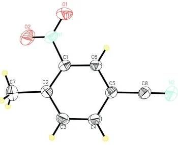
The crystal data show that in the title compound (Fig. 1), the benzene ring and the nitro group are not coplanar, they are
twisted with respect to each other by torsion angles of O1—N1—C1—C6 (-23.2 (4)°) and O2—N1—C1—C2
(-25.6 (3)°); the nitrile group C8≡N2 bond length of 1.144 (3) Å is within the normal range. The crystal structure is
stabilized only by van der Waals interactions.
S2. Experimental
The purchased p-methyl-m-nitrobenzonitrile (3 mmol, 486.44 mg) was dissolved in chloroform (20 ml) and evaporated in
air, affording colorless block crystals of this compound suitable for X-ray analysis.
S3. Refinement
All H atoms bonded to C atoms were fixed geometrically and treated as riding with C—H = 0.93 Å (aromatic), C—H =
Figure 1
A view of the molecular structure of the title compound, with the atomic numbering scheme. Displacement ellipsoids are
drawn at the 30% probability level. H atoms are represented by spheres of arbitrary radius.
(I)
Crystal data
C8H6N2O2
Mr = 162.15 Monoclinic, P21/c
Hall symbol: -P 2ybc
a = 3.9088 (8) Å
b = 13.576 (3) Å
c = 14.819 (4) Å
β = 99.13 (3)°
V = 776.4 (3) Å3
Z = 4
F(000) = 336
Dx = 1.387 Mg m−3
Mo Kα radiation, λ = 0.71073 Å Cell parameters from 1764 reflections
θ = 3.1–27.6°
µ = 0.10 mm−1
T = 298 K Block, colourless 0.35 × 0.30 × 0.1 mm
Data collection
Rigaku Mercury2 diffractometer
Radiation source: fine-focus sealed tube Graphite monochromator
Detector resolution: 13.6612 pixels mm-1
ω scans
Absorption correction: multi-scan
(CrystalClear; Rigaku/MSC, 2005)
Tmin = 0.965, Tmax = 0.990
7589 measured reflections 1761 independent reflections 1336 reflections with I > 2σ(I)
Rint = 0.037
θmax = 27.5°, θmin = 3.0°
h = −5→5
k = −17→17
Refinement
Refinement on F2 Least-squares matrix: full
R[F2 > 2σ(F2)] = 0.068
wR(F2) = 0.208
S = 1.10 1753 reflections 109 parameters 0 restraints
Primary atom site location: structure-invariant direct methods
Secondary atom site location: difference Fourier map
Hydrogen site location: inferred from neighbouring sites
H-atom parameters constrained
w = 1/[σ2(F
o2) + (0.1036P)2 + 0.2336P] where P = (Fo2 + 2Fc2)/3
(Δ/σ)max < 0.001 Δρmax = 0.33 e Å−3 Δρmin = −0.28 e Å−3
Special details
Geometry. All e.s.d.'s (except the e.s.d. in the dihedral angle between two l.s. planes) are estimated using the full covariance matrix. The cell e.s.d.'s are taken into account individually in the estimation of e.s.d.'s in distances, angles and torsion angles; correlations between e.s.d.'s in cell parameters are only used when they are defined by crystal symmetry. An approximate (isotropic) treatment of cell e.s.d.'s is used for estimating e.s.d.'s involving l.s. planes.
Refinement. Refinement of F2 against ALL reflections. The weighted R-factor wR and goodness of fit S are based on F2, conventional R-factors R are based on F, with F set to zero for negative F2. The threshold expression of F2 > σ(F2) is used only for calculating R-factors(gt) etc. and is not relevant to the choice of reflections for refinement. R-factors based on F2 are statistically about twice as large as those based on F, and R- factors based on ALL data will be even larger.
Fractional atomic coordinates and isotropic or equivalent isotropic displacement parameters (Å2)
x y z Uiso*/Ueq
O1 1.0921 (7) 0.79875 (18) 0.59741 (14) 0.1001 (9) O2 1.3658 (7) 0.89476 (18) 0.52048 (17) 0.0932 (8) N1 1.1627 (5) 0.82762 (16) 0.52533 (14) 0.0584 (6) N2 0.5214 (8) 0.45178 (18) 0.38967 (18) 0.0803 (8) C1 1.0021 (5) 0.77768 (15) 0.44045 (14) 0.0444 (5) C8 0.6195 (7) 0.53049 (18) 0.38358 (16) 0.0568 (6) C2 0.9631 (5) 0.82620 (16) 0.35611 (15) 0.0462 (5) C6 0.8914 (6) 0.68242 (16) 0.45126 (14) 0.0470 (5) H6 0.9185 0.6535 0.5088 0.056* C4 0.6963 (6) 0.67560 (17) 0.28898 (15) 0.0530 (6) H4 0.5942 0.6412 0.2374 0.064* C5 0.7388 (6) 0.63055 (16) 0.37444 (15) 0.0463 (5) C3 0.8057 (7) 0.77130 (18) 0.28090 (16) 0.0559 (6) H3 0.7738 0.8004 0.2234 0.067* C7 1.0660 (8) 0.93151 (18) 0.3406 (2) 0.0672 (7) H7A 1.1700 0.9601 0.3976 0.101* H7B 0.8641 0.9687 0.3157 0.101* H7C 1.2293 0.9326 0.2985 0.101*
Atomic displacement parameters (Å2)
U11 U22 U33 U12 U13 U23
N2 0.106 (2) 0.0562 (14) 0.0755 (16) −0.0203 (13) 0.0039 (14) −0.0061 (11) C1 0.0410 (10) 0.0476 (12) 0.0436 (11) 0.0013 (9) 0.0041 (8) −0.0064 (9) C8 0.0662 (15) 0.0501 (13) 0.0521 (14) −0.0036 (11) 0.0029 (11) −0.0063 (10) C2 0.0447 (11) 0.0456 (11) 0.0496 (12) 0.0061 (9) 0.0119 (9) 0.0026 (9) C6 0.0517 (12) 0.0478 (12) 0.0402 (11) 0.0010 (9) 0.0034 (9) 0.0011 (9) C4 0.0619 (14) 0.0521 (13) 0.0417 (12) 0.0062 (10) −0.0014 (10) −0.0048 (9) C5 0.0483 (12) 0.0446 (11) 0.0452 (12) 0.0015 (9) 0.0049 (8) −0.0036 (9) C3 0.0708 (16) 0.0543 (13) 0.0415 (12) 0.0083 (11) 0.0058 (10) 0.0059 (9) C7 0.0707 (17) 0.0504 (14) 0.0807 (19) −0.0005 (12) 0.0125 (14) 0.0096 (12)
Geometric parameters (Å, º)
O1—N1 1.210 (3) C6—C5 1.390 (3) O2—N1 1.218 (3) C6—H6 0.9300 N1—C1 1.478 (3) C4—C3 1.379 (3) N2—C8 1.144 (3) C4—C5 1.392 (3) C1—C6 1.381 (3) C4—H4 0.9300 C1—C2 1.400 (3) C3—H3 0.9300 C8—C5 1.450 (3) C7—H7A 0.9600 C2—C3 1.400 (3) C7—H7B 0.9600 C2—C7 1.513 (3) C7—H7C 0.9600
O1—N1—O2 122.4 (2) C3—C4—H4 120.0 O1—N1—C1 118.5 (2) C5—C4—H4 120.0 O2—N1—C1 119.1 (2) C6—C5—C4 119.7 (2) C6—C1—C2 123.55 (19) C6—C5—C8 120.1 (2) C6—C1—N1 115.43 (19) C4—C5—C8 120.2 (2) C2—C1—N1 121.0 (2) C4—C3—C2 122.4 (2) N2—C8—C5 178.9 (3) C4—C3—H3 118.8 C3—C2—C1 115.6 (2) C2—C3—H3 118.8 C3—C2—C7 118.4 (2) C2—C7—H7A 109.5 C1—C2—C7 125.9 (2) C2—C7—H7B 109.5 C1—C6—C5 118.75 (19) H7A—C7—H7B 109.5 C1—C6—H6 120.6 C2—C7—H7C 109.5 C5—C6—H6 120.6 H7A—C7—H7C 109.5 C3—C4—C5 119.9 (2) H7B—C7—H7C 109.5