Bis(5-phenyl-1
H
-1,2,4-triazol-3-yl)
disulfide dihydrate
Ai-Xin Zhu,a* Jun-Na Liu,bZhen Li,aHong-Can Wangaand Yuan-Chao Dua
aFaculty of Chemistry and Chemical Engineering, Yunnan Normal University, Kunming 650092, People’s Republic of China, andbSchool of Chemical Engineering, Henan University of Science and Technology, Luoyang 471003, People’s Republic of China
Correspondence e-mail: zaxchem@126.com
Received 2 April 2011; accepted 19 April 2011
Key indicators: single-crystal X-ray study;T= 293 K; mean(C–C) = 0.003 A˚; Rfactor = 0.042;wRfactor = 0.120; data-to-parameter ratio = 16.6.
A crystallographic twofold axis passing through the centre of the disulfide linkage in the title compound, C16H12N6S22H2O,
results in one-half of the molecule and one uncoordinated water molecule described in the asymmetric unit. In the molecule, the mean planes of the benzene and triazole rings are close to being coplanar and are separated by a dihedral angle of 2.08 (15). The triazole rings are twisted by a dihedral
angle of 37.67 (6) from the disulfide linkage. The crystal
packing is stabilized by intermolecular N—H O and O— H N hydrogen bonds with the water molecules, forming a three-dimensional supramolecular network.
Related literature
For applications of 1,2,4-triazole and its derivatives in coor-dination chemistry, see: Zhang et al. (2005); Ouelletteet al.
(2007); Zhuet al.(2009). For the related structure of a 1,2,4-triazole-based disulfide compound, see: Jianget al.(2007). For the previous synthesis of the title compound, see: El-Wareth & Sarhan (2000).
Experimental
Crystal data
C16H12N6S22H2O Mr= 388.47
Monoclinic,C2=c a= 12.3911 (13) A˚
b= 14.7125 (16) A˚
c= 10.2966 (11) A˚ = 104.125 (2)
V= 1820.4 (3) A˚3
Z= 4
MoKradiation = 0.32 mm1
T= 293 K
0.400.200.18 mm
Data collection
Bruker SMART APEX CCD diffractometer
Absorption correction: multi-scan (SADABS; Sheldrick, 1996)
Tmin= 0.884,Tmax= 0.945
7210 measured reflections 1953 independent reflections 1679 reflections withI> 2(I)
Rint= 0.023
Refinement
R[F2> 2(F2)] = 0.042
wR(F2) = 0.120
S= 1.06 1953 reflections
118 parameters
H-atom parameters constrained
max= 0.20 e A˚
3
min=0.18 e A˚
3
Table 1
Hydrogen-bond geometry (A˚ ,).
D—H A D—H H A D A D—H A
O1—H1C N3 0.90 2.02 2.9210 (19) 178 N1—H1B O1i
0.86 1.90 2.7077 (19) 156 O1—H1D N2ii
0.84 2.07 2.909 (2) 171
Symmetry codes: (i)xþ1 2;yþ
1 2;zþ
1
2; (ii)x;yþ1;zþ 1 2.
Data collection:SMART(Bruker, 2004); cell refinement:SAINT (Bruker, 2004); data reduction:SAINT; program(s) used to solve structure:SHELXS97(Sheldrick, 2008); program(s) used to refine structure: SHELXL97 (Sheldrick, 2008); molecular graphics: DIAMOND(Brandenburg, 1999); software used to prepare material for publication:SHELXTL(Sheldrick, 2008).
The authors thank the Youth Foundation of Yunnan Normal University (grant No. 10QZ02), the Science Founda-tion of the EducaFounda-tion Department of Yunnan Province (grant No. 2010Y004) and Henan University of Science and Tech-nology for supporting this work.
Supplementary data and figures for this paper are available from the IUCr electronic archives (Reference: JJ2087).
References
Brandenburg, K. (1999).DIAMOND. Crystal Impact GbR, Bonn, Germany. Bruker (2004).SMARTandSAINT. Bruker AXS Inc., Madison, Winconsin,
USA.
El-Wareth, A. & Sarhan, A. O. (2000).Heteroat. Chem.11, 399–402. Jiang, W.-Q., Liu, T.-B., Zou, J.-P. & Zhang, Y. (2007).Chin. J. Struct. Chem.26,
445–449.
Ouellette, W., Prosvirin, A. V., Valeich, J., Dunbar, K. R. & Zubieta, J. (2007).
Inorg. Chem.46, 9067–9082.
Sheldrick, G. M. (1996).SADABS. University of Go¨ttingen, Germany. Sheldrick, G. M. (2008).Acta Cryst.A64, 112–122.
Zhang, J.-P., Lin, Y.-Y., Huang, X.-C. & Chen, X.-M. (2005).J. Am. Chem. Soc.
127, 5495–5506.
Zhu, A.-X., Lin, J.-B., Zhang, J.-P. & Chen, X.-M. (2009).Inorg. Chem.48, 3882–3889.
Acta Crystallographica Section E Structure Reports Online
supporting information
Acta Cryst. (2011). E67, o1208 [doi:10.1107/S1600536811014607]
Bis(5-phenyl-1H-1,2,4-triazol-3-yl) disulfide dihydrate
Ai-Xin Zhu, Jun-Na Liu, Zhen Li, Hong-Can Wang and Yuan-Chao Du
S1. Comment
In the past few years, 1,2,4-triazole and its derivatives have attracted increasing attention as an N-heterocyclic aromatic
ligand, since they can combine both imidazoles and pyrazoles in their coordination geometry. In addition, metal-triazolate
frameworks can exhibit special luminescent, magnetic and favourable gas-adsorption abilities (Ouellette et al., 2007;
Zhang et al., 2005; Zhu et al., 2009). 1,2,4-triazole based thiols and disulfides are important 1,2,4-triazole derivatives,
and may exhibit a more diverse coordination geometry by combining heterocyclic nitrogen and sulfur donor atoms, and
therefore affect biological activity behaviour. However, only one example of a crystallographic study on organic
1,2,4-triazole based disulfide compounds is found in the literature (Jiang et al. 2007). Although the synthesis of the compound
1,2-bis(5-phenyl-1H-1,2,4-triazol-3-yl)disulfide has been reported by El-Wareth & Sarhan (2000), no crystallographic
study has been reported on the ligand and related metal coordination compounds. We reported herein another synthetic
method and the crystal structure of the title compound.
A crystallographic 2-fold axis passing through the centroid of the disulfide linkage in the title compound,
C16H12N6S2.2H2O, results in one-half of the molecule and one uncoordinated water molecule described in the asymmetric
unit (Fig. 1). In the molecule, the mean planes of the benzene and triazole rings are close to coplanar, separated by a
dihedral angle of 2.08 (15)°. The triazole rings are twisted by a dihedral angle of 37.67 (6)° from the disulfide linkage.
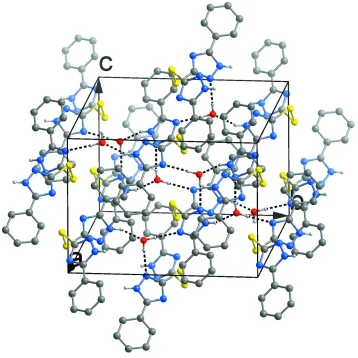
Crystal packing is stabilized by intermolecular N—H···O and O—H···N hydrogen bonds with the water molecules
forming a three-dimensional supramolecular network (Fig. 2).
S2. Experimental
A mixture of iron dichloride tetrahydrate (40 mg, 0.2 mmol), 3-phenyl-1H-1,2,4-triazole-5(4H)-thione (35 mg, 0.2
mmol), 8 ml methanol and 4 ml acetonitrile was stirred for 10 min, then filtered and allowed to stand at room temperature
for about two weeks. Yellow polyhedron crystals suitable for X-ray diffraction were obtained.
S3. Refinement
All H atoms were placed in idealized positions (O—H = 0.85 Å, N—H = 0.86 Å and C—H = 0.95 Å) and refined as
Figure 1
Molecular structure of the title compound, with atom labels and 30% probability displacement ellipsoids for non-H
atoms.
Figure 2
Packing diagram of the title compound viewed down the a axis. O—H···N and N—H···O hydrogen bonds with the water
[image:3.610.127.485.280.638.2]5-phenyl-3-[(5-phenyl-1H-1,2,4-triazol-3-yl)disulfanyl]-1H-1,2,4-triazole dihydrate
Crystal data
C16H12N6S2·2H2O Mr = 388.47 Monoclinic, C2/c
Hall symbol: -C 2yc
a = 12.3911 (13) Å
b = 14.7125 (16) Å
c = 10.2966 (11) Å
β = 104.125 (2)°
V = 1820.4 (3) Å3 Z = 4
F(000) = 808
Dx = 1.417 Mg m−3
Mo Kα radiation, λ = 0.71073 Å Cell parameters from 3134 reflections
θ = 2.7–26.4°
µ = 0.32 mm−1 T = 293 K
Polyhedron, yellow 0.40 × 0.20 × 0.18 mm
Data collection
Bruker SMART APEX CCD diffractometer
Radiation source: fine-focus sealed tube Graphite monochromator
ω scans
Absorption correction: multi-scan (SADABS; Sheldrick, 1996)
Tmin = 0.884, Tmax = 0.945
7210 measured reflections 1953 independent reflections 1679 reflections with I > 2σ(I)
Rint = 0.023
θmax = 27.0°, θmin = 2.2°
h = −15→15
k = −18→18
l = −13→13
Refinement
Refinement on F2
Least-squares matrix: full
R[F2 > 2σ(F2)] = 0.042 wR(F2) = 0.120 S = 1.06 1953 reflections 118 parameters 0 restraints
Primary atom site location: structure-invariant direct methods
Secondary atom site location: difference Fourier map
Hydrogen site location: inferred from neighbouring sites
H-atom parameters constrained
w = 1/[σ2(Fo2) + (0.0691P)2 + 0.5857P]
where P = (Fo2 + 2Fc2)/3
(Δ/σ)max < 0.001
Δρmax = 0.20 e Å−3
Δρmin = −0.18 e Å−3
Special details
Geometry. All e.s.d.'s (except the e.s.d. in the dihedral angle between two l.s. planes) are estimated using the full covariance matrix. The cell e.s.d.'s are taken into account individually in the estimation of e.s.d.'s in distances, angles and torsion angles; correlations between e.s.d.'s in cell parameters are only used when they are defined by crystal symmetry. An approximate (isotropic) treatment of cell e.s.d.'s is used for estimating e.s.d.'s involving l.s. planes.
Refinement. Refinement of F2 against ALL reflections. The weighted R-factor wR and goodness of fit S are based on F2,
conventional R-factors R are based on F, with F set to zero for negative F2. The threshold expression of F2 > σ(F2) is used
only for calculating R-factors(gt) etc. and is not relevant to the choice of reflections for refinement. R-factors based on F2
are statistically about twice as large as those based on F, and R-factors based on ALL data will be even larger.
Fractional atomic coordinates and isotropic or equivalent isotropic displacement parameters (Å2)
x y z Uiso*/Ueq
S1 0.43278 (4) 0.48004 (3) 0.16853 (5) 0.0605 (2)
N1 0.26234 (13) 0.68205 (9) 0.22572 (14) 0.0535 (4)
H1B 0.2373 0.7366 0.2112 0.064*
N3 0.28640 (12) 0.54543 (9) 0.30878 (14) 0.0501 (3)
C1 0.13764 (18) 0.58140 (14) 0.4862 (2) 0.0673 (5)
H1A 0.1698 0.5240 0.4897 0.081*
C2 0.0682 (2) 0.60203 (17) 0.5693 (2) 0.0792 (6)
H2A 0.0543 0.5586 0.6288 0.095*
C3 0.01979 (18) 0.68669 (16) 0.5640 (2) 0.0729 (6)
H3A −0.0284 0.6999 0.6180 0.087*
C4 0.0428 (2) 0.75119 (16) 0.4792 (2) 0.0723 (6)
H4A 0.0115 0.8088 0.4773 0.087*
C5 0.11194 (17) 0.73153 (13) 0.3963 (2) 0.0615 (5)
H5A 0.1270 0.7759 0.3388 0.074*
C6 0.15919 (13) 0.64584 (11) 0.39834 (16) 0.0478 (4)
C7 0.23446 (14) 0.62407 (11) 0.31284 (16) 0.0461 (4)
C8 0.34687 (14) 0.56106 (11) 0.21779 (17) 0.0503 (4)
O1 0.27936 (14) 0.35247 (9) 0.37303 (13) 0.0762 (5)
H1D 0.3018 0.3501 0.4572 0.091*
H1C 0.2833 0.4118 0.3533 0.091*
Atomic displacement parameters (Å2)
U11 U22 U33 U12 U13 U23
S1 0.0728 (4) 0.0462 (3) 0.0721 (3) −0.00574 (19) 0.0364 (3) −0.01332 (19)
N1 0.0701 (9) 0.0405 (7) 0.0563 (8) 0.0046 (6) 0.0276 (7) 0.0048 (6)
N2 0.0731 (9) 0.0448 (8) 0.0561 (8) −0.0012 (7) 0.0305 (7) 0.0014 (6)
N3 0.0584 (8) 0.0412 (7) 0.0564 (8) −0.0014 (6) 0.0248 (6) 0.0027 (6)
C1 0.0780 (13) 0.0589 (11) 0.0745 (12) 0.0101 (9) 0.0371 (10) 0.0135 (9)
C2 0.0917 (16) 0.0835 (15) 0.0757 (14) 0.0031 (12) 0.0463 (12) 0.0154 (11)
C3 0.0697 (13) 0.0864 (15) 0.0716 (13) 0.0043 (11) 0.0347 (10) −0.0062 (11)
C4 0.0757 (13) 0.0681 (13) 0.0813 (14) 0.0161 (10) 0.0347 (11) −0.0006 (10)
C5 0.0693 (12) 0.0539 (10) 0.0671 (11) 0.0098 (9) 0.0278 (9) 0.0082 (8)
C6 0.0478 (8) 0.0494 (9) 0.0477 (8) −0.0011 (7) 0.0142 (7) 0.0003 (7)
C7 0.0517 (9) 0.0405 (8) 0.0476 (8) −0.0031 (6) 0.0150 (7) 0.0014 (6)
C8 0.0596 (10) 0.0420 (8) 0.0543 (9) −0.0052 (7) 0.0233 (7) −0.0033 (7)
O1 0.1323 (14) 0.0421 (7) 0.0576 (8) −0.0052 (7) 0.0300 (8) −0.0033 (5)
Geometric parameters (Å, º)
S1—C8 1.7536 (17) C2—C3 1.378 (3)
S1—S1i 2.0556 (11) C2—H2A 0.9300
N1—C7 1.343 (2) C3—C4 1.366 (3)
N1—N2 1.346 (2) C3—H3A 0.9300
N1—H1B 0.8600 C4—C5 1.380 (3)
N2—C8 1.321 (2) C4—H4A 0.9300
N3—C7 1.330 (2) C5—C6 1.388 (2)
N3—C8 1.355 (2) C5—H5A 0.9300
C1—C6 1.381 (2) C6—C7 1.466 (2)
C1—C2 1.386 (3) O1—H1D 0.8434
C8—S1—S1i 101.08 (6) C3—C4—H4A 119.7
C7—N1—N2 110.73 (14) C5—C4—H4A 119.7
C7—N1—H1B 124.6 C4—C5—C6 120.28 (19)
N2—N1—H1B 124.6 C4—C5—H5A 119.9
C8—N2—N1 102.31 (13) C6—C5—H5A 119.9
C7—N3—C8 103.16 (14) C1—C6—C5 119.04 (17)
C6—C1—C2 120.15 (19) C1—C6—C7 119.78 (16)
C6—C1—H1A 119.9 C5—C6—C7 121.14 (16)
C2—C1—H1A 119.9 N3—C7—N1 109.05 (14)
C3—C2—C1 120.2 (2) N3—C7—C6 126.37 (15)
C3—C2—H2A 119.9 N1—C7—C6 124.58 (15)
C1—C2—H2A 119.9 N2—C8—N3 114.73 (15)
C4—C3—C2 119.78 (19) N2—C8—S1 121.06 (13)
C4—C3—H3A 120.1 N3—C8—S1 124.19 (13)
C2—C3—H3A 120.1 H1D—O1—H1C 104.4
C3—C4—C5 120.5 (2)
Symmetry code: (i) −x+1, y, −z+1/2.
Hydrogen-bond geometry (Å, º)
D—H···A D—H H···A D···A D—H···A
O1—H1C···N3 0.90 2.02 2.9210 (19) 178
N1—H1B···O1ii 0.86 1.90 2.7077 (19) 156
O1—H1D···N2iii 0.84 2.07 2.909 (2) 171