(3-Phenylsulfanyl-1-phenylsulfonyl-1
H
-indol-2-yl)methyl acetate
Alagappa Rammohan,aE. Govindan,aA. SubbiahPandi,a* R. Sureshbabuband A. K. Mohana Krishnanb
aDepartment of Physics, Presidency College (Autonomous), Chennai 600 005, India, andbDepartment of Organic Chemistry, University of Madras, Guindy Campus, Chennai 600 025, India
Correspondence e-mail: a_sp59@yahoo.in
Received 11 April 2011; accepted 20 April 2011
Key indicators: single-crystal X-ray study;T= 293 K; mean(C–C) = 0.003 A˚; Rfactor = 0.039;wRfactor = 0.104; data-to-parameter ratio = 19.2.
In the title compound, C23H19NO4S2, the indole ring system
makes dihedral angles of 89.6 (1) and 84.5 (8) with the
phenylsulfonyl and phenylsulfanyl rings, respectively. In the crystal, the molecules are linked into C(10) chains running along the c axis by an intermolecular C—H O hydrogen bond. In addition, the crystal packing is stabilized by C— H interactions.
Related literature
For biological activities of indole derivatives, see: Singhet al.
(2000); Andreani et al. (2001); Quetin-Leclercq (1994); Mukhopadhyayet al.(1981); Tayloret al.(1999); Williamset al.
(1993); Sivaraman et al. (1996). For related structures, see: Ravishankar et al.(2005); Chakkaravarthi et al.(2008). For graph-set notation of hydrogen bonds, see: Bernstein et al.
(1995).
Experimental
Crystal data
C23H19NO4S2
Mr= 437.51 Monoclinic,P21=c
a= 14.6530 (6) A˚
b= 9.4482 (4) A˚
c= 15.2461 (7) A˚
= 97.055 (3)
V= 2094.76 (16) A˚3
Z= 4
MoKradiation
= 0.29 mm1
T= 293 K
0.250.220.19 mm
Data collection
Bruker APEXII CCD area-detector diffractometer
Absorption correction: multi-scan (SADABS; Sheldrick, 1996)
Tmin= 0.981,Tmax= 0.985
19397 measured reflections 5235 independent reflections 3638 reflections withI> 2(I)
Rint= 0.027
Refinement
R[F2> 2(F2)] = 0.039
wR(F2) = 0.104
S= 1.03 5235 reflections
272 parameters
H-atom parameters constrained max= 0.26 e A˚
3
min=0.29 e A˚
[image:1.610.314.565.308.355.2]3
Table 1
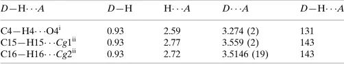
Hydrogen-bond geometry (A˚ ,).
Cg1 and Cg2 are the centroids of the N1/C1/C6–C8 and C1–C6 rings, respectively.
D—H A D—H H A D A D—H A
C4—H4 O4i 0.93 2.59 3.274 (2) 131
C15—H15 Cg1ii
0.93 2.77 3.559 (2) 143
C16—H16 Cg2ii
0.93 2.72 3.5146 (19) 143
Symmetry codes: (i)x;yþ3 2;zþ
1
2; (ii)x;yþ 1 2;z
3 2.
Data collection:APEX2(Bruker, 2004); cell refinement:APEX2; data reduction: SAINT (Bruker, 2004); program(s) used to solve structure:SHELXS97(Sheldrick, 2008); program(s) used to refine structure: SHELXL97 (Sheldrick, 2008); molecular graphics:
ORTEP-3(Farrugia, 1997); software used to prepare material for publication:SHELXL97andPLATON(Spek, 2009).
EG and ASP thank Dr Babu Vargheese, SAIF, IIT, Madras, India, for the X-ray intensity data collection.
Supplementary data and figures for this paper are available from the IUCr electronic archives (Reference: BT5514).
References
Andreani, A., Granaiola, M., Leoni, A., Locatelli, A., Morigi, R., Rambaldi, M., Giorgi, G., Salvini, L. & Garaliene, V. (2001).Anticancer Drug Des.16, 167–174.
Bernstein, J., Davis, R. E., Shimoni, L. & Chang, N.-L. (1995).Angew. Chem. Int. Ed. Engl.34, 1555–1573.
Bruker (2004).SAINTandSMART. Bruker AXS Inc., Madison, Wisconsin, USA.
Chakkaravarthi, G., Dhayalan, V., Mohanakrishnan, A. K. & Manivannan, V. (2008).Acta Cryst.E64, o542.
Farrugia, L. J. (1997).J. Appl. Cryst.30, 565.
Mukhopadhyay, S., Handy, G. A., Funayama, S. & Cordell, G. A. (1981).J. Nat. Prod.44, 696–700.
Quetin-Leclercq, J. (1994).J. Pharm. Belg.49, 181–192.
Ravishankar, T., Chinnakali, K., Arumugam, N., Srinivasan, P. C., Usman, A. & Fun, H.-K. (2005).Acta Cryst.E61, o2455–o2457.
Sheldrick, G. M. (1996).SADABS. University of Go¨ttingen, Germany. Sheldrick, G. M. (2008).Acta Cryst.A64, 112–122.
Singh, U. P., Sarma, B. K., Mishra, P. K. & Ray, A. B. (2000).Folia Microbiol. (Praha),45, 173–176.
Sivaraman, J., Subramanian, K., Velmurugan, D., Subramanian, E. & Seetharaman, J. (1996).J. Mol. Struct.385, 123–128.
Spek, A. L. (2009).Acta Cryst.D65, 148–155.
organic compounds
o1240
Rammohanet al. doi:10.1107/S1600536811014802 Acta Cryst.(2011). E67, o1240–o1241Acta Crystallographica Section E Structure Reports
Online
Falardeau, G., Lavallee, J. F., Brown, W., Rando, R. F. & Bowlin, T. (1999).
Antivir. Chem. Chemother.10, 79–86.
supporting information
sup-1
Acta Cryst. (2011). E67, o1240–o1241
supporting information
Acta Cryst. (2011). E67, o1240–o1241 [doi:10.1107/S1600536811014802]
(3-Phenylsulfanyl-1-phenylsulfonyl-1
H
-indol-2-yl)methyl acetate
Alagappa Rammohan, E. Govindan, A. SubbiahPandi, R. Sureshbabu and A. K. Mohana Krishnan
S1. Comment
Indole derivatives have been found to exhibit antibacterial, antifungal (Singh et al., 2000) and antitumour activities
(Andreani et al., 2001). Some of the indole alkaloids extracted from plants possess interesting cytotoxic, antitumour or
antiparasitic properties (Quetin-Leclercq, 1994; Mukhopadhyay et al., 1981). Pyrido[1,2-a]indole derivatives have been
identified as potent inhibitors of human immunodeficiency virus type 1 (Taylor et al., 1999), and
5-chloro-3-(phenyl-sulfonyl)indole-2-carboxamide is reported to be a highly potent non-nucleoside inhibitor of HIV-1 reverse transcriptase
(Williams et al.,1993). The interaction of phenylsulfonylindole with calf thymus DNA has also been studied by
spectroscopic methods (Sivaraman et al., 1996). Against this background, and in order to obtain detailed information on
molecular conformations in the solid state, X-ray studies of the title compound (I) have been carried out.
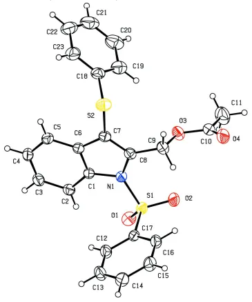
X-Ray analysis confirms the molecular structure and atom connectivity for (I), as illustrated in Fig. 1. The indole ring
system is essentially planar, with maximum deviation of 0.020 (2) Å for atom N1. The mean planes of the indole ring
system make a dihedral algles of 89.6 (1) and 84.5 (8)° with respect to the phenyl rings, it shows that both the phenyl
rings are perpendicular with respect to the indole ring system. The S—O, S—C, and S—N distances are 1.420 (12),
1.754 (17) and 1.676 (14) Å, respectively, these are comparable as observed in similar structures (Ravishankar et al.,
2005). As a result of the electron-withdrawing character of the phenylsulfonyl group, the N—Csp2 bond lengths, viz. N1
—C1 [1.422 (2) Å] and N1—C8 [1.418 (2) Å], are longer than the mean value of 1.355 (14) Å reported for N atoms with
planar configurations.
The S atom exhibits significant deviation from that of a regular tetrahedron, with the largest deviations being seen for
the O—S—O [O1—S1—O2 120.3 (7)°] and O—S—N angles [O1—S1—N1 105.4 (7)°]. The widening of the angles
may be due to repulsive interactions between the two short S═O bonds, similar to what is observed in related structures
(Chakkaravarthi et al., 2008). The atom C4 act as a donor to the atom O4 of the neighbouring molecule at (x, 3/2 - y, 1/2
+ z). This hydrogen bond is involved in a motif C(10) chain along b axis. In addition to van der Waals interaction, the
crystal packing is stabilized by C—H..O and C—H···π interactions.
S2. Experimental
To solution of 2-(bromomethyl)-1-(phenyl sulfonyl)-3-(phenylthio)-1H-indole (2.18 mmol) in dry dimethyl formamide
(10 ml), potassium acetate (4.36 mmol) was added under nitrogen atmosphere, the reaction mixture was stirred at room
temperature for 5 h, then it was poured over crushed ice (50 g) containing 1 ml of concentrated hydrochloric acid. The
obtained brown solid was filtered and dried. Single crystals of the title compound suitable for X-ray diffraction were
All H atoms were fixed geometrically and allowed to ride on their parent C atoms, with C—H distances fixed in the range
[image:4.610.123.488.118.556.2]0.93–0.97 Å with Uiso(H) = 1.5Ueq(C) for methyl H 1.2Ueq(C) for other H atoms.
Figure 1
View of the title molecule with the atom labeling scheme. The displacement ellipsoids are drawn at the 30% probability
supporting information
sup-3
[image:5.610.124.488.69.429.2]Acta Cryst. (2011). E67, o1240–o1241
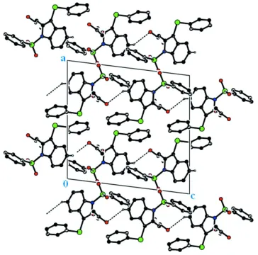
Figure 2
The molecular packing viewed down the b axis.
(3-Phenylsulfanyl-1-phenylsulfonyl-1H-indol-2-yl)methyl acetate
Crystal data
C23H19NO4S2
Mr = 437.51
Monoclinic, P21/c
Hall symbol: -P 2ybc
a = 14.6530 (6) Å
b = 9.4482 (4) Å
c = 15.2461 (7) Å
β = 97.055 (3)°
V = 2094.76 (16) Å3
Z = 4
F(000) = 912
Dx = 1.387 Mg m−3
Mo Kα radiation, λ = 0.71073 Å
Cell parameters from 5235 reflections
θ = 1.4–28.4°
µ = 0.29 mm−1
T = 293 K
Block, white
0.25 × 0.22 × 0.19 mm
Data collection
Bruker APEXII CCD area-detector diffractometer
Radiation source: fine-focus sealed tube Graphite monochromator
ω and φ scans
Absorption correction: multi-scan (SADABS; Sheldrick, 1996)
Tmin = 0.981, Tmax = 0.985
θmax = 28.4°, θmin = 1.4°
h = −19→19
l = −20→20
Refinement
Refinement on F2
Least-squares matrix: full
R[F2 > 2σ(F2)] = 0.039
wR(F2) = 0.104
S = 1.03
5235 reflections 272 parameters 0 restraints
Primary atom site location: structure-invariant direct methods
Secondary atom site location: difference Fourier map
Hydrogen site location: inferred from neighbouring sites
H-atom parameters constrained
w = 1/[σ2(F
o2) + (0.0433P)2 + 0.4791P]
where P = (Fo2 + 2Fc2)/3 (Δ/σ)max < 0.001
Δρmax = 0.26 e Å−3
Δρmin = −0.29 e Å−3
Special details
Geometry. All e.s.d.'s (except the e.s.d. in the dihedral angle between two l.s. planes) are estimated using the full covariance matrix. The cell e.s.d.'s are taken into account individually in the estimation of e.s.d.'s in distances, angles and torsion angles; correlations between e.s.d.'s in cell parameters are only used when they are defined by crystal symmetry. An approximate (isotropic) treatment of cell e.s.d.'s is used for estimating e.s.d.'s involving l.s. planes.
Refinement. Refinement of F2 against ALL reflections. The weighted R-factor wR and goodness of fit S are based on F2,
conventional R-factors R are based on F, with F set to zero for negative F2. The threshold expression of F2 > σ(F2) is used
only for calculating R-factors(gt) etc. and is not relevant to the choice of reflections for refinement. R-factors based on F2
are statistically about twice as large as those based on F, and R-factors based on ALL data will be even larger.
Fractional atomic coordinates and isotropic or equivalent isotropic displacement parameters (Å2)
x y z Uiso*/Ueq
C1 0.16888 (11) 0.62556 (16) 0.35882 (10) 0.0387 (4)
C2 0.09311 (12) 0.55131 (18) 0.38072 (11) 0.0463 (4)
H2 0.0349 0.5645 0.3500 0.056*
C3 0.10805 (13) 0.45722 (19) 0.44998 (12) 0.0530 (4)
H3 0.0586 0.4057 0.4660 0.064*
C4 0.19466 (14) 0.43671 (19) 0.49671 (12) 0.0543 (5)
H4 0.2019 0.3728 0.5434 0.065*
C5 0.26938 (13) 0.50947 (18) 0.47475 (11) 0.0482 (4)
H5 0.3273 0.4959 0.5061 0.058*
C6 0.25666 (11) 0.60461 (16) 0.40419 (10) 0.0410 (4)
C7 0.31844 (11) 0.69850 (17) 0.36612 (11) 0.0424 (4)
C8 0.27026 (11) 0.77336 (17) 0.30037 (11) 0.0422 (4)
C9 0.30417 (12) 0.89164 (18) 0.24908 (12) 0.0494 (4)
H9A 0.3708 0.8898 0.2536 0.059*
H9B 0.2795 0.8842 0.1872 0.059*
C10 0.28751 (13) 1.14037 (19) 0.24295 (13) 0.0534 (4)
C11 0.2490 (2) 1.2651 (2) 0.28528 (19) 0.0882 (8)
H11A 0.2619 1.3495 0.2540 0.132*
H11B 0.2765 1.2722 0.3456 0.132*
H11C 0.1837 1.2541 0.2836 0.132*
C12 0.07661 (13) 0.49985 (18) 0.14300 (12) 0.0522 (4)
supporting information
sup-5
Acta Cryst. (2011). E67, o1240–o1241
C13 0.08604 (15) 0.3989 (2) 0.08013 (13) 0.0622 (5)
H13 0.0656 0.3071 0.0880 0.075*
C14 0.12545 (14) 0.4326 (2) 0.00578 (13) 0.0600 (5)
H14 0.1311 0.3637 −0.0368 0.072*
C15 0.15663 (14) 0.5672 (2) −0.00607 (12) 0.0598 (5)
H15 0.1831 0.5894 −0.0567 0.072*
C16 0.14880 (12) 0.66998 (19) 0.05698 (11) 0.0506 (4)
H16 0.1703 0.7612 0.0495 0.061*
C17 0.10864 (10) 0.63531 (16) 0.13106 (10) 0.0391 (3)
C18 0.43670 (12) 0.7939 (2) 0.50554 (13) 0.0557 (5)
C19 0.39555 (14) 0.9229 (3) 0.51322 (15) 0.0681 (6)
H19 0.3656 0.9675 0.4634 0.082*
C20 0.39877 (17) 0.9870 (3) 0.59577 (18) 0.0861 (8)
H20 0.3692 1.0730 0.6018 0.103*
C21 0.4459 (2) 0.9222 (4) 0.66824 (17) 0.0938 (9)
H21 0.4489 0.9654 0.7233 0.113*
C22 0.48819 (18) 0.7960 (4) 0.66036 (17) 0.0896 (8)
H22 0.5203 0.7537 0.7099 0.108*
C23 0.48383 (15) 0.7297 (3) 0.57906 (15) 0.0736 (6)
H23 0.5123 0.6426 0.5739 0.088*
N1 0.17660 (9) 0.73225 (14) 0.29437 (9) 0.0405 (3)
O1 0.01013 (8) 0.74327 (13) 0.24414 (9) 0.0550 (3)
O2 0.11545 (9) 0.90024 (12) 0.17575 (8) 0.0561 (3)
O3 0.27314 (8) 1.02147 (12) 0.28681 (8) 0.0544 (3)
O4 0.32709 (11) 1.14258 (15) 0.17888 (9) 0.0712 (4)
S1 0.09482 (3) 0.76585 (4) 0.21008 (3) 0.04212 (12)
S2 0.43695 (3) 0.71174 (6) 0.40011 (3) 0.05871 (15)
Atomic displacement parameters (Å2)
U11 U22 U33 U12 U13 U23
C1 0.0493 (9) 0.0344 (8) 0.0324 (8) −0.0008 (7) 0.0050 (7) −0.0040 (7)
C2 0.0514 (10) 0.0450 (9) 0.0421 (10) −0.0070 (8) 0.0040 (8) −0.0032 (8)
C3 0.0662 (12) 0.0463 (10) 0.0473 (11) −0.0130 (9) 0.0105 (9) 0.0007 (8)
C4 0.0806 (13) 0.0408 (9) 0.0405 (10) −0.0069 (9) 0.0033 (9) 0.0048 (8)
C5 0.0604 (11) 0.0417 (9) 0.0404 (9) 0.0018 (8) −0.0027 (8) −0.0027 (8)
C6 0.0519 (9) 0.0353 (8) 0.0355 (9) 0.0005 (7) 0.0036 (7) −0.0066 (7)
C7 0.0454 (9) 0.0422 (9) 0.0397 (9) −0.0008 (7) 0.0057 (7) −0.0065 (7)
C8 0.0481 (9) 0.0408 (9) 0.0390 (9) −0.0030 (7) 0.0105 (7) −0.0067 (7)
C9 0.0580 (10) 0.0438 (9) 0.0487 (10) −0.0043 (8) 0.0162 (8) −0.0032 (8)
C10 0.0585 (11) 0.0451 (10) 0.0539 (12) −0.0092 (8) −0.0044 (9) 0.0015 (9)
C11 0.113 (2) 0.0501 (13) 0.102 (2) 0.0067 (12) 0.0184 (16) −0.0035 (13)
C12 0.0707 (12) 0.0442 (10) 0.0436 (10) −0.0074 (8) 0.0148 (9) 0.0013 (8)
C13 0.0913 (15) 0.0389 (10) 0.0581 (12) −0.0086 (10) 0.0158 (11) −0.0035 (9)
C14 0.0825 (14) 0.0500 (11) 0.0482 (11) 0.0101 (10) 0.0100 (10) −0.0079 (9)
C15 0.0806 (13) 0.0579 (12) 0.0446 (11) 0.0002 (10) 0.0220 (10) 0.0007 (9)
C16 0.0657 (11) 0.0422 (9) 0.0455 (10) −0.0056 (8) 0.0128 (9) 0.0043 (8)
C19 0.0653 (13) 0.0789 (15) 0.0596 (13) −0.0093 (11) 0.0061 (10) −0.0100 (11)
C20 0.0887 (17) 0.0921 (18) 0.0806 (18) −0.0225 (14) 0.0230 (14) −0.0285 (15)
C21 0.101 (2) 0.126 (3) 0.0559 (15) −0.0570 (19) 0.0151 (14) −0.0210 (17)
C22 0.0854 (18) 0.126 (2) 0.0537 (15) −0.0416 (17) −0.0083 (12) 0.0084 (16)
C23 0.0645 (13) 0.0873 (16) 0.0653 (15) −0.0191 (11) −0.0060 (11) 0.0076 (13)
N1 0.0465 (7) 0.0395 (7) 0.0350 (7) −0.0020 (6) 0.0034 (6) 0.0009 (6)
O1 0.0473 (7) 0.0625 (8) 0.0560 (8) 0.0126 (6) 0.0097 (6) −0.0015 (6)
O2 0.0733 (8) 0.0355 (6) 0.0579 (8) 0.0073 (6) 0.0010 (6) 0.0053 (6)
O3 0.0705 (8) 0.0421 (7) 0.0537 (7) −0.0073 (6) 0.0205 (6) −0.0047 (6)
O4 0.0962 (11) 0.0635 (9) 0.0545 (9) −0.0183 (8) 0.0113 (8) 0.0085 (7)
S1 0.0477 (2) 0.0375 (2) 0.0408 (2) 0.00722 (17) 0.00382 (18) 0.00013 (17)
S2 0.0446 (3) 0.0741 (3) 0.0573 (3) −0.0010 (2) 0.0058 (2) −0.0084 (3)
Geometric parameters (Å, º)
C1—C2 1.388 (2) C12—C17 1.383 (2)
C1—C6 1.398 (2) C12—H12 0.9300
C1—N1 1.422 (2) C13—C14 1.371 (3)
C2—C3 1.377 (2) C13—H13 0.9300
C2—H2 0.9300 C14—C15 1.371 (3)
C3—C4 1.391 (3) C14—H14 0.9300
C3—H3 0.9300 C15—C16 1.381 (3)
C4—C5 1.369 (2) C15—H15 0.9300
C4—H4 0.9300 C16—C17 1.376 (2)
C5—C6 1.397 (2) C16—H16 0.9300
C5—H5 0.9300 C17—S1 1.7532 (16)
C6—C7 1.440 (2) C18—C19 1.371 (3)
C7—C8 1.353 (2) C18—C23 1.382 (3)
C7—S2 1.7545 (17) C18—S2 1.785 (2)
C8—N1 1.418 (2) C19—C20 1.392 (3)
C8—C9 1.484 (2) C19—H19 0.9300
C9—O3 1.451 (2) C20—C21 1.373 (4)
C9—H9A 0.9700 C20—H20 0.9300
C9—H9B 0.9700 C21—C22 1.356 (4)
C10—O4 1.196 (2) C21—H21 0.9300
C10—O3 1.337 (2) C22—C23 1.383 (4)
C10—C11 1.488 (3) C22—H22 0.9300
C11—H11A 0.9600 C23—H23 0.9300
C11—H11B 0.9600 N1—S1 1.6763 (14)
C11—H11C 0.9600 O1—S1 1.4190 (12)
C12—C13 1.371 (3) O2—S1 1.4201 (12)
C2—C1—C6 121.60 (15) C12—C13—H13 119.8
C2—C1—N1 131.17 (15) C14—C13—H13 119.8
C6—C1—N1 107.21 (13) C15—C14—C13 120.33 (18)
C3—C2—C1 117.01 (17) C15—C14—H14 119.8
supporting information
sup-7
Acta Cryst. (2011). E67, o1240–o1241
C1—C2—H2 121.5 C14—C15—C16 120.22 (17)
C2—C3—C4 122.14 (17) C14—C15—H15 119.9
C2—C3—H3 118.9 C16—C15—H15 119.9
C4—C3—H3 118.9 C17—C16—C15 118.97 (16)
C5—C4—C3 120.78 (17) C17—C16—H16 120.5
C5—C4—H4 119.6 C15—C16—H16 120.5
C3—C4—H4 119.6 C16—C17—C12 120.98 (15)
C4—C5—C6 118.46 (17) C16—C17—S1 119.59 (13)
C4—C5—H5 120.8 C12—C17—S1 119.40 (12)
C6—C5—H5 120.8 C19—C18—C23 120.1 (2)
C5—C6—C1 120.00 (15) C19—C18—S2 120.85 (16)
C5—C6—C7 132.54 (16) C23—C18—S2 118.90 (18)
C1—C6—C7 107.41 (14) C18—C19—C20 119.8 (2)
C8—C7—C6 108.92 (14) C18—C19—H19 120.1
C8—C7—S2 126.09 (13) C20—C19—H19 120.1
C6—C7—S2 124.99 (13) C21—C20—C19 119.4 (3)
C7—C8—N1 108.51 (14) C21—C20—H20 120.3
C7—C8—C9 127.31 (15) C19—C20—H20 120.3
N1—C8—C9 123.79 (15) C22—C21—C20 120.7 (3)
O3—C9—C8 106.62 (12) C22—C21—H21 119.6
O3—C9—H9A 110.4 C20—C21—H21 119.6
C8—C9—H9A 110.4 C21—C22—C23 120.4 (3)
O3—C9—H9B 110.4 C21—C22—H22 119.8
C8—C9—H9B 110.4 C23—C22—H22 119.8
H9A—C9—H9B 108.6 C18—C23—C22 119.4 (3)
O4—C10—O3 123.03 (18) C18—C23—H23 120.3
O4—C10—C11 126.05 (19) C22—C23—H23 120.3
O3—C10—C11 110.92 (18) C8—N1—C1 107.94 (13)
C10—C11—H11A 109.5 C8—N1—S1 126.33 (11)
C10—C11—H11B 109.5 C1—N1—S1 123.64 (11)
H11A—C11—H11B 109.5 C10—O3—C9 115.82 (13)
C10—C11—H11C 109.5 O1—S1—O2 120.31 (7)
H11A—C11—H11C 109.5 O1—S1—N1 105.42 (7)
H11B—C11—H11C 109.5 O2—S1—N1 106.70 (7)
C13—C12—C17 119.13 (16) O1—S1—C17 109.01 (8)
C13—C12—H12 120.4 O2—S1—C17 109.15 (8)
C17—C12—H12 120.4 N1—S1—C17 105.16 (7)
C12—C13—C14 120.36 (17) C7—S2—C18 100.69 (8)
C6—C1—C2—C3 0.8 (2) C19—C20—C21—C22 −0.9 (4)
N1—C1—C2—C3 −177.37 (16) C20—C21—C22—C23 −0.5 (4)
C1—C2—C3—C4 0.2 (3) C19—C18—C23—C22 0.7 (3)
C2—C3—C4—C5 −0.6 (3) S2—C18—C23—C22 176.63 (16)
C3—C4—C5—C6 −0.1 (3) C21—C22—C23—C18 0.6 (3)
C4—C5—C6—C1 1.1 (2) C7—C8—N1—C1 −1.09 (17)
C4—C5—C6—C7 178.16 (17) C9—C8—N1—C1 −174.33 (14)
C2—C1—C6—C5 −1.5 (2) C7—C8—N1—S1 −165.03 (11)
N1—C1—C6—C7 −0.66 (16) C6—C1—N1—C8 1.06 (16)
C5—C6—C7—C8 −177.32 (17) C2—C1—N1—S1 −16.1 (2)
C1—C6—C7—C8 −0.01 (18) C6—C1—N1—S1 165.54 (11)
C5—C6—C7—S2 2.9 (3) O4—C10—O3—C9 −3.4 (3)
C1—C6—C7—S2 −179.79 (12) C11—C10—O3—C9 177.29 (17)
C6—C7—C8—N1 0.68 (18) C8—C9—O3—C10 −172.38 (15)
S2—C7—C8—N1 −179.54 (11) C8—N1—S1—O1 −163.03 (13)
C6—C7—C8—C9 173.61 (15) C1—N1—S1—O1 35.40 (14)
S2—C7—C8—C9 −6.6 (2) C8—N1—S1—O2 −34.03 (15)
C7—C8—C9—O3 −100.22 (19) C1—N1—S1—O2 164.40 (12)
N1—C8—C9—O3 71.71 (19) C8—N1—S1—C17 81.83 (14)
C17—C12—C13—C14 −1.0 (3) C1—N1—S1—C17 −79.75 (13)
C12—C13—C14—C15 0.6 (3) C16—C17—S1—O1 143.04 (14)
C13—C14—C15—C16 0.2 (3) C12—C17—S1—O1 −35.16 (16)
C14—C15—C16—C17 −0.6 (3) C16—C17—S1—O2 9.83 (16)
C15—C16—C17—C12 0.2 (3) C12—C17—S1—O2 −168.37 (14)
C15—C16—C17—S1 −177.98 (14) C16—C17—S1—N1 −104.33 (14)
C13—C12—C17—C16 0.6 (3) C12—C17—S1—N1 77.48 (15)
C13—C12—C17—S1 178.76 (15) C8—C7—S2—C18 110.04 (16)
C23—C18—C19—C20 −2.1 (3) C6—C7—S2—C18 −70.22 (16)
S2—C18—C19—C20 −177.95 (16) C19—C18—S2—C7 −58.87 (17)
C18—C19—C20—C21 2.2 (3) C23—C18—S2—C7 125.22 (16)
Hydrogen-bond geometry (Å, º)
Cg1 and Cg2 are the centroids of the N1/C1/C6–C8 and C1–C6 rings, respectively.
D—H···A D—H H···A D···A D—H···A
C4—H4···O4i 0.93 2.59 3.274 (2) 131
C15—H15···Cg1ii 0.93 2.77 3.559 (2) 143
C16—H16···Cg2ii 0.93 2.72 3.5146 (19) 143