Detection of nongroup A rotaviruses in faecal samples
of pigs in the Czech Republic
R. Smitalova, L. Rodak, B. Smid, I. Psikal
Department of Bacteriology and Virology, Veterinary Research Institute, Brno, Czech Republic
ABSTRACT: Besides group A rotaviruses, group B and C rotaviruses have been detected as the cause of diarrheal diseases in pigs. Of a set of 329 faecal samples from pigs, 16 samples were selected in which rotavirus was detected by electron microscopy and at the same time group A rotavirus was excluded by ELISA method. Rotaviruses were assayed using specific primers for detection of group B and C rotaviruses, and RT-PCR and semi-nested PCR methods. In one sample, no rotavirus of group B or C was detected; in the remaining 15 samples rotavirus group C was detected, in two samples together with group B rotavirus. Sequencing of the obtained PCR products and comparison with corresponding gene sequences revealed 80% nucleotide sequence identity between group B rota-viruses and available sequences of porcine isolates. A nucleotide sequence identity of 92% was obtained in group C rotaviruses as compared with the Cowden strain.
Keywords: group B rotavirus; group C rotavirus; nongroup A rotavirus; enteritis; RT-PCR, semi-nested PCR
Supported by the Ministry of Agriculture of the Czech Republic (Grants No. QF 4051 and No. MZE 0002716201).
Rotaviruses are important pathogens with a worldwide distribution and cause gastroenteritis in warm-blooded animals and humans. Rotaviruses are classified as RNA viruses belonging to the fam-ily Reoviridae, genus Rotavirus. The viruses are uncapsulated and are 65–75 nm in diameter. The genome of rotaviruses consists of 11 segments of double-stranded RNA; the RNA is surrounded by a triple layer icosahedral protein capsid (Estes, 1996). Each segment codes for at least one protein. The outer protein layer is composed of two major neu-tralizing antigens – viral protein 4 (VP4) and viral protein 7 (VP7). Viral protein 6 (VP6) of the sec-ond layer of the capsid is called the group antigen. It is used for rotavirus species identification and classification into seven groups designated A–G. Viral proteins of the inner capsid layer (VP1, VP2, VP3) determine RNA segment arrangement inside the capsid, and VP1 is an RNA-dependent RNA polymerase. Rotaviruses of group A–C infect both humans and animals, causing diarrhea, while rota-viruses of group D–G infect only animals.
In swine, group A rotaviruses have been detected most frequently. group B and C rotaviruses, also called rotavirus-like or pararotaviruses (Bridger, 1980; Saif et al., 1988) are morphologically identi-cal with group A rotaviruses; however, they differ in electropherotype profile of genomic RNA and antigenicity. Therefore, they cannot be detected by tests using monoclonal antibodies for the detection of the VP6 protein of group A rotaviruses.
while in humans the prevalence has been found to be lower (40%). Prevalence increases with age and is also higher in people living in the countryside (Nilsson et al., 2000; Itturiza-Gomara et al., 2004). These results are suggestive of a possible zoonotic potential of group C rotaviruses.
Group B rotaviruses designated as “adult diar-rhea rotaviruses” were in the 1980s the leading cause of acute diarrheic disease in adult humans in China (Chen et al., 1985). Antibodies to group B rotaviruses are very rarely encountered in humans except for the above affected region. In animals, infections caused by group B rotaviruses have been reported in pigs, cattle, sheep, and rats (Gouvea et al., 1991).
Detection of nongroup A rotaviruses is limited by some of the particular characters of rotavirus-es from throtavirus-ese groups. Cultivation of nongroup A rotaviruses is difficult as they require, as well as group A rotaviruses, the presence of proteolytic enzymes, a sensitive tissue culture or possibly a roller type of culture. Even if all this is provided, the whole procedure is time consuming with very uncertain results. Limited replication of group B
and C rotaviruses can also limit the development of specific monoclonal antibodies and ELISA tests for detection of both viruses and antiviral antibod-ies. For this reason sensitive methods of molecular virology (RT-PCR, semi nested PCR, sequencing) are now being implemented for direct detection of nongroup A rotaviruses in faecal samples and their detailed characterization.
The objective of our study was to confirm the presence of nongroup A rotaviruses, by means of the implemented RT-PCR method, in faecal samples collected from pig herds. The obtained partial sequences have been used for comparison with those available in the Gene Bank and identi-fication of the obtained products of group B and C rotaviruses.
MATERIAL AND METHODS
Samples
The tests were carried out using faecal specimens from piglets exhibiting signs of diarrhoea prior to
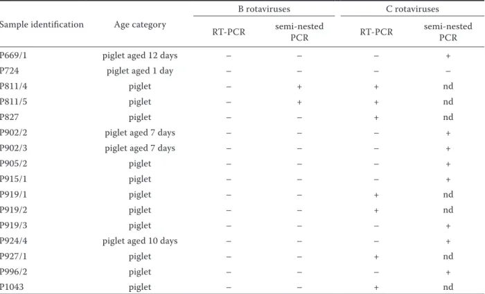
Table 1. Results of rotavirus B and C detection in fecal samples
Sample identification Age category
B rotaviruses C rotaviruses
RT-PCR semi-nestedPCR RT-PCR semi-nestedPCR
P669/1 piglet aged 12 days – – – +
P724 piglet aged 1 day – – – –
P811/4 piglet – + + nd
P811/5 piglet – + + nd
P827 piglet – – + nd
P902/2 piglet aged 7 days – – – +
P902/3 piglet aged 7 days – – – +
P905/2 piglet – – – +
P915/1 piglet – – – +
P919/1 piglet – – + nd
P919/2 piglet – – + nd
P919/3 piglet – – – +
P924/4 piglet aged 10 days – – – +
P927/1 piglet – – + nd
P996/2 piglet – – – +
P1043 piglet – – + nd
and post-weaning. A total of 329 fecal samples were examined by electron microscopy (EM) with nega-tive staining and the ELISA test for detection of group A rotaviruses, transmissible gastroenteritis virus (TGEV) and porcine epidemic diarrhoea virus (PEDV) (Rodak et al., 2004, 2005a,b). EM rotavi-rus positive but ELISA group A rotavirotavi-rus negative samples were selected for the detection of group B and C rotaviruses by PCR. Sample designation and age of animals (if specified) are shown in Table 1. The samples were processed as 20% suspension in Minimum Essential Medium Eagle (E MEM) sup-plemented with antibiotics: penicillin 20 IU/ml, streptomycin 20 µg/ml and amphotericin B 0.005 µg/ml of medium. The supernatant obtained after centrifugation was further used for RNA ex-traction.
RNA extraction
RNA extraction from the obtained supernatant was carried out using Trizol LS reagent (GibcoBRL, GrandIsland, N.Y., USA) according to the manufac-turer’s instructions. The extracted RNA was dis-solved in RNase-free water and stored at –80°C until further processing.
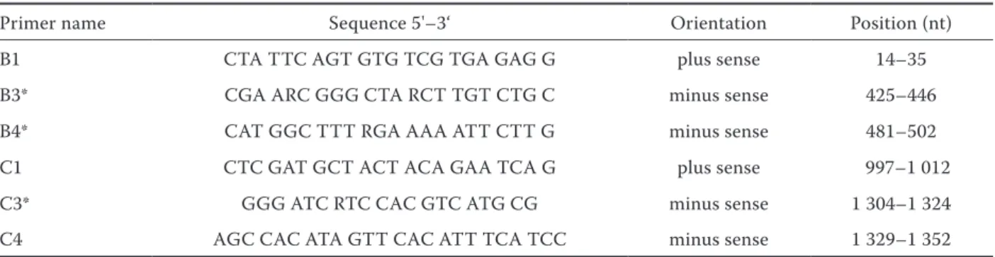
Primers for detection of group B and C rotaviruses
Modified primers described previously by Gouvea et al. (1991) were used in our experiments. After comparison of sequences for these genes of rotaviral groups available in the Gene Bank we modified se-quences of the primers B3, B4 and C3 (Table 2).
RT-PCR
Extracted dsRNA in a volume of 2 µl was de-natured with 1.4 µl of concentrated dimethyl-sul-phoxide (Sigma-Aldrich, USA) at 97°C for 5 min (Martella et al., 2001). The reverse transcription (RT) of dsRNA was carried out using StrataScript Reverse Transcriptase (StrataScript RT, Stratagene, USA) and PCR amplification was carried out with
Taq DNA Polymerase (Promega, USA). RT was per-formed in 20µl volume according to the manufac-turer’s recommendation. After synthesis of cDNA the RT mixture was brought up to a volume of 100 µl of PCR mixture containing 1.5mM MgCl2, 200µM dNTPs, 5U Taq DNA Polymerase and 250nM of both primers. PCR amplification was accomplished in 30 cycles (94°C for 1 min, 42°C for 2 min, 72°C for 1 min) followed by a final extension for 7 min at 72°C (Gouvea et al., 1991). Semi nested PCR was performed with primer pairs B1-B3 and C1-C3 for detection of group B and C rotaviruses and with 4 µl DNA of template from the first PCR. Conditions for reamplification were identical with the conditions used for the first round of PCR. When attempting to detect group C rotavirus, analysis of a positive control (strain P543/1) (Smitalova et al., 2006) was always a part of the examination. As a standard positive strain of group B rotavirus was not avail-able, inter-group primer specificity for detection of group B rotaviruses was tested using samples of positive group A and C rotaviruses.
Electrophoresis in agarose gel
PCR products (8 µl) were analyzed on 1.5% agar-ose containing 0.5 µg of ethidium bromide per ml.
Table 2. Primers used for the detection of Group B and C rotaviruses
Primer name Sequence 5'–3‘ Orientation Position (nt)
B1 CTA TTC AGT GTG TCG TGA GAG G plus sense 14–35
B3* CGA ARC GGG CTA RCT TGT CTG C minus sense 425–446
B4* CAT GGC TTT RGA AAA ATT CTT G minus sense 481–502
C1 CTC GAT GCT ACT ACA GAA TCA G plus sense 997–1 012
C3* GGG ATC RTC CAC GTC ATG CG minus sense 1 304–1 324
C4 AGC CAC ATA GTT CAC ATT TCA TCC minus sense 1 329–1 352
The gel was electrophoresed at 110 V for 60 min and photographed under UV light.
Sequence analysis
Products of the strains P811/4, P919/2 and P927/1 of 356 bp (group C), and strains P811/4 and P811/5 of 434 bp (group B) were used for sequence analy-sis. Sample purification was performed using the QIAquick PCR Purification Kit (QIAGEN GmbH, Hilden, Germany). Amplified C1-C4 products were sequenced using the MegaBACETM DNA Analysis
system (Amersham Biosciences), and the B1-B3 products using the ABI 310 Genetic Analyser sys-tem (Applied Biosyssys-tems). The obtained sequences of group C rotaviruses were compared with the VP6 sequence of the Cowden strain gene (acces-sion number M94157) and with the sequence of the previously described strain P543/1 (Smitalova et al., 2006). Sequences of strains where group B rotaviruses were expected based on product size, were compared with the sequences available in the Gene Bank for gene 8 of group B rotaviruses (ac-cession numbers AY539861, AY238393, AY238383, EF577259, EF577258 and EF577257).
RESULTS
PCR detection of group C rotaviruses
A total of 16 samples were examined by PCR. These samples were found positive by electron
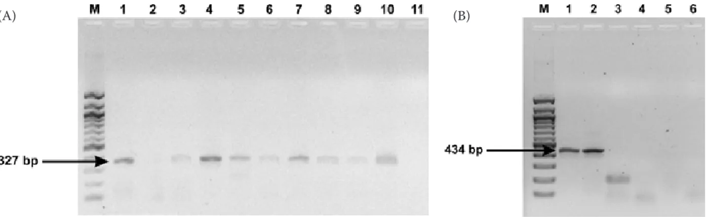
microscopy but negative in an ELISA test which detects group A rotaviruses. group C rotavirus was detected in 15 samples. The use of RT-PCR and the C1-C4 set of primers yielded a product of the expected 356 bp size in seven samples. A subsequent analysis of RT-PCR negative samples by semi-nested PCR (C1-C3m primers) detected a product of the expected 327 bp size in an additional eight samples (Figure 1A). Group C rotavirus was not detected in only one sample (Table 1).
PCR detection of group B rotaviruses
A product of the expected size corresponding to group B rotaviruses (434 bp) was successfully ampli-fied in two samples. A positive result was obtained by analysis using semi-nested PCR (B1-B3m prim-ers) (Figure 1B). No product of the corresponding size (489 bp) was detected by RT-PCR and B1-B4m primers. In the remaining samples, amplification did not detect products of the expected size. Both the samples positive for group B rotavirus were also PCR positive for the detection of group C ro-taviruses (Table 1).
Sequence analysis of group C and B rotaviruses
Nested PCR products were sequenced and the obtained sequences with a size of 317 bp (gene position 1022–1338 nt) of the P811/4, P919/2 and P927/1 strains, positive for group C rotavirus, were
Figure 1. (A) Results of Group C rotavirus detection by semi-nested PCR: M = size marker; 1 to 9 = corresponds to the samples P669/2, P724, P902/2, P902/3, P905/2, P915/1, P919/3, P924/3 and P996/2; 10 = positive control; 11 = negative control. (B) Results of Group B rotavirus detection by semi-nested PCR: M = size marker; 1 to 5 = corresponds to the samples P811/4, P811/5, P724, P669/2, P543/1; 6 = negative control
compared with the corresponding sequence of VP6 protein (gene 6) of the Cowden strain (porcine ro-tavirus group C) and the P543/1 strain. The se-quences of the P811/4, P919/2 and P927/1 strains showed 92% nucleotide sequence identity with the Cowden strain and 97% with the P543/1 strain. The mutual nucleotide sequence identity of these three sequences was 96–97%.
Two sequences of the 398 bp nested PCR products (gene position 46–443 nt) from samples positive for group B rotaviruses were compared with each other and also with the corresponding sequence of NSP2 protein (gene 8) of group B rotaviruses. Sequences obtained from two samples were identical. The nucleotide identity of the sequence obtained by comparison with three sequences of human strains ranged from 73 to 74%. Nucleotide sequence iden-tity obtained by comparison with three sequences from porcine isolates reached 79–80%.
Detection of other viral enteropathogens
The detection of coronaviruses (TGEV and PEDV) using an ELISA test was also attemted on the tested samples. Porcine epidemic diarrhea virus was detected in one sample (P919/2).
DISCUSSION
Our study had the aim of detecting group B and C rotaviruses in samples where the occurrence of nongroup A rotaviruses was expected based on pre-vious results (EM, ELISA). RT-PCR was used for the detection of both group B and C rotaviruses. Group C rotavirus was detected more frequently, which is in accordance with previous reports (Janke et al., 1990; Geyer et al., 1996). In contrast, group B rota-viruses were more frequently detected by Magar et al. (1991), together with other viral, bacterial and parasitic agents, while group C rotaviruses were detected as separate infections. In two samples, group B rotaviruses were detected together with group C rotaviruses. Another viral agent was de-tected in only one sample. Evidently, the presence of bacteria and parasites and their participation in the development of a clinical form of disease cannot be excluded. The selection of samples for the detection of group B and C rotaviruses based on preliminary findings contributed to their detec-tion in most of the samples; however, the presence
of nongroup A rotaviruses cannot be ruled out in samples in which group A rotaviruses as well as TGEV and PEDV were detected by ELISA tests.
In a group of samples which were used in differ-ent diagnostic tests for the detection of group A, B and C rotaviruses, sample P724 was the only one found positive by electron microscopy but nega-tive for the detection of group A, B and C rotavi-ruses. Due to the high sensitivity and specificity of ELISA and semi-nested PCR methods we assume that the negative results for group A, B and C ro-tavirus detection but a positive finding for typical morphological structure indicate the presence of rotaviruses belonging to other groups.
The nucleotide sequence identity of the VP6 pro-tein of group C rotavirus in samples from three different farms was 92% and 97% as compared with the strain Cowden and P543/1, respectively. The nucleotide sequence identity in sequenced samples from our herds is higher (96–97%) than in com-parison with the Cowden strain. A 100% nucle-otide identity of a partial sequence of the NSP2 protein (gene 8) of group B rotavirus found in two samples was probably down to their origin from the same herd. The nucleotide identity of 73–74% obtained by comparison of nucleotide sequences of these products with the corresponding sequence of gene 8 of human group B rotaviruses is relatively low. Rather surprisingly, comparison with three currently available sequences of gene 8 of group B rotaviruses isolated from pigs was not significantly higher than this (79–80%). Yang et al. (2004) per-formed a phylogenetic analysis of human group B rotavirus strain WH1 based on a comparison of selected genes with both human and animal strains. A comparison of the VP7 sequences from human strains revealed a considerable degree of nucleotide sequence identity (92–98%) while comparison with animal (bovine and murine) strains showed a sig-nificantly lower sequence identity (61–64%). The sequence identity of the NSP2 gene (gene 8) in mice and of the analysed strain WH1 was 79%, which is in agreement with the results of our comparison of porcine and human strains in this gene.
in pigs. Clarification of their significance, vari-ability and participation in possible inter-species transmission is at present the objective of other investigations.
Acknowledgment
The authors wish to thank Mrs. Farnikova and Mrs. Cebakova for their skillful technical assistance.
REFERENCES
Bohl E.H., Saif L.J., Theil K.W., Agnes A.G., Cross R.F. (1982): Porcine pararotavirus: detection, differentia-tion from rotavirus, and pathogenesis in gnotobiotic pigs. Journal of Clinical Microbiology, 15, 312–319. Bridger J.C. (1980): Detection by electron microscopy
of caliciviruses, astroviruses, and rotavirus-like par-ticles in the feces of piglets with diarrhea. Veterinary Record, 107, 532–533.
Bridger J.C., Pedley S., McCrae M.A. (1986): Group C rotaviruses in humans. Journal of Clinical Microbio-logy, 23, 760–763.
Chen G.M., Hung T., Bridger J.C., McCrae M.A. (1985): Chinese adult rotavirus is a Group B rotavirus. Lancet ii, 1123–1124.
Estes M. K.(1996): Rotaviruses and their replication. In: N.Eields B. Knipe D.M., Howley P.M. et al. (eds.): Fields Virology. Lippincott-Raven Publisher, Philadelphia. 1625–1655.
Geyer A., Sebata T., Peenze I., Steele A.D. (1996): Group B and C porcine rotaviruses identified for the first time in South Africa. Journal of the South African Veteri-nary Association, 67, 115–116.
Gouvea V., Allen J.R., Glass R.I., Fang Z.Y., Bremont M., Cohen J., McCrae M.A., Saif L.J., Sinarachatanant P., Caul E.O. (1991): Detection of group B and C rotavi-ruses by polymerase chain reaction. Journal of Clinical Microbiology, 29, 519–523.
Itturiza-Gomara M., Clarke I., Desselberger U., Brown D., Thomas D., Gray J. (2004): Seroepidemiology of group C rotavirus infection in England and Wales. European Journal of Epidemiology, 19, 589–595. Janke B.H., Nelson J.K., Benfield D.A., Nelson E.A.(1990):
Relative prevalence of typical and atypical strains among rotaviruses from diarrheic pigs in conventional swine herds. Journal of Veterinary Diagnostic Inves-tigation, 2, 308–311.
Kim, Y., Chang, K.O., Straw, B. and Saif, L.J. (1999): Cha-racterization of group C rotaviruses associated with
diarrhea outbreaks in feeder pigs. Journal of Clinical Microbiology, 37, 1484–1488.
Magar R., Robinson Y. Morin M. (1991): Identificatin of atypical rotaviruses in outbreaks of preweaning and postweaning diarrrhea in Quebec swine heards. Cana-dian Journal of Veterinary Research, 55, 260–263. Martella V., Pratelli A., Greco G., Tempesta M., Ferrari
M., Losio M.N., Buonavoglia C. (2001): Genomic characterization of porcine rotaviruses in Italy. Cli-nical and Diagnostic Laboratory Imunology, 8, 129– 132.
Morin M., Magar R., Robinson Y. (1990): Porcine group C rotavirus as a cause of neonatal diarrhea in a Quebec swine herd. Canadian Journal of Veterinary Reseasrch, 54, 385–389.
Nilsson M., Sigstam G., Svensson L. (2000): Antibody prevalence and specificity to group C rotavirus in Swe-dish sera. Journal of Medical Virology, 60, 210–215. Rodak L., Smid B., Nevorankova Z., Smitalova R., Valicek
L. (2004): Verification of sensitivity and specificity of group A rotavirus detection in piglets faeces with mo-noclonal blocking ELISA Methods. Journal of Veteri-nary Medicine B, 51, 160–165.
Rodak L., Smid B., Nevorankova Z., Valicek L., Smitalova R. (2005a): Use of monoclonal antibodies in blocking ELISA detection of transmissible gastroenteritis virus in faeces of piglets. Journal of Veterinary Medicine B, 52, 105–111.
Rodak, L., Valicek, L., Smid B., Nevorankova Z. (2005b): An ELISA optimized for porcine epidemic diarrhoea virus detection in faeces. Veterinary Microbiology, 105, 9–17.
Saif L.J., Jiang B. (1994): Nongroup A rotaviruses of hu-mans and animals. Current Topics in Microbiology and. Immunology, 185, 330–371.
Saif L.J., Terrett L.A., Miller K.L., Cross R.F. (1988): Se-rial propagation of porcine group C rotavirus (para-rotavirus) in a continuous cell line and characterization of the passaged virus. Journal of Clinical Microbiology, 1982, 1277–1282.
Smitalova R., Rodak L., Psikal I., Smid B. (2006): Isola-tion, immunochemical demonstration of field strains of porcine group A rotaviruses and electrophoretic analysis of RNA segments of group A and C rotaviru-ses. Veterinarni Medicina, 51, 288–295.
Terrett L.J., Saif L.J., Theil K.W., Kohler E.M. (1987): Physicochemical characterization of porcine pararo-tavirus and detection of virus and viral antibodies us-ing cell culture immunofluorescence. Journal of Clinical Microbiology, 25, 268–272.
and humans by enzyme-linked immunosorbent assays. Journal of Clinical Microbiology, 30, 2129–2134. Yang J.H., Kobayashi N., Wang Y.H., Zhou X., Li Y., Zhou
D.J., Hu Z.H., Ishino M., Alam M.M., Naik T.N., Ahmed M.U. (2004): Phylogenetic analysis of a human group
B rotavirus WH-1 detected in China in 2002. Journal of Medical Virology, 74, 662–667.
Received: 2008–01–29 Accepted after corrections: 2009–01–21
Corresponding Author:
MVDr. Radka Smitalova, Ph.D., Veterinary Research Institute, Department of Bacteriology and Virology, Hudcova 70, 621 00 Brno, Czech Republic