Isolation, cloning and sequence analysis of the lactate
dehydrogenase gene from
Theileria
annulata
may lead
to design of new antitheilerial drugs
A. Erdemir
1, M. Aktas
2, N. Dumanli
2, D. Turgut-Balik
11Faculty of Chemical and Metallurgical Engineering, Yildiz Technical University, Davutpasa
Campus, Istanbul, Turkey
2Faculty of Veterinary Medicine, Firat University, Department of Parasitology, Elazig, Turkey
ABSTRACT: Theileriosis is a serious animal disease that is transmitted by ticks. Theileria species are tick-borne obligate intracellular protozoan parasites that cause severe and mild infections in their hosts. Two of them,
Theileria annulata and Theileria parva, cause lymphoproliferative disease with high mortality and morbidity in cattle commonly known as tropical theileriosis and East Coast fever, respectively. Currently available antiparasitic drugs are effective in animals but animals may remain carriers and treatment is most effective in the early stages of the disease. The isolation, cloning and analysis of lactate dehydrogenase from T. annulata was the goal of the present study with the ultimate aim of designing new antiparasitic drugs that will hopefully have a wider mode of action in animals. Blood samples were taken from a four year-old Brown Swiss cow showing signs of acute tropical theileriosis and genomic DNA was extracted following the confirmation of the clinical diagnosis. For the first time, in this study, the lactate dehydrogenase sequence was isolated from from a Theileria species. Following extraction from genomic DNA by PCR the sequence was cloned into the vector pGEM-T easy. Sequencing of the whole gene from both directions indicated that the open reading frame was interrupted by two introns. Several single nucleotide exchanges, deletions and insertions were also observed in the T. annulata lactate dehydrogenase sequence compared to the host. The most remarkable difference between the parasite and host enzyme is a five residue insertion in the active site loop region that might be an attractive target for inhibitors of the enzyme. This study opens a new route to further kinetic and structural studies towards the development of novel inhibitors of
T. annulata lactate dehydrogenase.
Keywords: Theileria annulata; theileriosis; antitheilerials; lactate dehydrogenase; gene cloning
Supported by the Yildiz Technical University (Grant No. 2010-07-04-KAP01).
Theileriosis is a serious animal disease that is transmitted by ticks. It is caused by obligate
intra-cellular parasitic protozoa of the genus Theileria
classified in the phylum of apicomplexan
proto-zoa as Plasmodium and Toxoplasma (Bishop et al.
2004). Theileria parva and Theileria annulata are
globally the most important species causing East Coast fever and tropical theileriosis, respectively (Bishop et al. 2004).
Tropical theileriosis is an important tick-borne disease of cattle in tropical and sub-tropical coun-tries (Gardner et al. 2005; Mhadhbi et al. 2010). Two
treat-Original Paper Veterinarni Medicina, 57, 2012 (10): 559–567
ment of Theileria infections in cattle in subsequent
years without the development of any resistance to these drugs. However, resistance to buparvaquone has been recently reported for the first time in the literature (Mhadhbi et al. 2010) and this has very recently been followed by a new case (Sharifiyazdi et al. 2012). The first study was conducted in a farm located in Tunisia. Four out of seven cattle showing symptoms of acute tropical theileriosis died in spite of repeated buparvaquone injections (2.5 mg/kg). It was reported that buparvaquone and parvaquone have been the only available drugs for the treatment of tropical theileriosis to date and possibly for sev-eral more years (Mhadhbi et al. 2010). The second case was reported from Fars Province, southern Iran (Sharifiyazdi et al. 2012). In this study seven out of eight animals were reported to have died despite single (five cases) and double (two cases) injection of buparvaquone (2.5 mg/kg) as a result of the oc-currence of resistance to buparvaquone treatment. Resistance was explained by point mutations in the
mitochondrial cytochrome b gene in the parasite
(Sharifiyazdi et al. 2012). This emphasizes the need for the design of new anti-theilerial drugs that will have different modes of action then the currently available drugs. Mining of the genome and the char-acterization of genes has led to investigations which have focused on essential metabolic enzymes as po-tential new targets for the design of anti-parasitic drugs (Fernandez-Robledo and Vasta 2010).
Lactate dehydrogenase (LDH) is one of the gly-colytic enzymes targeted for the design of such drugs. This enzyme catalyses the interconversion of pyruvate and l-lactate with a concomitant
inter-conversion of NADH and NAD+ (Holbrooket al.
1975). On the basis of molecular cloning studies, kinetic characterisations and X-ray structure analy-sis, plasmodial LDH’s have been studied in most detail (Dunn et al. 1996; Turgut-Balik et al. 2001a; Chaikuad et al. 2005) among the apicomplexan parasites and have been identified as new enzyme targets for the development of novel antimalarial drugs. Determination of the key residues in the
ac-tive site of this enzyme from Plasmodium showed
that this site could be an attractive target for en-zyme inhibitors (Dunn et al. 1996; Turgut-Balik et al. 2001b). A series of azole-based compounds were reported to bind within the active site of the enzyme
and to preferentially bind Plasmodium falciparum
LDH vs. the human LDH (Cameron et al. 2004).
Comparative analysis of Theileria and Plasmodium
genome sequences for the metabolic potential of
parasites indicates that the complete TCA cycle may not be functional in both parasites (Gardner et al. 2005), and studies suggest glycolysis as the
main pathway for energy production in Theileria
(Kiama et al. 1999) as in Plasmodium. It is also
suggested that T. parva schizonts may be similar
to the erythrocytic stage of P. falciparum that uses
glycolysis as the main pathway for the production of its energy (Sherman 1991). In the light of all these studies, it is reasonable to study the same enzyme
from a Theileria species with the ultimate aim of
discovering enzyme inhibitors of the parasite, as drug resistance has now been reported (Mhadhbi et al. 2010; Sharifiyazdi et al. 2012). Because the struc-tural differences between host and parasite LDHs allow discrimination for preferential inhibition of the parasite enzyme (Cameron et al. 2004), LDH
was amplified and cloned from a Theileria species
for the first time in the literature in this study and the sequence was analysed in comparision to the host LDH and some other known LDH sequences.
MATERIAL AND METHODS
All general methods were applied according to Sambrook and Russel (2001) unless otherwise stated.
Bacterial strain, growth media, enzymes and vector
The host bacterial strain used to prepare DNA for cloning and sequencing in the pGEM-T Easy vector
(Promega, USA) was Escherichia coli JM105 {supE
hsdD5 thiD(lac-proAB) D(srl-recA) (306:Tn10)
(tetr) F’[traD36 proAB+ lacIq lacZDM15 d]}. The
E. coli JM105 cells were cultured in LB broth.
Twenty mg/ml IPTG, 20 mg/ml X-Gal and 100 mg/ml ampicillin were used in media for the selection
and growth of transformants where necessary. Taq
DNA Polymerase was obtained from (Fermentas, #Cat. No: EP0402, Vilnus, Lithuania).
Parasite isolate and genomic DNA
was confirmed by observation of T. annulata pi-roplasms and schizonts on Giemsa-stained blood and lymph node biopsy smears (immersion oil objective) at the Laboratory of Parasitology of the Veterinary School of Firat University, Elazig, Turkey. The Wizard genomic DNA purification system (Promega Corporation, #Cat. No: A1120, Madison, USA) was used to prepare DNA accord-ing to the manufacturer’s instructions.
Amplification of Theileria annulata LDH by PCR
The initial sequence of T. annulata LDH
(TaLDH) was obtained from NCBI with the
ac-cession number of XM_948495. This sequence was then used to design two specific oligonu-cleotide primers for the amplification of the LDH
gene from genomic DNA of T. annulata Elazig
strain. Two oligonucleotide primers were pre-pared, complementary to the forward and reverse
strands of the TaLDH gene. The forward primer
was Ta3, 5'-CGCGCGGGATCCATGGCAAGAA-
ATAATAAGAGG-3', and reverse primer was Ta4,
5'-TTTTCTGCAGTTAGTGATGGTGATGGTGA-TGTTTAATGAGTGCTTCTAAACG-3'.
The enzyme Taq DNA Polymerase was used to
amplify the DNA. This polymerase often adds a sin-gle deoxyadenosine to the 3’-end of the amplified fragments which makes these products suitable for use for T vector cloning. The reaction mixture
con-tained 5 µl of Taq DNA Polymerase buffer, which
is supplied with the enzyme, 1.5mM MgCl2, 5 µl
of a stock of dNTPs (10 µl of each 10mM dNTPs
and 10 µl of H2O). 50 pmol of each
oligonucleo-tide primer, 1 µl of genomic DNA from T.
an-nulata Elazig strain, 1 µl (2.5 units) of Taq DNA
Polymerase and H2O to a final volume of 50 µl.
DNA was pre-denatured at 95 °C for 5 min prior
to amplification. DNA was then denatured at 94 °C
for 1 min and 30 s, annealed at 44 °C for 2 min
and extended at 72 °C for 2 min for 45 cycles. The
reaction ended with a final extension at 72 °C for
10 min. Analysis of PCR products on a 1% agarose gel revealed the presence of a band of the expected size. After confirmation of the product size, PCR was set up using the same conditions again and the DNA band was extracted directly from the agarose gel using Wizard SV Gel and PCR Clean-Up System (Promega Corporation, #Cat. No: A9281, Madison, USA) prior to ligation.
Ligation and transformation
The pGEM-T easy vector system is used for the cloning of PCR products (Promega Corporation, #Cat. No: A1360, Madison, USA). This vector is
pre-pared by cutting with Eco RV and adding a 3' terminal
thymidine to both ends. The addition of single 3'-T overhangs at the insertion site contributes to the efficiency of ligation of the PCR product into the plasmid. The A-tailing procedure was applied to the PCR product to improve cloning efficiency prior to ligation according to the supplier’s instructions. Ligation and transformation were also performed according to the supplier’s instructions.
DNA sequencing
Inserts were initially checked by colony PCR
us-ing the Ta3 and Ta4 oligonucleotide primers and
obtained bands at the expected size indicated the amplification of the correct DNA locus. Plasmid DNA was then prepared using Wizard Plus SV Minipreps DNA Purification System (Promega Corporation, #Cat. No: A1460, Madison, USA) and submitted for sequencing from both directions. All of the cloning experiments and sequencing were repeated twice independently starting from the genomic DNA step, because of the lack of
proof-reading activity of Taq DNA polymerase.
Database analysis and molecular modelling
LDH sequences of apicomplexans were ob-tained from NCBI (http://www.ncbi.nlm.nih. gov/). Alignments of sequences at the nucleotide level were performed by using NCBI BLASTn tool (http://blast.ncbi.nlm.nih.gov/Blast.cgi). The po-sitions of exons and introns were determined by the GSDS utility (http://gsds.cbi.pku.edu.cn/index. php) (Guo et al. 2007). The molecular weight of pro-teins was calculated using the web based Peptide Properties Calculator (http://www.basic.north-western.edu/biotools/proteincalc.html). Amino acid sequence alignment was performed manually by using catalytic residues as reference points to set up residue numbering correctly. The Clustal W2 (http://www.ebi.ac.uk/Tools/msa/clustalw2/) (Chenna et al. 2003) tool was used to align LDH
from T. annulata, Bos taurus and some other
Original Paper Veterinarni Medicina, 57, 2012 (10): 559–567
was downloaded and adapted to MEGA to built the phylogenetic tree (Tamura et al. 2007) using the neighbour-joining method. Modelling studies
of B. taurus LDH-A and TaLDH was conducted
using SWISS-Model automated mode (http:// swissmodel.expasy.org/) (Arnold et al. 2006). The nucleotide sequences used in this study are avail-able in GenBank under the following accession
numbers: HM176661 for T. annulata, BC146210
for B. taurus (LDH A), XM_761610 for T. parva,
DQ198261 for Plasmodium falciparum, DQ060151
for Plasmodium vivax, AY437808 for Plasmodium
berghei, BM165756 for Plasmodium yoelii, U35118
for Toxoplasma gondii (LDH 1), U23207 for T.
gon-dii (LDH2), AF274310 for Cryptosporodium
par-vum, AY143389 for Eimeria tenella andFJ617009
for Eimeria acervulina.
RESULTS
Amplification, cloning and DNA sequencing of the Theileria annulata LDH gene
Theileria annulata genomic DNA was
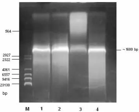
ampli-fied using two oligonucleotides a DNA fragment of about 1.6 kb in size was obtained. This product was then purified and inserted into the pGEM-T Easy plasmid vector prior to transformation into
E. coli JM105 cells. Cloning of the target sequence
was pre-checked and confirmed by PCR prior to sequencing (Figure 1). Sequence analysis of the in-sert demonstrated the presence of the full length
TaLDH. This sequence was deposited in GenBank
with the accession number of HM176661.
This cloned LDH sequence is the first sequence
to be cloned from a Theileria species to date, to our
knowledge. Cloning and DNA sequencing results indicated that the open reading frame (ORF) of
TaLDH consisted of 1591 bp starting with an AUG
codon and ending with a TAA codon. The ORF was interrupted by two introns conforming to the GT/AG rule at the splicing junctions. The main finding of this study is the presence of a 15 nucleotide inser-tion in the substrate specificity loop of the enzyme.
The deduced TaLDH protein sequence consisted of
322 amino acids with a calculated molecular weight of 35 220 g/mol.
Multiple amino acid sequence alignment and phylogenetic analysis of Theileria annulata LDH with some other known LDHs
The amino acid sequence of TaLDH cloned in this
study (HM176661) was aligned with the amino acid
sequences of some other species, including B. taurus
(LDH A: BC146210; LDH B: BC151427), T. parva
(XM_761610), P. falciparum (DQ198261), T. gondii
(LDH 1: U35118), E. tenella (AY143389) and
C. par-vum (AF274310) after removal of the two intron
sequences (Table 1). TaLDH showed 88% similarity
to T. parva, 50% to P. falciparum, 47% to T gondii,
47% to E. tenella and 45% to C. parvum, respectively
at the amino acid level. This alignment shows that the amino terminal extension observed in
mamma-lian LDHs is lacking in TaLDH. Residues involved
in catalysis perfectly matched with the residues and positions of the other known LDHs. Although many characteristic residues are conserved, amino acid
differences were observed between host B. taurus
LDH (BtLDH) and parasite LDH. One hundred
and sixty three, 250 and 246 are some important positions (Turgut-Balik et al. 2001b) where amino acid exchanges were observed. Comparison of the
TaLDH with BtLDH sequence revealed that TaLDH
has deletions at positions 48 and 283 and two in-sertions. The first insertion was at position 74. The second and most remarkable insertion was a penta peptide or 5 nucleotide insertion, between positions serine 108 and arginine 109 in the active site loop of the enzyme. This five amino acid insertion is present in Apicomplexan parasite LDH sequences,
[image:4.595.65.291.531.712.2]exclud-ing Cryptosporidium parvum (Table 1).
Figure 1. Confirmation of the cloning of TaLDH by PCR. Lines: M = marker, 1,2,4 = positive results, 3 = negative result
Ta
ble 1. Alig
nmen
t of
Ta
LDH amino ac
id s equenc e w ith L DH f rom Bo s ta ur us (L
DH A: B
C146210; L
DH B: B
C151427), Thei ler ia p ar va (XM761610), Pl as m odium fa lc ip ar um (D Q198261), To xopl as ma gond ii (L
DH 1: U35118),
Ei mer ia tenel la (A Y143389) and Cr ypto sp or id iu m p ar vu m (A
F274310). The five r
esidue ins er tion in apic om ple xan p ara sit e L DHs i s s how
n in b
Original Paper Veterinarni Medicina, 57, 2012 (10): 559–567
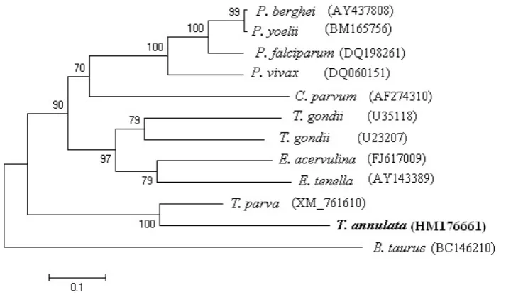
A phylogenetic tree was also constructed among
TaLDH, some apicomplexan LDHs and BtLDH.
Analysis of the results showed that TaLDH and
T. parva LDH are in a different clade than the other
LDHs (Figure 2).
Modelling of T. annulata and Bos taurus LDHs
The deduced TaLDH amino acid sequence obtained
after the removal of the two introns from the gene
[image:6.595.117.474.84.293.2]was used to model the 3D structure of TaLDH in
[image:6.595.65.531.490.724.2]the SWISS-MODEL workspace using the automatic modelling mode (Figure 3). Formation of an extended substrate specificity loop (101–109) caused by the insertion of five amino acids (NEEWN) is clearly
observed in TaLDH compared to its counterpart in
B. taurus.
DISCUSSION
To our knowledge, this study is the first to de-scribe the isolation, cloning and analysis of the
LDH gene from a Theileria species. The cloning of
TaLDH opens up the route to the application of
fur-ther structural and kinetic studies towards the de-velopment of enzyme inhibitors via structure-based drug design studies. The LDH sequence is known for some other apicomplexan parasites such as
Plasmodium (Turgut-Balik et al. 2004), Toxoplasma
(Yangand Parmley 1997) and Eimeria (Schaapet al.
2004) (Table 1). Characteristic residues responsible for catalytic efficiency of the LDHs (Clarke et al.
1989) are also present in TaLDH. This shows that
T. annulata LDH was cloned successfully in this
study.
Analysis of the genomic sequence of T. parva
in comparison with P. falciparum suggests that
Theileria parasites do not have a completely
func-tional TCA cycle similarly to plasmodial LDHs and rely heavily on glycolysis for the production of their energy (Kiama et al. 1999; Gardner et al. 2005). Therefore, inhibition of a crucial enzyme, like LDH, in the glycolytic pathway is a rational approach for the development of a novel antithei-lerial drug that will have a different mode of action than the existing drugs. Determination of differ-ences between plasmodial LDH and host LDH indicated that both enzymes are distinguishable in their structural and kinetic properties (Vander-Jagt et al. 1981; Royer et al. 1986; Makler and Hinrichs, 1993; Dunn et al. 1996; Turgut-Balik et al. 2001a). Insertion of five amino acids between
residues S108-R109 in the substrate specificity loop
(101–109) is characteristic for the apicomplexan parasite LDHs cloned to date. This insertion is
NEEWN in Theileria, DKEWN in Plasmodium,
DS/KEWS in Toxoplasma and DQEWS in Eimeria
(Table 1). This site has been well determined in
Plasmodium and suggested to be an attractive
tar-get for the design of drugs against malaria (Dunn
et al. 1996). The crystal structure of P. falciparum
LDH (Dunn et al. 1996) indicated that this five
amino acid insertion creates a distinctive cleft in the surface of the enzyme adjacent to the substrate binding site in contrast to the same region of the host LDH. Similar structures were also observed
for P. vivax (Chaikuad et al. 2005), P. berghei
(Winter et al. 2003) and T. gondii (Kavanagh et
al. 2004) LDHs. Several organic molecules have recently been designed and developed with the
aim of blocking LDH from P. falciparum (Granchi
et al. 2010). Gossypol is a polyphenolic binaphthyl disesquiterpene compound present in cottonseed
oil. Its derivatives have been tested against PfLDH
and reported to be strong inhibitors of the parasite enzyme (Royer et al. 1986). However, the cytotox-icity of gossypol has stopped any further
develop-ment with regard to PfLDH inhibition (Granchi et
al. 2010). A series of azole-based compounds have also been identified in high throughput enzymatic
screening and tested against PfLDH. These
com-pounds inhibited LDH activity at sub-micromolar
concentrations in vitro and displayed modest
anti-malarial activity in vivo on P. berghei in a rodent
model (Cameron et al. 2004). The same study also showed that these inhibitors form a network of interactions with residues within the active site of the enzyme. The present study indicates that the
active site of TaLDH is quite similar to the same
region from PfLDH. Therefore, the application of
similar approaches to TaLDH would be expected
to produce valuable results as in other apicompl-exan LDH studies.
Structure-based drug design studies often pro-ceed through multiple steps before clinical trials (Anderson 2003). The first cycle of the process includes the cloning of the target gene, purifica-tion of the protein and determinapurifica-tion of its struc-ture by X-ray crystallography, NMR or homology modelling (Anderson 2003). In this study, cloning and sequence analysis in combination with ho-mology modelling of the lactate dehydrogenase
gene from T. annulata was described with the
aim of facilitating structure-based drug design studies. The results show that the active site of
TaLDH, extended by five amino acids, is similar
to the same sequence from other apicomplexan parasites and may be expected to be similar in
character Plasmodial LDH’s. To the best of our
knowledge, this is the first report of the cloning
of the LDH gene from a Theileria species and
pro-vides valuable insight into the structure of LDH
from Theileria species that will greatly facilitate
Original Paper Veterinarni Medicina, 57, 2012 (10): 559–567
REfERENCES
Anderson CA (2003): The process of structure-based drug design. Chemistry and Biology 10, 787–797. Arnold K, Bordoli L, Kopp J, Schwede T (2006): The
SWISS-MODEL Workspace: A web-based environ-ment for protein structure homology modelling. Bio-informatics 22, 195–201.
Bishop R, Musoke A, Morzaria S, Gardner M, Nene V (2004): Theileria: intracellular protozoan parasites of wild and domestic ruminants transmitted by ixodid ticks. Parasitology 129, 271–283.
Cameron A, Read J, Tranter R, Winter VJ, Sessions RB, Brady RL, Vivas L, Easton A, Kendrick H, Croft SL, Barros D, Lavandera JL, Martin JJ, Risco F, Garcia-Ochoa S, Gamo FJ, Sanz L, Leon L, Ruiz JR, Gabarro R, Mallo A, de las Heras FG (2004): Identification and activity of a series of azole-based compounds with lactate dehydrogenase-directed anti-malarial activity. Journal of Biological Chemistry 279, 31429–31439. Chaikuad A, Fairweather V, Conners R, Joseph-Horne
T, Turgut-Balik D, Brady RL (2005): Structure of lac-tate dehydrogenase from Plasmodium vivax: Com-plexes with NADH and APADH. Biochemistry 44, 16221–16228.
Chenna R, Sugawara H, Koike T, Rodrigo L, Gibson TJ, Higgins DG, Thompson JD (2003): Multiple sequence alignment with the Clustal series of programs. Nucleic Acids Research 31, 3497–3500.
Clarke AR, Atkinson T, Holbrook JJ (1989): Substitution of the amino acid at position 102 with polar and aro-matic residues influences substrate specificity of lac-tate dehydrogenase. Trends. Biochemical Sciences 14, 101–105.
Darghouth MA, Bouattour A, Kilani M (1999): Tropical theileriosis in Tunisia: epidemiology and control. Par-assitologia 41, 33–36.
Dunn CR, Banfield MJ, Barker JJ, Higham CW, Moreton KM, Turgut-Balik D, Brady RL, Holbrook JJ (1996): The structure of lactate dehydrogenase from Plasmo-dium falciparum reveals a new target for anti-malarial design. Nature Structural Biology 3, 912–915. Fernandez-Robledo JA, Vasta GR (2010): Production of
recombinant proteins from protozoan parasites. Trends in Parasitology 26, 244–254.
Gardner MJ, Bishop R, Shah T, de Villiers EP, Carlton JM, Hall, N, Ren QH, Paulsen IT, Pain A, Berriman M, Wilson RJM, Sato S, Ralph SA, Mann DJ, Xiong ZK, Shallom SJ, Weidman J, Jiang LX, Lynn J, Weaver B, Shoaibi A, Domingo AR, Wasawo D, Crabtree J, Wort-man JR, Haas B, Angiuoli SV, Creasy TH, Lu C, Suh B, Silva JC, Utterback TR, Feldblyum TV, Pertea M,
Allen J, Nierman WC, Taracha ELN, Salzberg SL, White OR, Fitzhugh HA, Morzaria S, Venter JC, Fraser CM, Nene V (2005): Genome sequence of Theileria parva, a bovine pathogen that transforms lympho-cytes. Science 309, 134–137.
Granchi C, Bertini S, Macchia M, Minutolo F (2010): Inhibitors of lactate dehydrogenase isoforms and their therapeutic Potentials. Current Medicinal Chemistry 17, 672–697.
Guo AY, Zhu QH, Chen X, Luo JC (2007): GSDS: a gene structure display server. Yi Chuan 29, 1023–1026. Holbrook JJ, Liljas A, Steinde, SJ, Rossmann MG (1975):
In: Boyer PD (ed.): The Enzymes Lactate Dehydroge-nase. Academic Press, New York, San Fransisco, Lon-don. 191–292.
Kavanagh KL, Elling RA, Wilson DK (2004): Structure of Toxoplasma gondii LDH1: Active-site differences from human lactate dehydrogenases and the structural basis for efficient APAD+ use. Biochemistry 43, 879–889.
Kiama TN, Kiaira JK, Konji VN, Musoke AJ (1999): En-zymes of glucose and glycerol catabolism in in vitro-propagated Theileria parva schizonts. Veterinary Journal 158, 221–227.
Makler MT, Hinrichs DJ (1993): Measurement of the lac-tate dehydrogenase activity of Plasmodium falciparum as an assessment of parasitemia. American Journal of Tropical Medicine and Hygiene 48, 205–210.
McHardy N, Morgan DW (1985): Treatment of Theileria annulata infection in calves with parvaquone. Research in Veterinary Science 39, 1–4.
McHardy N, Wekesa LS, Hudson AT, Randall AW (1985): Antitheilerial activity of BW720C (buparvaquone): a comparison with parvaquone. Research in Veterinary Science 39, 29–33.
Mhadhbi M, Naouach A, Boumiza A, Chaabani MF, Ben-Abderazzak S, Darghouth MA (2010): In vivo evidence for the resistance of Theileria annulata to bu-parvaquone. Veterinary Parasitology 169, 241–247. Royer RE, Deck LM, Campos NM, Hunsaker LA, David
L, Vander Jagt DL (1986): Biologically active deriva-tives of gossypol: synthesis and antimalarial activities of peri-acylated gossylic nitriles. Journal of Medicinal Chemistry 29, 1799–1801.
Sambrook J, Russell DW (2001): Molecular Cloning A Laboratory Manual I-II-III. 3rd ed. CSHL Press, New
York.
Sharifiyazdi H, Namazi F, Oryan A, Shahriari R (2012): Point mutations in the Theileria annulata cytochrome b gene is associated with buparvaquone treatment failure, Veterinary Parasitology 431–435.
Sherman IW (1991): The biochemistry of malaria. An over-view. In: Coombs G, North M (eds.): Biochemical Pro-tozoology. Taylor and Francis Ltd., London. 605–621. Tamura K, Dudley J, Nei M, Kumar S (2007): MEGA4:
Molecular evolutionary genetics analysis (MEGA) software version 4.0. Molecular Biology and Evolution 24, 1596–1599.
Turgut-Balik D, Shoemark DK, Moreton KM, Sessions RB, Holbrook JJ (2001a): Over-production of lactate dehydrogenase from Plasmodium falciparum opens route to new antimalarials. Biotechnology Letters 23, 917–921.
Turgut-Balik D, Shoemark DK, Sessions RB, Moreton KM, Holbrook JJ (2001b): Mutagenic exploration of the active site of lactate dehydrogenase from Plasmo-dium falciparum. Biotechnology Letters 23, 923–927. Turgut-Balik, D, Akbulut E, Shoemark DK, Celik V, More-ton KM, Sessions RB, Holbrook JJ, Brady RL (2004): Cloning, sequence and expression of the lactate
dehy-drogenase gene from the human malaria parasite, Plas-modium vivax. Biotechnology Letters 26, 1051–1055. Vander-Jagt D, Hunsaker LA, Heidrich JE (1981): Partial
purification and characterization of lactate dehydro-genase from Plasmodium falciparum. Molecular and Biochemical Parasitology 4, 255–264.
Wilkie GM, Brown CGD, Kirvar E, Thomas M, William-son SM, Bell-Sakyi LJ, Sparagano O (1998): Chemo-prophylaxis of Theileria annulata and Theileria parva infections of calves with buparvaquone. Veterinary Parasitology 78, 1–12.
Winter VJ, Cameron A, Tranter R, Sessions RB, Brady RL (2003): Crystal structure of Plasmodium berghei lactate dehydrogenase indicates the unique structural differences of these enzymes are shared across the Plasmodium genus. Molecular and Biochemical Para-sitology 131, 1–10.
Yang S, Parmley SF (1997): Toxoplasma gondii expresses two distinct lactate dehydrogenase homologous genes during its life cycle in intermediate hosts. Gene 184, 1–12.
Received: 2011–08–08 Accepted after corrections: 2012–10–20
Corresponding Author:
Dilek Turgut-Balik, Yildiz Technical University, Faculty of Chemical and Metallurgical Engineering, Department of Bioengineering, Davutpasa Campus, 34220 Esenler, Istanbul, Turkey