Morgan, R. G., Chambers, A. C., Legge, D. N., Coles, S. J., Greenhough, A.,
& Williams, A. C. (2018). Optimized delivery of siRNA into 3D tumor
spheroid cultures in situ. Scientific Reports, 8(1), [7952].
https://doi.org/10.1038/s41598-018-26253-3
Publisher's PDF, also known as Version of record
License (if available):
CC BY
Link to published version (if available):
10.1038/s41598-018-26253-3
Link to publication record in Explore Bristol Research
PDF-document
This is the final published version of the article (version of record). It first appeared online via Nature Publishing
Group at https://www.nature.com/articles/s41598-018-26253-3 . Please refer to any applicable terms of use of
the publisher.
University of Bristol - Explore Bristol Research
General rights
This document is made available in accordance with publisher policies. Please cite only the published
version using the reference above. Full terms of use are available:
www.nature.com/scientificreports
Optimized delivery of siRNA into
3D tumor spheroid cultures
in situ
R. G. Morgan
1,2, A. C. Chamber
s
1, D. N.
Legge
1, S. J. Coles
3, A. Greenhough
1&
A. C.
Williams
13D tissue culture provides a physiologically relevant and genetically tractable system for studying normal and malignant human tissues. Despite this, gene-silencing studies using siRNA has proved difficult. In this study, we have identified a cause for why traditional siRNA transfection techniques are ineffective in eliciting gene silencing in situ within 3D cultures and proposed a simple method for significantly enhancing siRNA entry into spheroids/organoids. In 2D cell culture, the efficiency of gene silencing is significantly reduced when siRNA complexes are prepared in the presence of serum. Surprisingly, in both 3D tumour spheroids and primary murine organoids, the presence of serum during siRNA preparation rapidly promotes entry and internalization of Cy3-labelled siRNA in under 2 hours. Conversely, siRNA prepared in traditional low-serum transfection media fails to gain matrigel or spheroid/organoid entry. Direct measurement of CTNNB1 mRNA (encoding β-catenin) from transfected tumour spheroids confirmed a transient but significant knockdown of β-catenin when siRNA:liposome complexes were formed with serum, but not when prepared in the presence of reduced-serum media (Opti-MEM). Our studies suggest a simple modification to standard lipid-based transfection protocols facilitates rapid siRNA entry and transient gene repression, providing a platform for researchers to improve siRNA efficiency in established 3D cultures.
Three-dimensional (3D) cell culture using primary organoids or tumour spheroids is becoming increasingly pop-ular for modelling both normal and malignant tissue owing to its tractability, relative low cost (versus in vivo
modelling) and increased physiological relevance1. Despite the rapid and widespread uptake of this technique,
gene modulation methods using non-viral transfection methods in situ have proven technically challenging2–5.
Stable and long-term target gene silencing in 3D organoid culture has required shRNA, CRISPR/Cas9 or inducible knockout systems. Well established protocols for adeno-6 retro-7 and lentiviral8 transduction of 3D
cul-tures already exist and CRISPR/Cas9 editing techniques have also proven successful9,10. However, experimental
design may demand only a transient silencing of target genes, for which siRNA has been enormously successful in traditional 2D culture. siRNAs remain popular because they are simple, convenient, well-established, econom-ical and routinely used for high-throughput screening. Previous attempts using siRNA in 3D cell culture have involved mechanical disruption of established organoids in order to obtain the single-cell suspension necessary for transfection, followed by reseeding into matrigel11. Another approach has involved mixing siRNA directly into
the matrigel used for seeding human embryonic stem cells into 3D culture12. Although these approaches have
shown some success, to our knowledge, there are no reports of in situ siRNA delivery to 3D organoids/spheroids using lipofection-based techniques despite the existence of multiple commercial products.
We report here that siRNA prepared with traditional reduced-serum transfection media (Opti-MEM) are excluded at the matrigel boundary with limited spheroid/organoid internalization. Instead, siRNA formed and delivered using standard serum-containing culture medium gains rapid entry to both matrigel and spheroids/ organoids, and elicits a transient gene knockdown.
Results
Uptake of siRNA into tumour spheroids/primary organoids is enhanced by the presence of
serum.
To assess the efficiency of siRNA uptake by SW1463 3D tumour spheroids we used a fluorescent Cy3-conjugated non-targeting control siRNA and formed complexes with Lipofectamine®
RNAiMAX in either1School of Cellular and Molecular Medicine, University of Bristol, Biomedical Sciences Building, University Walk, Bristol, BS8 1TD, UK. 2School of Life Sciences, University of Sussex, Falmer, Brighton, BN1 9QG, UK. 3Institute of Science and the Environment, University of Worcester, Worcester, WR2 6AJ, UK. R. G. Morgan, A. C. Chambers, A. Greenhough and A. C. Williams contributed equally to this work. Correspondence and requests for materials should be addressed to R.G.M. (email: rhys.morgan@sussex.ac.uk) or A.G. (email: a.greenhough@bristol.ac.uk)
Received: 22 July 2016 Accepted: 4 May 2018 Published: xx xx xxxx
www.nature.com/scientificreports/
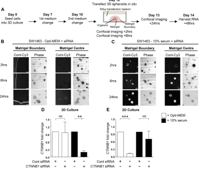
Opti-MEM, or DMEM containing 10% serum. The experimental timeframe is briefly summarized in Fig. 1A. Confocal imaging revealed that at 2 and 6-hour time points, siRNA complexes formed in Opti-MEM were almost completely excluded from spheroids and retained at the matrigel boundary (Fig. 1B). By 24 hours, some low-level fluorescence was detected in spheroids but the majority of fluorescence remained concentrated at the matrigel periphery. Surprisingly, the siRNA prepared with 10% serum had completely penetrated the matrigel and was internalized by spheroids in less than 2 hours (Fig. 1C). At the 6 and 24-hour time points the fluorescence level intensified within spheroids with no evidence of fluorescent accumulation at the matrigel periphery.
To assess whether fluorescence observations translated into gene knockdown, we used a non-fluorescent siRNA targeted to CTNNB1 under the same conditions and assessed mRNA levels direct from 3D culture using qRT-PCR. In agreement with fluorescence observations, Fig. 1D demonstrates that siRNA prepared in 10% serum exhibited a significantly more efficient CTNNB1 knockdown in 3D culture (81.1% ± 6%, p= 0.006) versus Opti-MEM prepared siRNA (3.6% ± 42.1%, p = 0.93). We did however observe that the CTNNB1 knockdown induced in 3D culture using 10% serum formed siRNA complexes was transient with significant knockdown demonstrated at 48 hours only by qRT-PCR. Consequently, we did not observe any phenotypic differences (data not shown) suggesting this approach is not suitable for sustained knockdown (where inducible shRNA systems are preferable). We hypothesized that serum-derived nucleases could be detrimental to siRNA integrity and so employed a nuclease-resistant siSTABLE
®
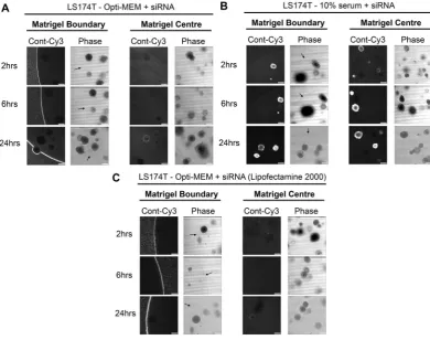
(GE Dharmacon, Lafayette, CO) targeted to CTNNB1 but still observed no significant β-catenin protein reduction (data not shown). As expected for 2D culture of SW1463 cells, Opti-MEM formed siRNA was more efficient forCTNNB1 knockdown (86.1% ± 1.3%, p< 0.0001) versus 10% serum (28.3% ± 31.7%, p= 0.26) (Fig. 1E). We ruled out cell line specific phenomena for these observations since similar results were observed using 3D LS174T tumour
Figure 1. Spheroid uptake of siRNA is more efficient when complexes are formed with serum. (A) Workflow
[image:3.595.156.553.50.387.2]www.nature.com/scientificreports/
spheroids (Fig. 2A and B). We also confirmed that these results were not transfection reagent-specific since siRNA complexes formed with Lipofectamine
®
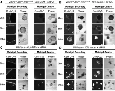
2000 reagent and Opti-MEM were also inhibited from entering LS174T tumour spheroids (Fig. 2C). We next investigated whether this method could be extended to the transfection of primary organoids. As observed in Fig. 3, organoids derived from the intestine of both VilCreERApcfl/flKrasG12D/+(Fig. 3A and B) and wild type mice (Fig. 3C and D) exhibited enhanced siRNA uptake when complexes were formed with the presence of 10% serum versus Opti-MEM.
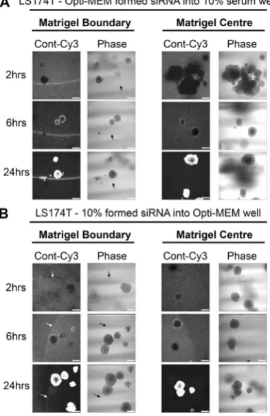
Finally, we examined whether the restricted uptake Opti-MEM-formed siRNA complexes could be relieved through co-incubation with 10% serum. Figure 4A demonstrates how the presence of 10% serum in the well greatly improved both the matrigel and spheroid uptake of Opti-MEM-formed siRNA complexes. At 24 hours however, there did remain a considerable proportion of fluorescence excluded at the matrigel boundary, sug-gesting siRNA complexes formed with Opti-MEM have a persistently decreased capacity for matrigel entry. We also performed the complementary experiment; does co-incubation with Opti-MEM restrict the uptake of serum-formed siRNA complexes. The presence of Opti-MEM appeared to delay full spheroid uptake of 10% serum formed siRNA until after 6 hours by the presence of Opti-MEM (Fig. 4B) but at 24 hours no fluorescence was observed at the matrigel boundary. Taken together, these data suggest siRNA formed in the presence of 10% serum has an enhanced capacity for matrigel penetration and spheroid/organoid uptake.
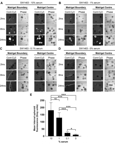
1–10% serum is optimal for siRNA uptake.
To confirm serum as the critical component of culture medium that facilitates siRNA uptake, and ascertain the optimal serum concentration, we formed siRNA com-plexes using a dose-response of serum from 10%, 1%, 0.1% and 0%. Figure 5A shows that siRNA prepared in DMEM containing 10% serum behaved as previous, with rapid uptake of fluorescence siRNA into the matrigel and spheroids in under 2 hours. As the concentration of serum dropped, a dose-dependent decrease in internal spheroid fluorescence occurred over 24 hours and fluorescence instead accumulated at the matrigel boundary (Fig. 5B–D). Figure 5E summarises how fluorescence internalization was optimal when siRNA complex for-mation was formed in the presence of 1–10% serum. At this point, we ruled out siRNA-specific phenomena since fluorescently tagged GAPDH-siRNA exhibited the same pattern of internalization during serum titration (Supplementary Figure S1). These data suggest a concentration of between 1–10% serum during siRNA complex formation is optimal for uptake by 3D spheroids.Figure 2. Failure of Opti-MEM prepared siRNA to penetrate matrigel or organoids is not cell line or
[image:4.595.157.548.46.354.2]www.nature.com/scientificreports/
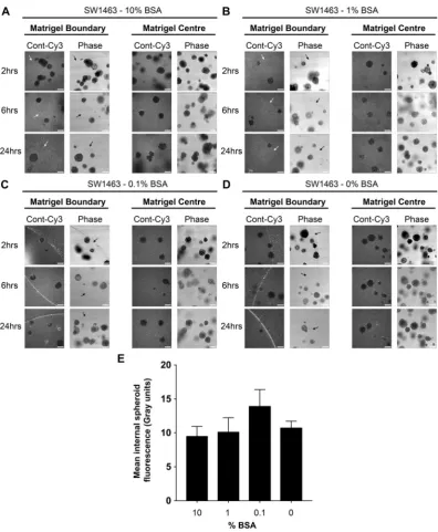
BSA alone can facilitate matrigel entry of siRNA but not spheroid uptake.
Since albumin is a major protein within serum and has carrier properties13,14, we examined the efficiency of BSA alone (by doseresponse, no serum present) during siRNA complex formation for matrigel/spheroid penetration. Enhanced matrigel penetration by fluorescent siRNA could be seen with as little as 0.1% BSA, but interestingly no concen-tration of BSA was sufficient to promote internal spheroid fluorescence suggesting additional serum components are necessary for siRNA uptake (Fig. 6A–E).
Serum molecules less than 10 kDa are dispensable for siRNA uptake.
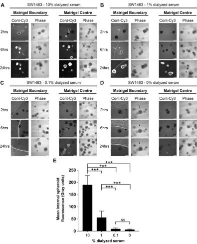
Primary organoid cultures are grown in strictly defined medium containing specialized growth factor supplements such as R-Spondin and Wnt3A15,16. Although supplemental growth factors are not required for the growth of SW1463 cells in 3D, weassessed the ability of siRNAs formed in basic 3D organoid medium (Advanced DMEM/F12 + NAC/B27/N2) to gain matrigel/spheroid entry. Similar to previous observations with reduced serum conditions, we found little siRNA uptake into matrigel and organoids at all time-points assessed (Supplemental Fig. 2). When culturing primary organoids, the presence of serum components such as small growth factors, cytokines and hormones maybe undesirable and so we assessed the ability of dialyzed serum to promote siRNA uptake. We observed that siRNA uptake by spheroids was similarly efficient when complex formation occurred in 10 kDa dialyzed serum (Fig. 7A–D). As summarized in Fig. 7E, internal spheroid fluorescence was again optimal between 1–10%, albeit a lower level of internal fluorescence was obtained at both the 1% and 0.1% concentrations of dialyzed serum (versus normal serum Fig. 5E). These data demonstrate that dialyzed serum was similarly efficient for matrigel penetration and/or spheroid uptake, and any soluble factors facilitating these processes are likely to be more than 10 kDa in molecular weight.
Discussion
Gene silencing through siRNA is a well-established technique owing to its relative simplicity, convenience, cost-effectiveness and routine use in high-throughput screens. In spite of this, transient transfections of estab-lished organoids in situ with siRNA at a specific time-point during 3D culture is not routinely performed despite the demand, and previous attempts have been met with poor efficiency. Bhise and colleagues used nanoparticle
Figure 3. Primary organoid uptake of siRNA is more efficient when complexes are formed with serum.
Representative confocal images of VilCreERApcfl/flKrasG12D/+ primary mouse organoids showing localization of
[image:5.595.157.553.44.358.2]www.nature.com/scientificreports/
technology to improve the transfection of gene delivery vectors into both 2D and 3D cultures2. Transfection
efficiencies using commercially available reagents such as FuGENE HD and Lipofectamine 2000 were generally low, but even the best performing polymeric nanoparticles transfected 57 ± 6% of the cells in 2D culture and only 6 ± 1% of the cells in 3D culture, confirming the difficulty in targeting 3D cultures. Mellor et al., also used poly-plexes, cationic lipids Lipofectamine and DCChol:DOPE, and the physical approach of tissue electroporation to optimise non-viral gene delivery to multicellular tumour spheroids (MCTS)3. Similar to us they used a colorectal
cancer cell line (DLD-1) but used agarose instead of matrigel as the extra-cellular matrix. They observed spatially restricted transfection as fluorescent complexes could only penetrate the outer 3–5 proliferating cell layers, how-ever it would be interesting to see if there was any accumulation of fluorescent complexes at the agarose boundary. Observations in this study are consistent with the difficulty of performing gene silencing in situ with 3D cul-tures using siRNA:lipofection-based techniques. Fluorescent siRNA has revealed that reduced serum media like Opti-MEM is inefficient at delivering siRNA to cells in matrigel. Critically, it is the presence of serum (1–10%) during the siRNA complex formation stage that significantly enhances matrigel penetration and spheroid/organoid uptake. Although previous studies show localization of fluorescently tagged siRNA correlates with gene silencing in 2D culture systems17–19, we observed only a transient reduction in CTNNB1 mRNA level (and consequently no
detectable protein decrease by Western blotting; data not shown). Such transient gene knockdown renders pro-tein reduction difficult to observe, particularly for the longer half-life β-catenin protein forms tethered at mem-brane complexes. Ly and colleagues recently trialled the use of a novel class of asymmetric, chemically stabilized, cholesterol-modified siRNAs referred to as sdrxRNAs
®
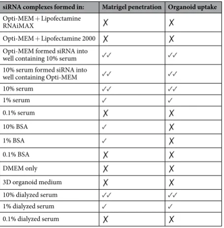
, which can enter cells and tissues without prior formula-tion20. Uptake and repression of gene expression by sdrxRNAs was efficient in 2D culture of HeLa cells, however,gene knockdown efficiency in 3D-matrigel based culture systems was not assessed. Data shown here suggests the addition of serum might improve the efficiency of this siRNA class for 3D culture use. The conditions assessed in this study, and their effect on fluorescent siRNA localization in 3D cultures, are summarized in Table 1 below.
The stark contrast between 2D and 3D transfection efficiencies (Fig. 1) highlight the key differences that exist during transfection of 2D versus 3D culture model systems. Rieux and co-workers assessed gene delivery using lipoplexes either encapsulated within or seeded onto hydrogel5. They found that the transgene expression
increased over 11 days of culture but was significantly higher in 2D compared with 3D cultures. Interestingly, data from encapsulated complexes showed that the hydrogel component fibrin interacts with and sequesters the
Figure 4. Matrigel and spheroid uptake of Opti-MEM-formed siRNA can be improved in the presence of 10%
[image:6.595.157.354.50.353.2]www.nature.com/scientificreports/
lipoplexes thus limiting cellular internalization and causing a delay in expression. Matrigel does not contain fibrin to our knowledge, but it does raise the prospect of other gel components sequestering siRNA complexes at the matrigel surface. Our BSA data show that even when the siRNA complexes gain matrigel entry they are not taken up by the spheroids, implying other serum factors promote this process. Marked differences in gene expression have already been noted between 2D and 3D systems21–23, and it would be interesting to see if proteins critical for
siRNA uptake, such as connexin-43 (Cx43)24, are differentially expressed between 2D and 3D cultures. Finally,
it may be useful to laboratories that use primary human/mouse organoids that dialyzed serum (with 10 kDa threshold) is works well for siRNA uptake since this will limit the presence of undesirable serum-derived small molecules such as growth factors, cytokines and hormones in carefully defined media.
Our study has increased understanding of why gene silencing studies in situ using siRNA with lipofection-based methods has proved ineffective in 3D culture to date. Moreover, we have been able to
Figure 5. Optimal siRNA uptake by matrigel and spheroids occurs with a 1–10% serum concentration.
Representative confocal images of SW1463 spheroids showing localization of control-Cy3 siRNA at 2, 6 and 24 hours post transfection with siRNA formed with (A) 10% (B) 1% (C) 0.1% or (D) 0% serum using RNAiMax. (E) Summary graph showing the mean internal spheroid fluorescence post transfection using siRNA formed with a dose-response of serum. Data represents mean ± 1 s.d (n= 3). Statistical significance is denoted by
[image:7.595.156.552.41.532.2]www.nature.com/scientificreports/
dramatically increase the efficiency of siRNA penetration through matrigel and into spheroid culture through the addition of serum during the transfection procedure. These developments will be useful for experimental approaches that require transient gene silencing in established 3D cultures and can be developed further to improve efficiency.
Methods
Cell culture.
LS174T and SW1463 cells were seeded at approximately 500 single cells per well (24-well plate) into 50 μl growth factor reduced and phenol red-free matrigel (Becton Dickinson, Oxford, UK). 3D tumour sphe-roids were maintained in Advanced Dulbecco’s Modified Eagle’s medium (DMEM)/F12 (Gibco, Paisley, UK), 2 mM glutamine, 10 mM HEPES, 1 mM N-acetyl-cysteine, 0.1% BSA (Sigma-Aldrich, Dorset, UK), penicillin (100 U/ml), streptomycin (100 U/ml), 1 × N2, and 1 × B27 supplements (Invitrogen, Paisley, UK). Primary intes-tinal organoids derived from wild type and VilCreERApcfl/flKrasG12D/+ mice were a kind gift from the SansomFigure 6. BSA can facilitate matrigel entry of siRNA, but not spheroid uptake. Representative confocal images
[image:8.595.156.553.41.523.2]www.nature.com/scientificreports/
laboratory (Beatson Institute, Glasgow, UK) and were maintained as previously described15. For 2D experiments,
SW1463 were maintained as previously described for carcinomas in this laboratory25.
siRNA transfection.
Following 12 days of 3D culture, tumour spheroids were transfected with siRNA. For imaging experiments, organoids received either 25 nM Ambion®
Silencer®
Cy™
3-labeled Negative Control orSilencer
®
Cy™
3-labeled GAPDH siRNA (Invitrogen). For mRNA assessment, spheroids received either 25 nMCTNNB1 or non-targeting control siRNA (unlabelled) as previously described26. siRNA complexes were formed
using either Lipofectamine 2000 or RNAiMAX (Invitrogen), in either Opti-MEM (reduced serum) or DMEM (Gibco) containing either 10% normal/dialyzed FBS (Gibco). In all conditions antibiotics were omitted as rec-ommended by the manufacturer to preserve viability during the transfection process. 500 μl of formed siRNA
Figure 7. 10 kDa dialyzed serum is also efficient for matrigel and spheroid uptake of siRNA. Representative
confocal images of SW1463 spheroids showing localization of control-Cy3 siRNA at 2, 6 and 24 hours post transfection with siRNA formed with (A) 10% (B) 1% (C) 0.1% or (D) 0% dialyzed serum using RNAiMax. (E) Summary graph showing the mean internal spheroid fluorescence post transfection using siRNA formed with a dose-response of dialyzed serum. Data represents mean ± 1 s.d (n= 3). Statistical significance is denoted by
[image:9.595.156.552.41.531.2]www.nature.com/scientificreports/
complex medium was then bathed over 50 μl matrigel contained within a single well of a 24-well plate overnight and replaced the following morning with normal spheroid/organoid medium as outlined above.
Confocal laser scanning microscopy.
Cy3 immunofluorescence in tumor spheroids/primary organoids was analyzed in situ at 2/6/24 hours post transfection using a Leica DM I6000 inverted epifluorescence micro-scope (Leica, Buckinghamshire, UK), with the resonant scanning head of a SP5-AOBS confocal laser micromicro-scope (Leica) and a x10 dry objective as previously described27. Images of the matrigel boundary were captured andZ-stacks were acquired through spheroids/organoids at their widest diameter to ensure a full cross-sectional view of the structure. Internal spheroid fluorescence was calculated by drawing regions of interest (ROI) around the entire circumference of the spheroid followed by measurement of mean Cy3 channel fluorescence intensity within this designated area using LAS AF software v2.6.0 (Leica). TIFFs were generated using Volocity confocal software v6.3.0 (Perkin-Elmer, Waltham, MA, USA) and post-acquisition processing performed using Photoshop CS6 v13.0 (Adobe, Berkshire, UK) with Cy3 channel levels adjusted (equal changes applied across the entire figure).
RNA extraction from 3D culture and qRT-PCR.
48 hours proceeding siRNA transfection of organoids, 3D culture medium was aspirated and matrigel scaffolds washed with ice cold PBS. Following wash, 500 μl of Tri-reagent®
(Sigma-Aldrich) was added to each culture and incubated for 5 minutes on ice. RNA was purified from Tri-reagent®
and CTNNB1 mRNA assessed by qRT-PCR performed as previously described26.Statistics.
All statistical analyses were performed using GraphPad Prism v7.0 (GraphPad Software, Inc., San Diego, CA). Significance of difference was assessed using a one-sample or students t test with significance defined at P< 0.05.References
1. Huch, M. & Koo, B. K. Modeling mouse and human development using organoid cultures. Development 142, 3113–3125, https://doi. org/10.1242/dev.118570 (2015).
2. Bhise, N. S. et al. The relationship between terminal functionalization and molecular weight of a gene delivery polymer and transfection efficacy in mammary epithelial 2-D cultures and 3-D organotypic cultures. Biomaterials 31, 8088–8096, https://doi. org/10.1016/j.biomaterials.2010.07.023 (2010).
3. Mellor, H. R. et al. Optimising non-viral gene delivery in a tumour spheroid model. J Gene Med 8, 1160–1170, https://doi. org/10.1002/jgm.947 (2006).
4. Oishi, M. et al. Enhanced growth inhibition of hepatic multicellular tumor spheroids by lactosylated poly(ethylene glycol)-siRNA conjugate formulated in PEGylated polyplexes. ChemMedChem 2, 1290–1297, https://doi.org/10.1002/cmdc.200700076 (2007). 5. des Rieux, A., Shikanov, A. & Shea, L. D. Fibrin hydrogels for non-viral vector delivery in vitro. J Control Release 136, 148–154,
https://doi.org/10.1016/j.jconrel.2009.02.004 (2009).
6. Wang, N. et al. Adenovirus-mediated efficient gene transfer into cultured three-dimensional organoids. PloS one 9, e93608, https:// doi.org/10.1371/journal.pone.0093608 (2014).
7. Koo, B. K., Sasselli, V. & Clevers, H. Retroviral gene expression control in primary organoid cultures. Current protocols in stem cell biology 27, Unit 5A 6, https://doi.org/10.1002/9780470151808.sc05a06s27 (2013).
8. Van Lidth de Jeude, J. F., Vermeulen, J. L., Montenegro-Miranda, P. S., Van den Brink, G. R. & Heijmans, J. A protocol for lentiviral transduction and downstream analysis of intestinal organoids. Journal of visualized experiments: JoVE, https://doi.org/10.3791/52531 (2015).
siRNA complexes formed in: Matrigel penetration Organoid uptake
Opti-MEM + Lipofectamine
RNAiMAX ✗ ✗
Opti-MEM + Lipofectamine 2000 ✗ ✗ Opti-MEM formed siRNA into
well containing 10% serum ✓✓ ✓✓ 10% serum formed siRNA into
well containing Opti-MEM ✓✓ ✓✓
10% serum ✓✓ ✓✓
1% serum ✓ ✓
0.1% serum ✗ ✗
10% BSA ✓ ✗
1% BSA ✓ ✗
0.1% BSA ✗ ✗
DMEM only ✗ ✗
3D organoid medium ✗ ✗ 10% dialyzed serum ✓✓ ✓✓ 1% dialyzed serum ✓ ✓ 0.1% dialyzed serum ✗ ✗
Table 1. Summary of all conditions used to prepare siRNA and their subsequent effect upon siRNA uptake
[image:10.595.156.384.45.277.2]www.nature.com/scientificreports/
9. Matano, M. et al. Modeling colorectal cancer using CRISPR-Cas9-mediated engineering of human intestinal organoids. Nature medicine 21, 256–262, https://doi.org/10.1038/nm.3802 (2015).
10. Schwank, G. et al. Functional repair of CFTR by CRISPR/Cas9 in intestinal stem cell organoids of cystic fibrosis patients. Cell stem cell 13, 653–658, https://doi.org/10.1016/j.stem.2013.11.002 (2013).
11. Zhang, Q. et al. Commensal bacteria direct selective cargo sorting to promote symbiosis. Nature immunology 16, 918–926, https:// doi.org/10.1038/ni.3233 (2015).
12. Zoldan, J. et al. Directing human embryonic stem cell differentiation by non-viral delivery of siRNA in 3D culture. Biomaterials 32, 7793–7800, https://doi.org/10.1016/j.biomaterials.2011.06.057 (2011).
13. Baker, M. E. Albumin, steroid hormones and the origin of vertebrates. J Endocrinol 175, 121–127 (2002).
14. Fasano, M. et al. The extraordinary ligand binding properties of human serum albumin. IUBMB Life 57, 787–796, https://doi. org/10.1080/15216540500404093 (2005).
15. Sato, T. et al. Single Lgr5 stem cells build crypt-villus structures in vitro without a mesenchymal niche. Nature 459, 262–265, https:// doi.org/10.1038/nature07935 (2009).
16. Sato, T. et al. Long-term expansion of epithelial organoids from human colon, adenoma, adenocarcinoma, and Barrett’s epithelium.
Gastroenterology 141, 1762–1772, https://doi.org/10.1053/j.gastro.2011.07.050 (2011).
17. Harborth, J. et al. Sequence, chemical, and structural variation of small interfering RNAs and short hairpin RNAs and the effect on mammalian gene silencing. Antisense Nucleic Acid Drug Dev 13, 83–105, https://doi.org/10.1089/108729003321629638 (2003). 18. Wittrup, A. et al. Visualizing lipid-formulated siRNA release from endosomes and target gene knockdown. Nature biotechnology 33,
870–876, https://doi.org/10.1038/nbt.3298 (2015).
19. Chiu, Y. L., Ali, A., Chu, C. Y., Cao, H. & Rana, T. M. Visualizing a correlation between siRNA localization, cellular uptake, and RNAi in living cells. Chem Biol 11, 1165–1175, https://doi.org/10.1016/j.chembiol.2004.06.006 (2004).
20. Ly, S. et al. Visualization of self-delivering hydrophobically modified siRNA cellular internalization. Nucleic acids research 45, 15–25, https://doi.org/10.1093/nar/gkw1005 (2017).
21. Zschenker, O., Streichert, T., Hehlgans, S. & Cordes, N. Genome-wide gene expression analysis in cancer cells reveals 3D growth to affect ECM and processes associated with cell adhesion but not DNA repair. PloS one 7, e34279, https://doi.org/10.1371/journal. pone.0034279 (2012).
22. Price, K. J. et al. Matrigel basement membrane matrix influences expression of microRNAs in cancer cell lines. Biochemical and biophysical research communications 427, 343–348, https://doi.org/10.1016/j.bbrc.2012.09.059 (2012).
23. Edmondson, R., Broglie, J. J., Adcock, A. F. & Yang, L. Three-dimensional cell culture systems and their applications in drug discovery and cell-based biosensors. Assay Drug Dev Technol 12, 207–218, https://doi.org/10.1089/adt.2014.573 (2014).
24. Brink, P. R., Valiunas, V., Gordon, C., Rosen, M. R. & Cohen, I. S. Can gap junctions deliver? Biochimica et biophysica acta 1818, 2076–2081, https://doi.org/10.1016/j.bbamem.2011.09.025 (2012).
25. Moore, A. E. et al. HGF/Met signalling promotes PGE(2) biogenesis via regulation of COX-2 and 15-PGDH expression in colorectal cancer cells. Carcinogenesis 30, 1796–1804, https://doi.org/10.1093/carcin/bgp183 (2009).
26. Petherick, K. J. et al. Autolysosomal beta-catenin degradation regulates Wnt-autophagy-p62 crosstalk. The EMBO journal 32, 1903–1916, https://doi.org/10.1038/emboj.2013.123 (2013).
27. Morgan, R. G. et al. Nutrient stress alters the glycosylation status of LGR5 resulting in reduced protein stability and membrane localization in colorectal tumour cells: implications for targeting cancer stem cells. British journal of cancer 112, 714–719, https:// doi.org/10.1038/bjc.2015.4 (2015).
Acknowledgements
We are grateful to the Wolfson Bioimaging Facility at University of Bristol for aiding confocal microscopy experiments. Thanks to Prof. Owen Sansom and David Gay of the Cancer Research UK Beatson Institute for sharing primary organoid cultures. This work was funded by a Cancer Research UK Programme Grant (C19/ A11975) and the John James Bristol Foundation. ACC is supported by a Clinical Research Training Fellowship from the Medical Research Council and DL is funded by a Bowel & Cancer Research studentship.
Author Contributions
R.M. and A.C.C. designed and conducted the experiments and wrote the manuscript. D.L. performed 2D experiments and S.J.C. provided technical advice. A.G. and A.C.W. designed the study, provided experimental guidance, coordinated the project, and helped write the manuscript.
Additional Information
Supplementary information accompanies this paper at https://doi.org/10.1038/s41598-018-26253-3.
Competing Interests: The authors declare no competing interests.
Publisher's note: Springer Nature remains neutral with regard to jurisdictional claims in published maps and
institutional affiliations.
Open Access This article is licensed under a Creative Commons Attribution 4.0 International
License, which permits use, sharing, adaptation, distribution and reproduction in any medium or format, as long as you give appropriate credit to the original author(s) and the source, provide a link to the Cre-ative Commons license, and indicate if changes were made. The images or other third party material in this article are included in the article’s Creative Commons license, unless indicated otherwise in a credit line to the material. If material is not included in the article’s Creative Commons license and your intended use is not per-mitted by statutory regulation or exceeds the perper-mitted use, you will need to obtain permission directly from the copyright holder. To view a copy of this license, visit http://creativecommons.org/licenses/by/4.0/.