1,3,3-Trimethyl-5-nitro-1-phenylindane
Xiao-Yan Ma,aDi-Feng Wu,bYang Wang,bGuo-Wei Gaob and Jian Menb*
a
College of Material and Chemical Engineering, Chengdu University of Technology, Chengdu 610059, People’s Republic of China, andbDepartment of Chemistry, Sichuan University, Chengdu 610064, People’s Republic of China
Correspondence e-mail: [email protected]
Received 30 January 2010; accepted 10 February 2010
Key indicators: single-crystal X-ray study;T= 292 K; mean(C–C) = 0.004 A˚; Rfactor = 0.057;wRfactor = 0.195; data-to-parameter ratio = 12.8.
In the title compound, C18H19NO2, the five-membered ring of the indane fragment adopts an envelope conformation with the unsubstituted carbon atom at the flap displaced by 0.412 (3) A˚ from the plane formed by the other four atoms. The nitro group forms a dihedral angle of 5.3 (2) with the indane benzene ring while the dihedral angle between the phenyl ring and the indane benzene ring is 76.74 (9).
Related literature
For general background to the synthesis, properties and applications of indane and its derivatives, see: Clark et al.
(1998); Numata et al. (1976); Aliakbar et al. (2007). For a related structure, see: Menet al.(2008).
Experimental
Crystal data
C18H19NO2 Mr= 281.34
Monoclinic,P21=c
a= 8.306 (3) A˚ b= 17.600 (3) A˚ c= 12.090 (4) A˚
= 120.50 (3)
V= 1522.8 (9) A˚3
Z= 4
MoKradiation
= 0.08 mm 1 T= 292 K
0.580.480.42 mm
Data collection
Enraf–Nonius CAD-4 diffractometer 3123 measured reflections 2750 independent reflections 1600 reflections withI> 2(I)
Rint= 0.009
3 standard reflections every 200 reflections
intensity decay: 2.1%
Refinement
R[F2> 2(F2)] = 0.057
wR(F2) = 0.195 S= 1.12 3524 reflections
275 parameters
H-atom parameters constrained
max= 0.52 e A˚ 3 min= 0.40 e A˚ 3
Data collection:DIFRAC(Gabe & White, 1993); cell refinement:
DIFRACdata reduction:NRCVAX(Gabeet al., 1989); program(s) used to solve structure: SHELXS97(Sheldrick, 2008); program(s) used to refine structure: SHELXL97 (Sheldrick, 2008); molecular graphics:ORTEP-3 for Windows(Farrugia, 1997); software used to prepare material for publication:SHELXL97.
The authors gratefully thank Mr Zhi-Hua Mao of Sichuan University for the X-ray data collection.
Supplementary data and figures for this paper are available from the IUCr electronic archives (Reference: RZ2417).
References
Aliakbar, T., Abdelkhalek, R. & Jacques, M. (2007).Catal. Commun.8, 1153– 1155.
Clark, W. M., Tickner-Eldridge, A. M., Huang, G. K.,Pridgen, L. N., Olsen, M. A., Mills, R. J., Lantos, I. & Baine, N. H. (1998).J. Am. Chem. Soc.120, 4550–4551.
Farrugia, L. J. (1997).J. Appl. Cryst.30, 565.
Gabe, E. J., Le Page, Y., Charland, J.-P., Lee, F. L. & White, P. S. (1989).J. Appl. Cryst.22, 384–387.
Gabe, E. J. & White, P. S. (1993). DIFRAC. American Crystallographic Association Meeting, Pittsburgh, Abstract PA 104.
Men, J., Yang, M.-J., Jiang, Y., Chen, H. & Gao, G.-W. (2008).Acta Cryst.E64, o847.
Numata, S., Tsutomu, T. & Toshio, T. (1976). US Patent 3985818. Sheldrick, G. M. (2008).Acta Cryst.A64, 112–122.
Acta Crystallographica Section E
Structure Reports
Online
supporting information
Acta Cryst. (2010). E66, o645 [doi:10.1107/S1600536810005647]
1,3,3-Trimethyl-5-nitro-1-phenylindane
Xiao-Yan Ma, Di-Feng Wu, Yang Wang, Guo-Wei Gao and Jian Men
S1. Comment
Indane has found wide industrial applications in rubber industry and as aviation fuel, lubricant, stabilizer and plasticizer
(Clark et al., 1998; Numata et al., 1976). Indane derivatives are important intermediates for biomedical and organic
synthesis. The title compound can efficiently be synthesized from 1,1,3-trimethyl-3-phenylindane by nitration (Men et al.,
2008; Aliakbar et al., 2007), but no report on the crystal structure has been found. We report therefore herein the crystal
structure of the title compound.
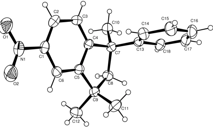
In the molecule of the title compound (Fig. 1), the bond lengths and angles of the phenylindane moiety are comparable
with those observed in 1,1,3-trimethyl-3-phenyl-2,3-dihydro-1H-indane (Men et al., 2008). The C4, C5, C8, C7, C9
atoms in the indane fragment are not coplanar, atom C8 deviating by 0.412 (3) Å from the plane formed by the other four
atoms. The indane benzene ring (C1—C6) and the phenyl ring (C13—18) form a dihedral angle of 76.74 (9)°. The nitro
group is twisted by 5.3 (2)° with respect to the indane benzene ring. The O2—N1—C1—C2 and O1—N1—C1—C2
torsion angles are -175.0 (2)° and 4.8 (4)°, respectively.
S2. Experimental
1,1,3-Trimethyl-3-phenylindane (23.6 g, 0.10 mol) was dissolved in a solution of acetic anhydride (120 ml) and
chloro-form (30 ml) in a three-necked flask. After stirring, the mixture was cooled down to 278 K, and concentrated nitric acid
(8.2 ml, 0.12 mol) was added dropwise in 30 min. Then, the mixture was stirred for 1 h at 283-289 K and poured into
water (200 ml). The organic layer was washed with 10% NaOH (20 ml) and water (150 ml), then dried over anhydrous
magnesium sulfate. After the solvent was removed under reduced pressure, the shallow yellow residue was recrystallized
from a methanol/ethyl solution (2:1 v/v) to give a colourless solid (16.8 g, yield 59.7%, m.p. 402-404 K). Single crystals
suitable for X-ray diffraction were obtained at room temperature by slow evaporation of a methanol solution over a
period of several days.
S3. Refinement
H atoms were positioned geometrically and refined using a riding model, with C—H = 0.93–0.97 Å and with Uiso(H) =
Figure 1
The molecular structure of the title compound, with displacement ellipsoids drawn at the 30% probability level.
1,3,3-trimethyl-5-nitro-1-phenylindane
Crystal data
C18H19NO2 Mr = 281.34 Monoclinic, P21/c
Hall symbol: -P 2ybc
a = 8.306 (3) Å
b = 17.600 (3) Å
c = 12.090 (4) Å
β = 120.50 (3)°
V = 1522.8 (9) Å3
Z = 4
F(000) = 600
Dx = 1.227 Mg m−3
Mo Kα radiation, λ = 0.71073 Å Cell parameters from 20 reflections
θ = 5.4–6.2°
µ = 0.08 mm−1 T = 292 K Block, colourless 0.58 × 0.48 × 0.42 mm
Data collection
Enraf–Nonius CAD4 diffractometer
Radiation source: fine-focus sealed tube Graphite monochromator
ω/2–θ scans
3123 measured reflections 2750 independent reflections 1600 reflections with I > 2σ(I)
Rint = 0.009
θmax = 25.4°, θmin = 1.7°
h = −9→10
k = −21→0
l = −8→14
3 standard reflections every 200 reflections intensity decay: 2.1%
Refinement
Refinement on F2
Least-squares matrix: full
R[F2 > 2σ(F2)] = 0.057 wR(F2) = 0.195 S = 1.12 3524 reflections 275 parameters 0 restraints
Primary atom site location: structure-invariant direct methods
Secondary atom site location: difference Fourier map
Hydrogen site location: inferred from neighbouring sites
w = 1/[σ2(Fo2) + (0.1033P)2]
where P = (Fo2 + 2Fc2)/3
(Δ/σ)max < 0.001
Δρmax = 0.52 e Å−3
Δρmin = −0.40 e Å−3
Special details
Geometry. All esds (except the esd in the dihedral angle between two l.s. planes) are estimated using the full covariance matrix. The cell esds are taken into account individually in the estimation of esds in distances, angles and torsion angles; correlations between esds in cell parameters are only used when they are defined by crystal symmetry. An approximate (isotropic) treatment of cell esds is used for estimating esds involving l.s. planes.
Refinement. Refinement of F2 against ALL reflections. The weighted R-factor wR and goodness of fit S are based on F2,
conventional R-factors R are based on F, with F set to zero for negative F2. The threshold expression of F2 > σ(F2) is used
only for calculating R-factors(gt) etc. and is not relevant to the choice of reflections for refinement. R-factors based on F2
are statistically about twice as large as those based on F, and R- factors based on ALL data will be even larger.
Fractional atomic coordinates and isotropic or equivalent isotropic displacement parameters (Å2)
x y z Uiso*/Ueq
C15 0.7731 (5) 0.45205 (18) 0.6572 (3) 0.0657 (9) H15 0.7272 0.4936 0.6801 0.079* C16 0.8742 (5) 0.39802 (18) 0.7466 (3) 0.0649 (9) H16 0.8993 0.4030 0.8305 0.078* C17 0.9385 (4) 0.33629 (17) 0.7115 (3) 0.0619 (8) H17 1.0052 0.2986 0.7714 0.074* C18 0.9053 (4) 0.32970 (15) 0.5890 (3) 0.0536 (8) H18 0.9512 0.2876 0.5672 0.064*
Atomic displacement parameters (Å2)
U11 U22 U33 U12 U13 U23
O1 0.105 (2) 0.0546 (14) 0.0831 (18) 0.0145 (13) 0.0427 (16) 0.0192 (12) O2 0.0538 (15) 0.114 (2) 0.0807 (18) 0.0204 (14) 0.0243 (14) 0.0379 (15) N1 0.0687 (19) 0.0662 (17) 0.0489 (15) 0.0182 (15) 0.0348 (15) 0.0129 (13) C1 0.0531 (18) 0.0503 (16) 0.0419 (15) 0.0122 (13) 0.0290 (14) 0.0097 (12) C2 0.066 (2) 0.0441 (15) 0.0586 (18) 0.0014 (14) 0.0355 (17) 0.0093 (14) C3 0.0448 (17) 0.0513 (16) 0.0500 (17) −0.0111 (13) 0.0189 (15) −0.0031 (13) C4 0.0422 (16) 0.0455 (14) 0.0407 (15) 0.0016 (12) 0.0240 (14) 0.0024 (11) C5 0.0432 (15) 0.0465 (14) 0.0385 (14) −0.0023 (12) 0.0241 (13) −0.0024 (12) C6 0.0435 (16) 0.0579 (17) 0.0416 (15) 0.0000 (13) 0.0232 (13) 0.0009 (13) C7 0.0430 (16) 0.0428 (14) 0.0445 (16) 0.0007 (11) 0.0225 (14) 0.0013 (12) C8 0.0589 (19) 0.0421 (14) 0.0493 (17) −0.0005 (13) 0.0281 (16) −0.0057 (13) C9 0.0499 (17) 0.0459 (15) 0.0461 (16) −0.0061 (12) 0.0240 (14) −0.0021 (12) C10 0.0440 (17) 0.0702 (19) 0.0536 (18) 0.0084 (14) 0.0255 (15) 0.0057 (14) C11 0.063 (2) 0.0612 (18) 0.073 (2) −0.0036 (15) 0.0405 (18) 0.0111 (16) C12 0.070 (2) 0.0606 (19) 0.0594 (19) −0.0165 (16) 0.0203 (18) −0.0095 (16) C13 0.0376 (15) 0.0455 (14) 0.0427 (15) −0.0034 (11) 0.0194 (13) −0.0001 (12) C14 0.0540 (18) 0.0528 (16) 0.0495 (16) 0.0063 (13) 0.0254 (15) −0.0006 (13) C15 0.071 (2) 0.074 (2) 0.0593 (19) 0.0067 (18) 0.0385 (18) −0.0074 (17) C16 0.070 (2) 0.086 (2) 0.0445 (17) −0.0004 (18) 0.0338 (17) 0.0021 (17) C17 0.064 (2) 0.070 (2) 0.0500 (18) 0.0018 (16) 0.0277 (17) 0.0146 (15) C18 0.0608 (19) 0.0491 (16) 0.0548 (18) 0.0069 (14) 0.0323 (16) 0.0074 (14)
Geometric parameters (Å, º)
C5—C9 1.522 (3) C14—C15 1.385 (4) C6—H6 0.9300 C14—H14 0.9300 C7—C13 1.531 (3) C15—C16 1.363 (4) C7—C10 1.540 (4) C15—H15 0.9300 C7—C8 1.551 (4) C16—C17 1.370 (4) C8—C9 1.535 (4) C16—H16 0.9300 C8—H8A 0.9700 C17—C18 1.366 (4) C8—H8B 0.9700 C17—H17 0.9300 C9—C12 1.524 (4) C18—H18 0.9300