R E V I E W
Open Access
Methods for evaluating cervical range of motion
in trauma settings
Sarah Voss
1*, Michael Page
2and Jonathan Benger
1Abstract
Immobilisation of the cervical spine is a common procedure following traumatic injury. This is often precautionary as the actual incidence of spinal injury is low. Nonetheless, stabilisation of the head and neck is an important part of pre-hospital care due to the catastrophic damage that may follow if further unrestricted movement occurs in the presence of an unstable spinal injury. Currently available collars are limited by the potential for inadequate
immobilisation and complications caused by pressure on the patient’s skin, restricted airway access and
compression of the jugular vein. Alternative approaches to cervical spine immobilisation are being considered, and the investigation of these new methods requires a standardised approach to the evaluation of neck movement. This review summarises the research methods and scientific technology that have been used to assess and measure cervical range of motion, and which are likely to underpin future research in this field. A systematic search of international literature was conducted to evaluate the methodologies used to assess the extremes of movement that can be achieved in six domains. 34 papers were included in the review. These studies used a range of
methodologies, but study quality was generally low. Laboratory investigations and biomechanical studies have gradually given way to methods that more accurately reflect the real-life situations in which cervical spine
immobilisation occurs. Latterly, new approaches using virtual reality and simulation have been developed. Coupled with modern electromagnetic tracking technology this has considerable potential for effective application in future research. However, use of these technologies in real life settings can be problematic and more research is needed.
Keywords:Cervical range of motion, Cervical vertebrae injuries, Immobilisation methods, Evaluation, Assessment, Measurement
Introduction
Serious injuries, particularly those resulting from road traffic collisions, are increasing worldwide with more than one million deaths annually [1]. In many countries with developed pre-hospital and in-hospital trauma sys-tems it is usual practice to apply a cervical collar to the neck of patients who have sustained blunt trauma, par-ticularly those with head injury, to reduce neck move-ment prior to definitive assessmove-ment. This practice is supported by widely accepted international trauma
guidelines, such as those promoted in the “Advanced
Trauma Life Support”(ATLS) course [2]. The theory
be-hind the use of cervical collars is that the trauma patient may have sustained an unstable cervical spine injury,
and that the application of a collar reduces the risk of additional neurological damage before the presence or absence of such an injury can be reliably determined, ei-ther clinically or radiologically.
However, the proposed benefits of cervical collars are theoretical only, and have never been shown to exist in clinical practice [3], with one study concluding that spinal immobilisation had little or no effect on neurological out-come for blunt trauma injury patients [4]. On the con-trary, spinal immobilisation following penetrating trauma has been associated with increased mortality [5]. Further-more, cervical collars are known to cause a number of complications including respiratory compromise, pressure necrosis and raised intracranial pressure [3]. As new approaches to potential cervical spine injury are intro-duced, along with new immobilisation devices, it is im-portant to consider ways in which these can be usefully assessed and compared, particularly in relation to their * Correspondence:[email protected]
1
Faculty of Health and Life Sciences, University of the West of England, Glenside Campus, Blackberry Hill, Bristol, UKBS16 1DD
Full list of author information is available at the end of the article
ability to limit cervical movement. Furthermore, with the application of improved technology to the measurement of neck movement it becomes possible to address a num-ber of related questions. Shafer and Naunheim [6] recently reported a preliminary study in which a six-camera motion capture system was used to examine cer-vical movement during mock extrications from a vehicle, suggesting that for conscious patients neck movement may be minimised by applying a cervical collar and invit-ing the patient to remove themselves from the car, rather than employing a more complex and time-consuming ex-trication technique.
Further research is therefore needed both to evaluate the effectiveness of existing and new immobilisation devices, and also to investigate alternative approaches to cervical spine management in trauma patients. In order to carry out this research, there is a need to identify and employ standardised methods of assessing and measur-ing cervical range of motion. This is the subject of the following review.
Aim
This review aims to provide a brief overview of relatively recent research in the evaluation of cervical spine range of motion (CROM), with particular reference to the test-ing of neck immobilisation devices. It is divided into two main sections:
1) Research methods that have been used to assess CROM, and
2) Scientific methods that have been used to measure and record CROM.
Methods
Data sources and search strategy
A systematic search of international literature was con-ducted in AMED, Cochrane Library, CINAHL Plus, EMBASE, MEDLINE, PubMed, and SPORTDiscuss. Boolean/Phase mode was applied using the terms cer-vical motion OR cercer-vical range of motion OR cercer-vical movement OR cervical range of movement AND eva-luat* OR compar* OR assess*.
Inclusion and exclusion criteria
Due to a lack of high quality research in the area all study types were included. Peer reviewed articles published in English between January 2001 and January 2012 were included in the search. Articles were excluded if the pri-mary focus of the evaluation was to assess neck pain, chronic disorders or range of motion in children. Studies were included only if they were considered relevant to the study objectives. A summary of papers included in the study is available in Additional file 1: Appendix. Informa-tion was extracted from each study on: methods used to assess CROM (11 papers included) [6-16]; technologies used to evaluate and quantify CROM (29 papers included) [7-9,11,13,16-39]; the statistics supporting the main find-ings of the study.
Results
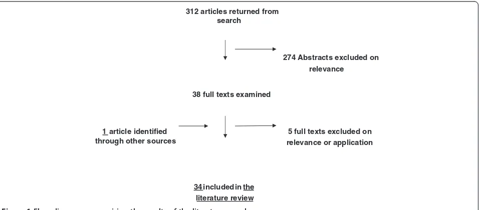
A flow diagram summarising the results of the literature search is shown in Figure 1.
CROM domains
CROM is generally described in the following terms:
312 articles returned from search
274 Abstracts excluded on relevance
38 full texts examined
1 article identified through other sources
5 full texts excluded on relevance or application
[image:2.595.61.539.512.722.2]34 included in the literature review
Extension (E)
Flexion (F)
Right rotation (RR)
Left Rotation (LR)
Left lateral flexion (LLF)
Right lateral flexion (RLF)
Research methods to assess CROM
There has been much research to assess CROM; this has been conducted to test both the efficacy of different devices and also to compare CROM during different movement procedures.
When comparing devices, most authors compare CROM in all the domains, before and after application of a cervical collar [7-9]. Volunteers are instructed to ac-tively move their necks as much as possible in the direc-tions of flexion, extension, rotation, and lateral flexion while lying supine or seated.
Hoppenbrouwers et al [10] used both passive and ac-tive methods to assess CROM using two independent examiners. For the passive tests, the subject was instructed to allow the tester to move the subject's neck through the motions without resisting. The authors do not specify the amount of force applied for the passive testing, but each movement was carried out twice by each examiner. For the active tests, the subject was instructed to rotate his or her head as far as possible in the direction of the motion being tested. The authors conclude that reproducibility for active and passive range of motion is good for extension but poor for lateral flexion.
Numerous investigations, similar to those described above, have attempted to assess the general ability of cervical collars to restrict or reduce both segmental and overall spinal motion. However, these studies were designed to evaluate the ability of collars to prevent the cervical spine from moving (be it actively or passively) through large ranges of extreme motion.
Del Rossi et al [11] suggest that the results from such studies cannot be used to establish the relative useful-ness of cervical collars:
“Normally, the purpose of an extrication-type collar is to help minimize or control the (presumably) small amount of spinal motion that may be unintentionally generated during the pre-hospital stages of emergency care. Extrication collars are thus more aptly studied if the experimental protocol that is developed includes an external loading condition that results in the production of spinal motion that approximates the quality and
quantity of motion generated in real-life situations.”
(Pg. 620).
Earlier research by Richter et al [12] addressed this issue by using a 50 N load to test the ability of four col-lars to control the amount of motion produced between
unstable cervical spine segments. The authors surgically created an experimental lesion at C1-2 and C2-3 in four cadavers. However, it would not be practical to apply this methodology in vivo; the authors recognise that fac-tors such as patient comfort and compliance are beyond the limits of a biomechanical study.
Del Rossi et al [11] also used cadavers of differing morphologies with surgically created spinal lesions (complete segmental injury resulting in complete in-stability) to compare 3 collars. They recruited 6 partici-pants to execute log rolls and lift-and-slide transfer techniques and assessed CROM using electromagnetic tracking. Their findings indicated that there were no sig-nificant differences in the 3 collars in limiting CROM during either procedure (the three devices had similar characteristics). Moreover, there were no significant dif-ferences between any of the collar treatments and the control condition (no collar). The authors acknowledge a number of limitations in their methodology. These in-clude issues with the use of cadavers; muscle changes resulting in dissimilarity in motion to living patients and the surgical creation of one type of injury reducing the generalisability of findings. They recommend that repli-cation of research of this nature should use a larger sample size and cervical devices with different character-istics. The latter recommendation is derived from their observation that significantly greater CROM was gener-ated by log roll than the lift-and-slide procedure.
was more precise than conventional assessment. One point to note is that LLF and RLF were not assessed. However, this VR method has also been found to be valid and reliable in the clinical assessment of deficits in movement control [14].
The characteristics of different devices will vary according to their purpose; rehabilitation collars require different qualities from trauma collars. There is a need to evaluate the effectiveness of cervical orthoses in the environment in which they will be employed; i.e. outside of the laboratory [15]. James et al [7] completed a study to quantify CROM during the application of four differ-ent rigid collars in a simulated, athletic- related, spine-board situation. Similarly, Krell et al [16] examined differences in spinal movement whilst healthy subject were being placed onto a traditional long backboard and a commercially available scoop stretcher.
As outlined in the introduction, Shafer and Naunheim [6] used a mock extrication scenario to compare differ-ent methods of extrication. They developed a mock-up vehicle with the roof removed and compared 4 extrica-tion methods, evaluating CROM with a camera moextrica-tion capture system. They recruited 3 paramedics and one acted as driver and was extricated by the other two, for each of the methods being investigated. The main limita-tions acknowledged by the authors were: problems with the use of the camera system and placement of markers resulting in a loss of data; use of a mock-up vehicle (no further detail given); use of medically trained personnel as participants (their medical knowledge was considered a drawback). The limitations of the measurement record-ing in this study were unfortunate, and the authors en-courage future studies to consider other methods.
Scientific methods for measuring and recording CROM Measurement of cervical motion provides substantial in-formation for clinicians about the severity of motion limitation as well as being applicable to the evaluation of new immobilization devices and techniques [17]. Conse-quently, there has been much research to evaluate the reliability and validity of different measurement techniques.
Radiography Radiographs are used to examine segmen-tal motion confined to a single plane; therefore multiple exams are needed to assess the different domains [18,19]. This is ethically problematic due to the asso-ciated radiation exposure, and is not appropriate for de-velopmental research with volunteers.
Goniometers and inclinometers Various commercial measurement devices have been used to measure CROM in the clinical setting [20,21]. Hostler et al [9] used a goniometer and tape measure to compare three cervical
immobilization devices on healthy volunteers in a la-boratory setting.
The Cervical Range of Motion device (CROM) is a validated piece of equipment for measuring cervical spine range of movement [22-25] and has been used in a number of cervical movement studies [8,26,27]. The de-vice consists of a plastic frame mounted over the nose and ears and secured with a strap. Flexion, extension and lateral flexion are recorded by two gravity goni-ometers. However, the CROM does not measure rota-tion and can only be used on participants in an upright position as the measurement system relies on gravity. A study by Schneider et al [28] compared cervical range of movement in seven different orthoses. The authors em-phasise the importance of effective immobilisation in both supine and upright positions; it is essential to be able to assess this in order to evaluate a cervical collar.
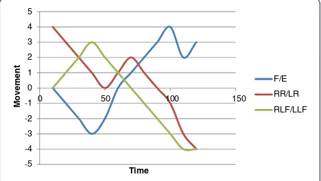
Electromagnetic tracking devices In recent years there has been increasing interest in more accurate and reli-able methods of measuring CROM. This has involved the development and use of non-invasive methods of 3D motion analysis. Sensors are fitted at bony anchor points (e.g. head and sternum) and computer software is used to measure relative movement. These methods produce data to show movements over time in the relative domains. This can be represented graphically, with Figure 2 offering an example, and there are further illustrations in other papers [13]. This form of tracking offers great promise for evaluating CROM when comparing immobilisation devices in both simulated and real conditions.
Published literature concerned with the evaluation of CROM reports on a variety of electromagnetic systems. The Flock of Birds (Ascension, Burlington, USA) is a sophisticated electromagnetic tracking device, but it requires extensive calibration and is not portable [29,30]. Motion Star (Ascension, Burlington, USA) has been used in research involving simulated scenarios [7,16,31]
5 4 3 2 1 0 1 2 3 4 5
0 50 100 150
Movement
Time
F/E
RR/LR
[image:4.595.305.539.556.688.2]RLF/LLF
and the Liberty device (Polhemus Inc., USA) and the Optotrak (Northern Digital, USA) have been used in a variety of cavaderic biomechanical studies [32-36].
An increasingly popular method of electromagnetic tracking is the 3D Fastrak measurement system (Polhe-mus Inc., USA) [11,13,37]. Rechtine et al [38] used Fas-trak to assess movement during hospital bed transfers with different collars. Some recent studies considered Fastrak to be a criterion standard instrument for evalu-ation of cervical ROM, and have used it for the valid-ation of other methods [23,39].
Discussion
Assessing CROM with sufficient accuracy to evaluate dif-ferent neck collars is problematic. In order to compare the efficacy of devices, the physical characteristics of those wearing them (e.g. BMI, height, neck circumference) need to be controlled. There is no commonly accepted method-ology for standardising movement, and there is debate about whether subjects should be instructed to move in all directions as far as they can or if the movement should be passive. In terms of active movement, strength and size of the subject as well as interpretation of the instructions given by an assessor are likely to confound the findings. For passive movements there is no accepted degree of force that should be applied, and the reliability of findings to date has been based on the use of a consistent force and assessor for all subjects.
In clinical use, cervical collars need to minimise or control the small amount of spinal motion that may be unintentionally generated during the pre-hospital stages of emergency care. Thus there is a need for an experi-mental protocol that approximates the quality and quan-tity of motion generated in real-life situations.
Recent research has focussed on evaluating CROM in more authentic situations using virtual reality or mock ex-trication. This has the potential to provide a useful func-tional comparison of different immobilisation devices and techniques, particularly when combined with modern electromagnetic tracking systems which are proving to have both the reliability and accuracy required to usefully compare different approaches to cervical immobilisation. However, a drawback to these technologies is the fact that electromagnetic waves can be distorted by proximity to metal objects [39]. This has the potential to limit their usefulness in both simulated and clinical settings. Thus, there is a need to explore further portable or multiple-modality technology that can be used to accurately evalu-ate devices and procedures that are designed to protect the cervical spine in trauma care.
Conclusion
The effective comparison of various devices and techni-ques to immobilise the cervical spine following trauma
requires robust methods to evaluate cervical range of motion in a range of static, dynamic, simulated and clin-ical situations. None of the current systems are perfect, and controversy remains over the best way to undertake a systematic evaluation. Electromagnetic tracking tech-nology is emerging as the preferred and most reliable approach for laboratory and biomechanical studies. How-ever, electromagnetic measurement may be less useful in simulated and clinical environments as a result of the interference caused by the close proximity of metal. Therefore, the identification of reliable and valid protocols and technologies to measure CROM in these most im-portant settings is a priority for future research. This will in turn facilitate the comparison of different devices and techniques for cervical immobilisation in trauma patients, thereby improving future standards of clinical care.
Additional file
Additional file 1: Appendix.Summary of papers included in the review.
Competing interests
The authors declare that they have no competing interests.
Authors’contributions
SV conducted the review and drafted the manuscript. MP assisted with the review and inclusion and exclusion of relevant studies. JB helped draft the manuscript and provided clinical context for the aim of this research. All authors read and approved the final manuscript.
Acknowledgements
The authors would like to thank Pauline Shaw for advice on the search strategy and providing access to the papers.
Author details
1Faculty of Health and Life Sciences, University of the West of England,
Glenside Campus, Blackberry Hill, Bristol, UKBS16 1DD.2Great Western Ambulance Service NHS Trust, Chippenham, UK.
Received: 28 March 2012 Accepted: 23 June 2012 Published: 2 August 2012
References
1. World Health Organization:Global status report on road safety: time for action. Geneva: WHO; 2009. http://whqlibdoc.who.int/publications/2009/ 9789241563840_eng.pdf.
2. American College of Surgeons Committee on Trauma:Advanced Trauma Life Support (7th Edition). Chicago: American College of Surgeons; 2007. 3. Benger J, Blackham J:Why do we put cervical collars on conscious
trauma patients?Scand J Trauma, Resuscitation Emerg Med2009,17:44. doi:10.1186/1757-7241-17-44.
4. Hauswald M, Ong G, Tandberg D, Omal Z:Out-of-hospital spinal immobilization: its effect on neurologic injury.Acad Emerg Med1998,5 (3):214–219.
5. Haut ER, Kalish BT, Efron DT, Haider AH, Stevens KA, Kieninger AN, Cornwell EE, Chang DC:Spine immobilization in penetrating trauma: more harm than good?J Trauma-Injury Infect Crit Care2010,68(1):115–121. 6. Shafer JS, Naunheim RS:Cervical spine motion during extrication: a pilot
study.West J Emerg Med2009,10(2):74–8.
8. Tescher AN, Rindflesch AB, Youdas JW, Jacobson TM, Downer LL, Miers AG, Basford JR, Cullinane DC, Stevens SR, Pankratz VS, Decker PA: Range-of-motion restriction and craniofacial tissue-interface pressure from four cervical collars.J Trauma2007,63(5):1120–1126.
9. Hostler D, Colburn D, Seitz SR:A comparison of three cervical immobilization devices.Prehospital Emerg Care2009,13(2):256–260. 10. Hoppenbrouwers M, Eckhardt M, Verkerk K, Verhagen A:Reproducibility of
the measurement of active and passive cervical range of motion.J Manip Physiol Ther2006,29(5):363–367.
11. Del Rossi G, Horodyski M, Heffernan TP, Powers ME, Siders R, Brunt D, Rechtine GR:Spine-board transfer techniques and the unstable cervical spine.Spine2004,29(7):E134–8.
12. Richter D, Latta L, Milne E, Varkarakis G, Biedermann L, Ekkernkamp A, Ostermann P:The stabilizing effects of different orthoses in the intact and unstable upper cervical spine: a cadaver study.J Trauma Inj, Infect Crit Care2001,50(5):848–854.
13. Sarig-Bahat H, Weiss PL, Laufer Y:Cervical motion assessment using virtual reality.Spine2009,34(10):1018–1024.
14. Kristjansson E, Oddsdottir GL:"The fly": a new clinical assessment and treatment method for deficits of movement control in the cervical spine: reliability and validity.Spine2010,35(23):E1298–305.
15. Swartz EE, Nowak J, Shirley C, Decoster LC:A comparison of head movement during back boarding by motorized spine-board and log-roll techniques.J Athl Train2005,40(3):162–168.
16. Krell JM, Mccoy MS, Sparto PJ, Fisher GL, Stoy WA, Hostler DP:Comparison of the Ferno Scoop Stretcher with the long backboard for spinal immobilization.Prehospital Emerg Care2006,10(1):46–51.
17. Prushansky T, Dvir Z:Cervical motion testing: methodology and clinical implications.J Manip Physiol Ther2008,31(7):503–508.
18. Wolfenberger VA, Bui Q, Batenchuk DC:A comparison of methods of evaluating cervical range of motion.J Manip Physiol Ther2002,25(3):154–160. 19. Lador R, Ben-Galim P, Hipp JA:Motion within the unstable cervical spine
during patient maneuvering: the neck pivot-shift phenomenon.J Trauma
2011,70(1):247–251.
20. Yankai A, Manosan P:Reliability of the universal and invented gravity goniometers in measuring active cervical range of motion in normal healthy subjects.Int J Appl Biomed Eng2009,2(1):49–53.
21. Prushansky T, Deryi O, Jabarreen B:Reproducibility and validity of digital inclinometry for measuring cervical range of motion in normal subjects. Physiother Res Int2010,15(1):42–48.
22. Tousignant M, Smeesters C, Breton A, Breton E, Corriveau H:Criterion validity study of the cervical range of motion (CROM) device for rotational range of motion on healthy adults.J Orthop Sports Phys Ther
2006,36(4):242–248.
23. Audette I, Dumas J, Cote JN, De Serres S:Validity and between-day reliability of the cervical range of motion (CROM) device.J Orthop Sports Phys Ther2010,40(5):318–323.
24. Whitcroft KL, Massouh L, Amirfeyz R, Bannister G:Comparison of methods of measuring active cervical range of motion.Spine2010,35(19):E976–80. 25. Williams MA, Mccarthy CJ, Chorti A, Cooke MW, Gates S:A systematic
review of reliability and validity studies of methods for measuring active and passive cervical range of motion.J Manip Physiol Ther2010,33 (2):138–155.
26. Lark S, McCarthy P:Active cervical range of motion recovery following the rugby off-season.J Sports Med Phys Fit2010,50(3):318–325. 27. Whitcroft KL, Massouh L, Amirfeyz R, Bannister GC:A comparison of neck
movement in the soft cervical collar and rigid cervical brace in healthy subjects.J Manip Physiol Ther2011,34(2):119–122.
28. Schneider AM, Hipp JA, Nguyen L, Reitman CA:Reduction in head and intervertebral motion provided by 7 contemporary cervical orthoses in 45 individuals.Spine2007,32(1):E1–6.
29. Assink N, Bergman G, Knoester B, Winters JC, Dijkstra PU:Assessment of the cervical range of motion over time, differences between results of the Flock of Birds and the EDI-320: a comparison between an electromagnetic tracking system and an electronic inclinometer.Man Ther2008,13(5):450–455.
30. Gelalis ID, Defrate LE, Stafilas KS, Pakos EE, Kang JD, Gilbertson LG: Three-dimensional analysis of cervical spine motion: reliability of a computer assisted magnetic tracking device compared to inclinometer.Eur Spine J
2009,18(2):276–281.
31. Agarwal S, Allison GT, Singer KP:Validation of the Spin-T goniometer, a cervical range of motion device.J Manip Physiol Ther2005,28(8):604–609. 32. Dipaola CP, Sawers A, Conrad BP, Horodyski M, Dipaola MJ, Del Rossi G,
Rechtine G:Comparing cervical spine motion with different halo devices in a cadaveric cervical instability model.Spine2009,34(2):149–155. 33. Dipaola CP, Conrad BP, Horodyski M, Dipaola MJ, Sawers A, Rechtine G:
Cervical spine motion generated with manual versus Jackson table turning methods in a cadaveric c1-c2 global instability model.Spine
2009,34(26):2912–2918.
34. Horodyski M, Conrad BP, Del Rossi G, Dipaola CP, Rechtine G:Removing a patient from the spine board: Is the lift and slide safer than the log roll? J Trauma2011,70(5):1282–1285.
35. Ivancic PC, Telles CJ:Neck motion due to the halo-vest in prone and supine positions.Spine2010,35(10):E400–6.
36. Conrad BP, Rechtine G, Weight M, Clarke J, Horodyski M:Motion in the unstable cervical spine during hospital bed transfers.J Trauma2010,69 (2):432–436.
37. Amiri M, Jull G, Bullock-Saxton J:Measuring range of active cervical rotation in a position of full head flexion using the three-dimensional Fastrak measurement system: an intratester reliability study.Man Ther
2003,8(3):176–9.
38. Rechtine G, Del Rossi G, Conrad B, Horodyski M:Motion generated in the unstable spine during hospital bed transfers.J Trauma-Inj Infect Crit Care
2004,57(3):609–612.
39. Jasiewicz JM, Treleaven J, Condie P, Jull G:Wireless orientation sensors: their suitability to measure head movement for neck pain assessment. Man Ther2007,12(4):380–385.
doi:10.1186/1757-7241-20-50
Cite this article as:Vosset al.:Methods for evaluating cervical range of motion in trauma settings.Scandinavian Journal of Trauma, Resuscitation and Emergency Medicine201220:50.
Submit your next manuscript to BioMed Central and take full advantage of:
• Convenient online submission
• Thorough peer review
• No space constraints or color figure charges
• Immediate publication on acceptance
• Inclusion in PubMed, CAS, Scopus and Google Scholar
• Research which is freely available for redistribution