Original Article
H
2S improves myocardial fibrosis
caused by hyperthyroidism by downregulating stat
pathways and regulating mir-21 and mir-29a expression
Wenting Tan1, Liangui Nie1, Fen Tang1, Shengquan Liu1, Junrong Long1, Chun Chu2, Jun Yang1
1Department of Cardiology, The First Affiliated Hospital of University of South China, Hengyang 421001, Hunan,
China; 2Department of Pharmacy, The Second Affiliated Hospital of University of South China, Hengyang 421001,
Hunan, China
Received September 16, 2018; Accepted February 8, 2019; Epub May 15, 2019; Published May 30, 2019
Abstract: Objective: The aim of this study was to explore the effects of H2S on myocardial fibrosis induced by hy-perthyroxine and expression of STAT signal pathways, miR-21, and miR-29a in the myocardium. Methods: A total of 40 rats were selected and randomly stratified into 4 groups, including the control group (Control), HCM model group (Thy), H2S-intervention model group (Thy+H2S), and H2S-intervention normal group (H2S). Hyperthyroidism rat models were prepared by intraperitoneal injections of L-thyroxine. Rats in the control group received intraperi-toneal injections of normal saline. NaHS solution was added to rats in the Thy+H2S group and H2S group. LVs, LVd, LVPWs, and EF values were measured by echocardiography, Pathological changes in myocardial cells and myocardial interstitium were observed by H&E staining. Collagen deposition in the myocardium was also observed by Masson’s staining. Protein expression of STAT1, STAT3, AKT1, TGF-β1, NFKB p65, PI3K, TIMP1, TIMP4, MMP12, MMP13, MMP16, and MMP24 was detected by Western blotting and expression of miR-21 and miR-29a in myo-cardial tissues was measured by real-time fluorescence quantitative PCR (RT-qPCR). Results: Compared with the control group, rats in the Thy group showed significant decreases in weight and rapid acceleration in heart rates (P<0.05). Findings of pathological examinations revealed that myocardial fibers were in disordered arrangement. Collagen deposition in the myocardium was significantly increased and values of LVs and LVd were significantly increased (P<0.05). Moreover, protein expression of STAT1, STAT3, AKT1, TGF-β1, PI3K, TIMP1, TIMP4, MMP13, and MMP24 in myocardial tissues was significantly upregulated (P<0.05). Expression of MMP12 and MMP16 was significantly downregulated (P<0.05). Comparisons between the Thy group and Thy+H2S group suggested that the weight of rats in Thy+H2S was significantly increased, accompanied by a reduction in heart rates (P<0.05). Also, the pathological examination showed that some improvement was identified in myocardial fibers in disordered arrange-ment and collagen deposition in the myocardium was significantly reduced. Moreover, values of LVs and LVd were significantly decreased (P<0.05), protein expression of STAT1, STAT3, AKT1, TGF-β1, PI3K, TIMP1, TIMP4, MMP13, and MMP24 was obviously downregulated (P<0.05), and expression of MMP12 and MMP16 was significantly up-regulated (P<0.05). Conclusion: H2S can ameliorate myocardial fibrosis induced by hyperthyroxine. Relevant mecha-nisms might be correlated with regulation of STAT3 signal pathways, downregulation of expression of miR-21, and upregulation of expression of miR-29a.
Keywords: H2S, hyperthyroid cardiomyopathy, STAT, miR-21, miR-29a
Introduction
Hyperthyroid cardiomyopathy (HCM), a type of endocrinological cardiomyopathy, is character-ized by a series of cardiovascular systems and vital signs caused by direct or indirect toxicity on the heart generated by excessive thyroid hormones. It can lead to variations, such as hypertension and arrhythmia or even structural
of p-STAT in rats of the diabetic cardiomyopathy group was significantly upregulated, with obvi-ous fibrosis in the myocardial interstitium [5]. Some studies have shown that STAT signaling pathway can promote the activation of PI3K/ AKT signaling pathways [6] and inflammatory factors were found overexpressed. It was found that micro-RNAs, a type of short non-coding endogenous RNAs, are involved in the regula-tion of myocardial fibrosis [7]. A study found that miR-29 might be involved in the expression of collagen-I via Smad3-dependent TGF-β1 pathways [8]. Hydrogen sulfide (H2S) is a type of newly found endogenous gaseous signaling molecule. Its multiple biological effects include resistance to oxidative stress, fibrosis, and my- ocardial remodeling. Whether H2S can exert its protective effects in hyperthyroid cardiomyopa-thy or function in regulation via STAT pathways or miRNA remains unclear. Thus, the hyperthy-roid cardiomyopathy rat models were built in this study to investigate the effects of H2S on myocardial fibrosis induced by hyperthyroidism and expression of STAT, miRNA-21, and miR- 29a.
Experimental material
Experimental animals
A total of 40 adult male SD rats, with an aver-age weight of (180±40) g, were purchased from Laboratory Animal Center of University of South China. Rats were fed in separate cages in a clean laboratory at (23±1)°C, during which the day and night were set to 12 hours, respective-ly. All rats could freely access water and food.
Major experimental reagents
NaHS, L-thyroxine, EDTA, Tris, and DEPC were purchased from Sigma, USA; BCA protein quan-titative kit was purchased from Solarbio Co., Ltd., China; RIPA was provided by Beyotime Biotech Co., Ltd., China; Horseradish peroxi-dase-labelled goat anti-mouse IgG was pur-chased from Proteintech Group, USA; Primary antibodies of GAPDH, T1MP1, TIMP4, MMP12, MMP13, MMP16, and MMP24 were provided by Boster Biological Technology Co., Ltd., China; STAT1, STAT3, AKT, NFKB p65, PI3K, and TGF-β1 were purchased from Cell Signaling Co., Ltd., USA; Reverse transcription kit of miRNA and reagent for extracting the free RNA were pur-chased from Cwbiotech, China; Taq enzyme,
DL2000 DNA Marker and dNTP were purchased from Genestar, China; Design and synthesis of primers were performed by Sangon Biotech, Shanghai, China.
Experimental methods
Establishing the animal models and grouping
After being fed for 1 week, SD rats were ran-domly stratified into 4 groups, the control group (Control), HCM model group (Thy), H2S-inter- vention model group (Thy+H2S), and H2S-inter- vention normal group (H2S). Hyperthyroidism rat models were prepared by intraperitoneal injections of L-thyroxine (100 µg/kg/day). Rats in the control group underwent intraperitoneal injections of normal saline. NaHS solution (100 umol/kg) was given to rats for 4 weeks in the Thy+H2S group and H2S group, then left ven-tricular posterior wall thickness (LVPWs), left ventricular end diastolic diameter (LVd), left ventricular end systolic diameter (LVs), and ejection fraction (EF) were measured by echo-cardiography. After the continuous measure-ment of three cardiac cycle parameters, the mean value was calculated.
HE staining detection
Myocardial tissues in left ventricle were col-lected for rats in each group. They were then fixed in 4% paraformaldehyde solution, followed by rinsing with running water, dehydration, clearing with xylene, and embedding with par-affin. Subsequently, sections were then regu-larly dewaxed and hydrated for staining with hematoxylin. After free hematoxylin was re- moved by rinsing, 1% hydrochloric-alcohol solu-tion was added onto the secsolu-tion for differentia-tion. Next, the sections were washed by warm water until turning blue. Moreover, 1% eosin was applied for counterstaining. Sections were dried and sealed using neutral balsam, after being rinsed, dehydrated by gradient alcohol, and cleared. Sections were observed under the light microscope.
Detecting collagen deposition via Masson’s staining
dehydra-tion with gradient alcohol, clearing with xylene, and embedding with paraffin. Subsequently, sections were regularly dewaxed and hydrated for staining with hematoxylin. Next, sections were placed under running water for rinsing, then 1% hydrochloric-alcohol solution was add- ed onto the section for differentiation. After- ward, ponceau fuchsin acid was used for stain-ing, followed by rinsing. Next, 1% phosphomo-lybdic acid solution was used for intervention for 5 minutes. Next, sections were counter-stained with aniline blue or green, treated with glacial acetic acid, dehydrated with anhydrous ethanol, cleared with xylene, and sealed by neutral balsam. Sections were placed under a microscope for observation.
Detecting target protein expression via Western blotting
From the -80°C refrigerator, rat hearts in each group were taken out to extract proteins. After quantification using BCA method, protein sam-ples were boiled for 5 minutes at 100°C. Next, 10% SDS-PAGE separation gel was prepared strictly following the proportions in instructions and then placed into the buffer for electropho-resis. Targeted stripes were transferred onto a membrane, which was then blocked with TBST. Next, primary antibodies of STAT1, STAT3, AKT, TGF-β1, NFKB p65, PI3K, TIMP1, TIMP4, MMP12, MMP13, MMP16, and MMP24 and GAPDH (1:1000) were added onto the mem-brane for incubation at 37°C. This was followed by incubation at 4°C overnight. Membranes were then washed with TBST and incubated with secondary antibody (1:2000) at 37°C. Subsequently, membranes were washed with TBST and treated by ECL for color development, followed by exposure and scanning. In this sec-tion, the primary antibody of GAPDH was select-ed as internal reference.
Detecting expression of miR-21 and miR-29a via RT-Qpcr
Tissues preserved in TRIzol were ground and cracked. Subsequently, tissues were trans-ferred into the chloroform for vibration, followed by standing and centrifugation. This study pre-pared the reaction solutions in accordance with the requirements of kit and according to the instructions on reverse transcription. Moreover, cRNA was synthesized with the total RNA as the template. Also, negative control was set. Re- verse transcription was performed as follows: 37°C for 15 minutes and 85°C for 5 seconds; cDNA reaction solution for synthesis was direct-ly employed for fluorescence quantitative de- tection, in which the sequences of primers were as follows. Quantitative PCR reaction pro-cedures were set as follows: 95°C for 10 min-utes for 1 cycle; 95°C for 10 seconds, 59°C for 50 seconds, for a total of 40 cycles. Temperature was collected in real-time with the melting curve: 60°C-95°C. Results were studied.
Statistical analysis
SPSS 18.0 software was employed for statisti-cal analysis. Data of observation indexes are denoted as mean ± SEM. LSD-t test was per-formed for intergroup comparisons and one-way ANOVA was conducted for analysis of ran-domized grouping design. P<0.05 suggests that differences are statistically significant.
Results
General conditions of rats in each group
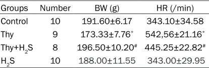
Four weeks later, compared with the control group, rats in Thy group showed significant decreases in weight and rapid increases in heart rates (P<0.05). Compared with the Thy group, the weights and heart rates of rats in the Thy+H2S group were respectively increased and slowed down (P<0.05). By comparing the con-trol group and H2S group, differences in weights and heart rates of rats showed no statistical significance (P>0.05) (Table 1). These results indicate that H2S could increase BM and de- crease HR caused by hyperthyroidism.
Detection results of echocardiography
LVs and LVd rose significantly in the Thy group and EF decreased significantly (P<0.05),
com-Table 1. Body weight and heart rate in each group (mean ± SD)
Groups Number BW (g) HR (/min)
Control 10 191.60±6.17 343.10±34.58
Thy 9 173.33±7.76* 542,56±21.16*
Thy+H2S 8 196.50±10.20# 445.25±22.82#
H2S 10 188.00±11.55 343.00±29.95
Mean ± SD: *P<0.05 vs. the Control group; #P<0.05 vs. the
[image:3.612.92.299.96.164.2]pared with those of the Control group. Thy+H2S group rats LVs and LVd were significantly lower (P<0.05). EF had no significant difference (P>0.05), compared with Thy group. The LVPWs value in each group showed no significant dif-ferences (P>0.05). Control group and H2S group showed no significant changes (P>0.05) (Table
2). These results indicate that H2S can improve heart function caused by hyperthyroidism.
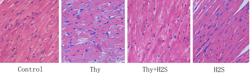
Analysis of H&E staining results for myocardial tissues
According to H&E staining results, in the control group, myocardium was in regular morphology and the structure was visible. In the Thy group, the diameter of myocardial cell increased and myocardial cells were disordered. Disordered arrangement in myocardial cells in the Thy+H2S group was somehow ameliorated, compared with that in the Thy group (Figure 1). Results showed that the improvement of heart fibrosis caused by hyperthyroidism after H2S interven-tion was significantly improved.
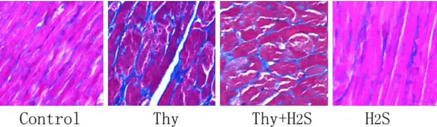
Analysis of Masson’s staining results for myo-cardial tissues
Masson’s staining results revealed that myo-cardial cells of rats in the Thy group were in dis-ordered arrangement, blue stained collagen fibers were significantly augmented, and
obvi-ous fibrosis was observed in the myocardial interstitium, compared with those of the con-trol group. However, compared with the Thy group, the ameliorations in disordered arrange-ment of myocardium, a significant decrease in blue stained collage fibers, and an improve-ment in myocardial fibrosis were found (Figure
2).
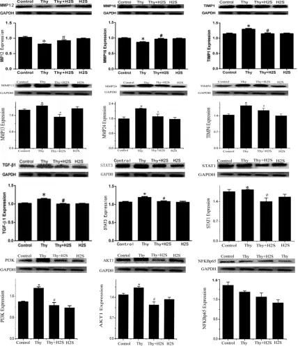
Effects of H2S on protein expression of STAT3, TGF-β1, TIMP1, MMP12, and MMP16 in myo-cardium in rats
Western blotting detection revealed that, com-pared with the control group, significant upreg-ulation was seen in the protein expression of STAT3, TGF-β1, and TIMP1 in myocardium in ra- ts in the Thy group (P<0.05). Protein expression of MMP12 and MMP16 was significantly down-regulated (P<0.05). Compared with the Thy group, significant downregulation was seen in the protein expression of STAT3, TGF-β1, and TIMP1 in myocardium in rats in the Thy group (P<0.05). Protein expression of MMP12 and MMP16 was significantly up-regulated (P<0.05) (Figure 3).
Detecting expression of miR-21 and miR-29a in myocardium of rats in all groups
[image:4.612.91.524.84.153.2]According to RT-qPCR results, miR-21 expres-sion in myocardium of rat in the Thy group was
Table 2. Comparison of echocardiographic parameters in each group (mean ± SD)
Groups Number LVd (mm) LVs (mm) LVPW (mm) EF (%)
Control 10 4.33±0.09 2.67±0.12 0.90±0.05 69.90±6.14
Thy 9 5.63±0.03* 4.10±0.12* 1.00±0.08 65.44±5.55
Thy+H2S 8 4.93±0.09# 3.50±0.06# 1.03±0.03 67.88±6.45
H2S 10 4.30±0.11 2.40±0.07 0.87±0.33 68.80±5.75
Mean ± SD: *P<0.05 vs. the Control group; #P<0.05 vs. the Thy group.
[image:4.612.92.525.188.314.2]significantly higher than that in the control group (P<0.05) and expression of miR-29a was significantly down-regulated (P<0.05). However, when compared with the Thy group, expression of miR-21 in myocardium of rats in the Thy+H2S group showed a significant decrease (P<0.05) and expression of miR-29a was up-regulated (P<0.05). Comparing the H2S group and the con- trol group, there existed no statistically signifi-cant differences in expression of miR-21 and miR-29a (P>0.05) (Figure 4). Results indicate that H2S could down-regulate miR-21 and up-regulate miR-29a to improve myocardial fibrosis.
Discussion
In general, pathological changes of hyperthy-roid cardiomyopathy include cardiac hypertro-phy, myocardial fibrosis, and left ventricular dysfunction, which are mainly associated with the toxicity of excessive thyroid hormones on myocardium. During the disease course of hyperthyroidism, renin-angiotensin-aldoste-rone system is activated to induce the prolifer-ation of cardiac fibroblasts and synthesis and deposition of collagen, thus resulting in fibrosis in myocardial interstitium [9]. Matrix metallo-proteinases are a Zn2+-dependent endopepti-dase family and TIMPs are specific inhibitors of MMPs. Disproportionate MMPs/TIMPs will ca- use disorders in extracellular matrix, further leading to myocardial fibrosis. Existing studies have confirmed that disproportionate MMPs/ TIMPs will induce myocardial fibrosis [10]. TGF-β1, a member of transforming growth factor family, can affect the synthesis and metabo-lism of extracellular matrix and bring about the excessive aggregation of extracellular matrix and potent pro-fibrosis effects. Occurrence of
myocardial fibrosis is also associated with dis-proportionate MMPs/TIMPs and regulation of TGF-β1 signal pathways. Thus, TGF-β1 can induce myocardial fibrosis through facilitating the deposition of extracellular matrix. PI3K/ AKT (phosphodylinsitol-3-kinase-series/thru ki- nase) signaling pathways play an important role in the prevention of myocardial ischemia-reper-fusion injury and inhibition of apoptosis [11]. At the same time, it was found that STAT signaling pathways can promote the activation of PI3K/ AKT signaling pathways. NFKB p65 (nuclear transcription factor-kb p65) is a group of nucleoprotein factors that regulate expression of a wide range of genes. NFKB p65 is vital for the regulation of gene transcription related to inflammatory response, cell proliferation, dif-ferentiation and apoptosis, immune response, and tumor formation [12]. In this study, signifi-cant upregulation was seen in the protein expression of STAT1, STAT3, TGF-β1, PI3K, AKT, TIMP1, TIMP4, MMP13, and MMP24 in myocar-dium in rats in the Thy group. Protein expres-sion of MMP12 and MMP16 was significantly down-regulated, suggesting that myocardial fi- brosis in rats with hyperthyroid cardiomyopathy might be correlated with the upregulation of TGF-β1 and disproportionate MMPs/TIMPs.
[image:5.612.94.521.71.196.2]MMPs/TIMPs proportion [15]. In addition, a study reported the down-regulation of expres-sion of miR-29 in the marginal zone of acute myocardial infarction, which can also induce the deposition of extracellular matrix and myo-cardial fibrosis [16]. In a study by Yuqing Huang et al., they further verified that, in patients with
[image:6.612.92.520.73.570.2]lation in myocardial energy metabolism [18]. STAT can deliver the extracellular signals to cell nucleus, thereby affecting the transcription of targeted genes and regulating cell proliferation, differentiation, apoptosis and immunoregula-tion. Thus, it is one of the key downstream sig-nal pathways of cytokines. Existing studies have found that STAT3 is involved in the regula-tion of miRNA expression, including miR-21, and participates in the fibrosis mechanism through affecting the activity of miR-21 to regu-late MMPs/TIMPs levels [19, 20]. Results here revealed that, besides the significant myocar-dial fibrosis and disproportionate MMPs/ TIMPs, rats in the Thy group also exhibited obvi-ous upregulation in expression of miR-21 in myocardium and the downregulation in miR-29a. Yet, there was no significant increase in protein expression of STAT3, suggesting th- at STAT signal pathways and STAT-mediated changes in expression of miR-21 and miR-29a are probably involved in the pathogenesis of myocardial fibrosis in HCM rats.
H2S is a type of newly found endogenous gas-eous signaling molecule and its multiple bio-logical effects include resistance to oxidative stress, fibrosis, and myocardial remodeling. Some studies have shown that, in myocardial ischemia reperfusion injury, H2S can dramatica- lly suppress the activity of STAT, thereby allevi-ating myocardial injuries [21]. By regulallevi-ating the expression of miR-21, myocardial injuries caused by ischemia or inflammatory responses can now be ameliorated [22]. In the present study, it was found that, in comparison with the Thy group, in the Thy+H2S group, diameter of myocardial cells of rats was reduced, disorder arrangement and deposition of myocardial fi- bers were improved, protein expression of STAT1, STAT3, TGF-β1, PI3K, AKT, TIMP1, TIMP4, MMP13, and MMP24 and expression of miR-21 was significantly decreased, and ex- pression of miR-29a and protein expression of MMP12 and MMP16 was obviously up-regulat-ed. Results suggest that H2S can amelior- ate myocardial fibrosis and disproportionate MMPs/TIMPs induced by hyperthyroidism, which might be correlated with downregulation of miR-21 and upregulation of miR-29a via reg-ulating STAT signal pathways. Current results provide new evidence for investigating the pathogenesis of hyperthyroid cardiomyopathy and new intervention targets. However, further
studies are needed to clarify its intrinsic regula-tion mechanisms.
Acknowledgements
This study was supported by grants from the National Natural Science Foundation of China, Hunan Provincial Graduate Research and In- novation Project, and the Science Foundation Project for Postgraduate student of the Un- iversity of South China (Grant No. 81202830, Grant No. 81270181, Grant No. CK2018B635, and Grant No. 2018KYZ063).
Disclosure of conflict of interest
None.
Address correspondence to: Jun Yang, Department of Cardiology, The First Affiliated Hospital of Uni- versity of South China, Hengyang 421001, Hunan, China. E-mail: [email protected]; Chun Chu, Department of Pharmacy, The Second Affiliated Hospital of University of South China, Hengyang 421001, Hunan, China. E-mail: [email protected]
References
[1] Freitas F, Estato V, Carvalho VF, Torres RC, Les-sa MA, Tibiriçá E. Cardiac microvascular rar-efaction in hyperthyroidism-induced left ven-tricle dysfunction. Microcirculation 2013; 20: 590-8.
[2] Dillmann W. Cardiac hypertrophy and thyroid hormone signaling. Heart Fail Rev 2010; 15: 125-32.
[3] Kaminski G, Makowski K, Michałkiewicz D, Kowal J, Ruchala M, Szczepanek E, Gielerak G. The influence of subclinical hyperthyroidism on blood pressure, heart rate variability, and prev-alence of arrhythmias. Thyroid 2012; 22: 454-60.
[4] Haghikia A, Stapel B, Hoch M, Hilfiker-Kleiner D. STAT3 and cardiac remodeling. Heart Fail Rev 2011; 16: 35-47.
[5] Wang L, Li J, Li D. Losartan reduces myocardial interstitial fibrosis in diabetic Cardiomyop-athy rats by inhibiting JAK/STAT signaling pathway. Int J Clin Exp Pathol 2015; 8: 466-73.
[6] Okada K, Nogami A, Ishida S, Akiyama H, Chen C, Umezawa Y, Miura O. FLT3-ITD induces ex-pression of Pim kinases through STAT5 to con-fer resistance to the PI3K/Akt pathway inhibi-tors on leukemic cells by enhancing the mTORC1/Mcl-1 pathway. Oncotarget 2017; 9: 8870-8886.
medi-ated via MCP-1 production and induction of a novel zinc-finger protein MCPIP. Cardiovasc Res 2010; 87: 665-74.
[8] Zhang Y, Huang XR, Wei LH, Chung AC, Yu CM, Lan HY. miR-29b as a therapeutic agent for an-giotensin ii-induced cardiac fibrosis by target-ing tgf-β/smad3 signaltarget-ing. Mol Ther 2014; 22: 974-85.
[9] Baker KM, Singer HA. Identification and char-acterization of guinea pig angiotensin II ven-tricular and atrial receptors: coupling to inosi-tol phosphate productionp. Circ Res 1988; 62: 896-904.
[10] Liu Y, Liu K. Effects of spironolactone and losartan on the early neovascularization of acute myocardial infarction. Exp Ther Med 2014; 8: 978-982.
[11] Lu C, Wang X, Ha T, Hu Y, Liu L, Zhang X, Yu H, Miao J, Kao R, Kalbfleisch J, Williams D, Li C. Attenuation of cardiac dysfunction and remod-eling of myocardial infarction by microRNA-130a are mediated by suppression of PTEN and activation of PI3K dependent signaling. J Mol Cell Cardiol 2015; 89: 87-97.
[12] Kis A, Yellon DM, Baxter GF. Role of nuclear factor-kappa B activation in acute ischaemia-reperfusion injury in myocardium. Br J Pharma-col 2003; 138: 894-900.
[13] Rubiś P, Totońżurańska J, Wiśniowskaśmiałek S, Holcman K, Kołton-Wróż M, Wołkow P, Wypasek E, Natorska J, Rudnicka-Sosin L, Pawlak A, Kozanecki A, Podolec P. Relations between circulating microRNAs (21, miR-26, miR-29, miR-30 and miR-133a), extracel-lular matrix fibrosis and serum markers of fi-brosis in dilated cardiomyopathy. Int J Cardiol 2017; 231: 201-206.
[14] Neumann J, Janssen W, Kojonazarov B, et al. Role of microRNA-21 in right ventricular hyper-trophy and fibrosis. Pneumologie 2012; 66: A701.
[15] Chen M, Liu Y, Varley P, Chang Y, He XX, Huang H, Tang D, Lotze MT, Lin J, Tsung A. High mobil-ity group box-1 promotes hepatocellular carci-noma progression through miR-21-mediated matrix metalloproteinase activity. Cancer Res 2015; 75: 1645-56.
[16] van Rooij E, Sutherland LB, Thatcher JE, Di-Maio JM, Naseem RH, Marshall WS, Hill JA, Ol-son EN. Dysregulation of microRNAs after myo-cardial infarction reveals a role of miR-29 in cardiac fibrosis. Proc Natl Acad Sci U S A 2008; 105: 13027-32.
[17] Huang Y, Tang S, Huang C, Chen J, Li J, Cai A, Feng Y. Circulating miRNA29 family expression levels in patients with essential hypertension as potential markers for left ventricular hyper-trophy. Clin Exp Hypertens 2017; 39: 119-125. [18] Hilfiker-Kleiner D, Kaminski K, Podewski E,
Bonda T, Schaefer A, Sliwa K, Forster O, Quint A, Landmesser U, Doerries C, Luchtefeld M, Poli V, Schneider MD, Balligand JL, Desjardins F, Ansari A, Struman I, Nguyen NQ, Zschemisch NH, Klein G, Heusch G, Schulz R, Hilfiker A, Drexler H. A cathepsin D-cleaved 16 kDa form of prolactin mediates postpartum cardiomyop-athy. Cell 2007; 128: 589-600.
[19] Zhou X, Li YJ, Gao SY, Wang XZ, Wang PY, Yan YF, Xie SY, Lv CJ. Sulindac has strong antifibrot-ic effects by suppressing STAT3-related miR-21. J Cell Mol Med 2015; 19: 1103-13. [20] Sun SS, Zhou X, Huang YY, Kong LP, Mei M,
Guo WY, Zhao MH, Ren Y, Shen Q, Zhang L. Tar-geting STAT3/miR-21 axis inhibits epithelial-mesenchymal transition via regulating CDK5 in head and neck squamous cell carcinoma. Mol Cancer 2015; 14: 213.