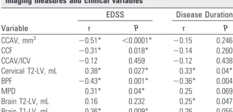
Comparison of Three Different Methods for Measurement of Cervical Cord Atrophy in Multiple Sclerosis
Full text
Figure


Related documents
are further organized via OÐH O hydrogen bonds invol- ving the water molecules and the nitrate ions, leading to the formation of a supramolecular ladder, with thiabendazole
The raw data from fatigue tests on notched and plain specimen conducted in various. heated environments are given in, Table 11 and Table
In sum, the analysis above has demonstrated the breadth of mechanisms that UV partners may use to exert effective control. Various control tools are applied to serve positive or
Presence of tested secondary metabolites in the acetone extract of M.puidca root are in line. with earlier reports [11] .The phytoconstituents detected in the roots
(2015) Access to services by children with intellectual disability and mental health problems : population- based evidence from the UK.. Journal of Intellectual and
AAN ⫽ American Academy of Neurology; DBS ⫽ deep brain stimulation; ED ⫽erectile dysfunction; EDS ⫽ excessive daytime somnolence; ESS ⫽ Epworth Sleepiness Score; FDA ⫽ Food
Figure 3.5 Concept of how the historical wetland inventory, enhanced potential wetland inventory, restorable wetland inventory, and existing wetland inventory can be compared
The above figure shows membrane and bending stresses developed in modified generator frame for spectrum analysis in Z-direction. The combination of bending
