Original Article
Biomechanical comparison of dynamic hip screw and
Gamma nail for the treatment of unstable trochanteric
fractures: a finite element study
Xiaowei Huang2*, Baoqing Yu1*, Yong Gu2, Zexiang Li1
1Department of Orthopedic Surgery, Shanghai Pudong Hospital; Fudan University Pudong Medical Center, 2800
Gongwei Road, Pudong, Shanghai 201399, China; 2Department of Orthopedic Surgery, The First Affiliated
Hospi-tal of Soochow University, Suzhou, Jiangsu 215000, China. *Equal contributors.
Received December 22, 2016; Accepted February 7, 2017; Epub May 15, 2017; Published May 30, 2017
Abstract: Objectives: To investigate the biomechanical behavior of the femur with two different implant configu -rations, namely dynamic hip screw and Gamma nail, in the treatment of two types of unstable intertrochanteric
femoral fracture. Methods: A 3D finite element model of the intact proximal femur was constructed, from which two types of fractured models (AO classification 31-A2 and 31-A3) were produced. Then the fracture models were
loaded with the peak force during a single gait cycle. The effect of stress and displacement of femurs with three
different implant configurations were recorded and analyzed. Results: The stress induced on the two different im -plants demonstrated a similar pattern; stress concentration was mainly located in the intersection area between lag screw and the nail or between lag screw and the lateral plate. Gamma nail in both types of fractures shared the
smaller displacement. The displacement of femurs fixed by Gamma nail showed smaller displacement. The stress
in the calcar region which was the main load-bearing area of the intact femoral neck was decreased markedly in all
of the models, especially in model fixed with Gamma nail. Conclusions: In conclusion, for the 31-A3 fracture, DHS
may be not suitable because it failed to maintain the integrity of the calcar region and showed larger displacement of fractured fragment. Generally, the Gamma nail has mechanical superiority over DHS, but the shielding effect accompanied may increase the risk of intra-operative and later fracture around or below the implant. When the integrity of the calcar region fails to maintain, the Gamma nail is preferred.
Keywords: Finite element analysis, intertrochanteric fracture, comparative study
Introduction
Fractures of the hip are common injuries. And approximately 50% of hip fractures occur in the intertrochanteric region, the prevalence of which increases exponentially with age [1]. It is also associated with a high rate of morbidity and mortality [2]. Beyond preventing avoidable deaths, the goal of the treatment is to restore patients to their pre-injury level of mobility. In order to allow immediate return to unrestricted weight-bearing, secure fixation is often required [3]. The implants chosen for the operative inter-vention are mainly categorized into two types, namely extra-medullary and intramedullary. For the treatment of stable intertrochanteric femo-ral fracture, dynamic hip screw (DHS) has been proven to be the most widely acceptable
Materials and methods
This study was approved by the local institu-tion. One of the co-authors (a 29-year-old male with a weight of 70 kg and a height of 170 cm) volunteered to be the subject of this study. Pelvic and lower extremity fractures, osteoar-thritis and other bone diseases were excluded by X-ray before undergoing a CT scan of his right lower extremity.
Experimental model
Three dimensional (3D) model of the right proxi-mal femur was reconstructed from CT images,
isotropic, linear, elastic material [10]. Elastic moduli and Poisson’s ratios of cortical and can-cellous bone and implants were obtained from published literatures [8, 11], as listed in Table 1.
Nomenclature
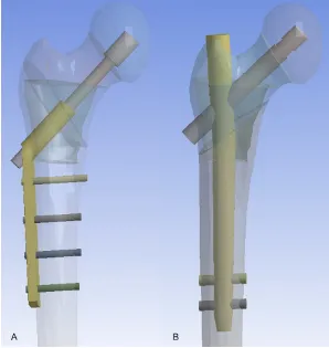
[image:2.612.89.387.72.387.2]In this study, AO classification 31-A2 and 31-A3 were designated as “S” and “R” respectively. Arabic numerals “1”, “2” stood for DHS and Gamma nail respectively. For example, 31-A2 fracture fixed with DHS was labeled as ‘S1’. And 31-A3 fracture fixed with Gamma nail was labeled as ‘R2’.
Figure 1. A: CAD model of AO 31-A2.1 fracture fixed with DHS. B: CAD model of AO 31-A3.1 fracture fixed with Gamma nail.
using efilm software (MER-GE Co., Milwaukee, WI, USA), with 1 mm cuts and in the form of the DICOM format. After the proce-dures of segmentation, re- fining and rendering, non-uniform rational B-spline (NURBS) surface recon-struction was performed via Pro/Engineer2001 soft-ware (PTC Co., MA, USA), to build the entire CAD model. Then the CAD model of the proximal femur was import-ed to ANSYS software (ver-sion 8.0, Ansys, Inc., Canon- sburg, PA, USA) which was employed to establish the finite element model with 1.0 mm sized tetrahedral mesh of both the bones and implants. AO classifica -tion 31-A2.1 femoral frac-ture and 31-A3.1 fracfrac-ture were created respectively on the basis of the intact femur model [8, 9]. Given the data provided by manu-facturers, CAD and finite element models of dynamic hip screw (DHS) and Gam- ma nail (asian type) were also created, as shown in
Figure 1.
Material properties
In this study, the material of either the bone or the implant was regarded as
Table 1. Material properties assigned
Materials Tensile moduli (MPa) Poisson’s ratio Cancellous bone in femoral head 900 0.29 Cancellous bone in femoral neck 620 0.29 Cancellous bone in intertrochanteric region 260 0.29
Cortical bone 17000 0.30
Loading and boundary condition
The distal end of the femur was fixed in all degrees of freedom to prevent rigid body motions during the analysis. Forces were load-ed to simulate the maximal loading in a single gait cycle of a 70 kg weight man, as shown in
Table 2 and Figure 2, which included joint reac-tion forces and abductor forces [11, 12]. For the screw part of DSH, it was assumed that they were completely bonded by femoral head without possible motion, while in the other part, they were partially fixed with a friction factor of 0.3 [13, 14]. The contact of distal locking screws for Gamma nail with femoral cortex was regarded as completely fixed without move -ment. And the effect of load sharing between the lag screws and sleeves was simulated by linking the screw and sleeve with virtual mechanical rigid links.
Analysis
The analysis was done using ANSYS software (version 8.0, Ansys, Inc., Canonsburg, PA, USA) with the equivalent Von Mises stress (EVMS)
[15-17], displacement of the model relative to the intact femur used as the output measures.
Results
Stress distribution on implants
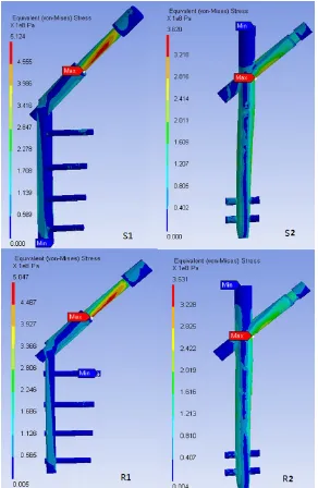
The stress induced on the two different implants demonstrated a similar pattern, as shown in Figure 3. The maximum stress regions of the devices were located in the intersection area between lag screw and the nail or between lag screw and the lateral plate. Both in model S1 and S2, the stress concentrated at the mid-dle part of the lag screw. As for 31-A3.1 frac-ture, the stress distribution in R1 and R2 were similar to those in S1 and S2, which indicated that, for DHS and Gamma nail, the stress distri-bution basically remained the same, as showed in Table 3.
Stress distribution on femurs
The peak stress of femurs in two types of frac-ture was listed in Table 3. It was found that the stress concentration was mainly located where the distal part of the lateral plate or distal lock screw was contacted. As demonstrated in Table 3, the peak stress decreased by 24.27% from S1 to R1. The decreasing ratio was larger than that in S2 (16.33%). As shown in Table 3, stress in the calcar region which was the main load-bearing area of the intact femoral neck was decreased markedly in all of the models, espe-cially in model R2 and S2.
Displacement pattern on proximal femurs The maximal displacement of the proximal femur was compared. The displacement of femurs fixed by Gamma nail showed smaller displacement. As demonstrated in Table 3, the displacement of femurs decreased by 9.5% from S1 to R1, and decreased by 25% from S2 to R2.
Displacement pattern on implants
The displacements of two types of implants were compared in Table 3. The displacement
Table 2. Decomposition of loading forces
Loading forces Decomposition of loading forces in three axises (N) Resulant force (N)
X Y Z
Joint reaction force 616 2800 171 2872
Abductor force 430 1160 0 1237
patterns were similar in two implants. Gamma nail in both types of fractures shared the small-er displacement. This is consistent with dis-placement pattern of femurs aforementioned.
Discussion
Intertrochanteric fractures have been treated with numerous implants, intramedullary or extra-medullary. However, the optimal manage-ment of intertrochanteric fractures is still con-troversial. Fracture patterns, osteoporosis, and aging process make the selection of implants a difficult problem [1, 2], especially in unstable
trated where the lateral plate contacted the fracture line, which can be explained by the anti-sliding effect of DHS to prevent the proxi-mal fractured fragment from moving laterally and the distal fractured fragment from moving medially. The stress concentration also oc- curred at the intersection area between the most distal screw and the plate, which indicat-ed potential implant failure in this region, as illustrated in Figure 3.
[image:4.612.89.381.71.519.2]Comparing with the intact femur, unlike S1, the stress distribution of femur in S2 was similar to that of the intact femur. And in both S2 and R2, Figure 3. Stress distribution of implants in different models.
concen-Table 3. Stress and displacement pattern on the femurs and implants
Model Maixmal stress on implants (MPa)
Maximal stress on femurs
(MPa)
Maximal displace-ment on implants
(mm)
Maximal displacement on proximal femurs
(mm)
Stress on the medial cortex of fracture end
(MPa)
Stress on the anterior cortex of fracture end
(MPa)
Stress on the posterior cortex of fracture end
(MPa)
Stress on the calcar region
(MPa)
S1 512 128 3.85 10.5 49 31 36 66
S2 362 57 3.15 9.6 30 20 32 32
R1 504 103 3.53 9.5 18 17 5 62
R2 363 49 2.53 7.22 22 27 22 26
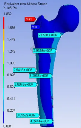
[image:5.792.88.709.86.176.2]the stress distribution of the femur was similar and the stress in the calcar area was much smaller than that in S1 and R1, which indicated that, for unstable fractures involving the calcar area, Gamma nail can maintain the loading transmission pathway similar to the intact femur and the fractured medial calacr region can be effectively protected in 31-A3.1 frac-tures. It also seemed that the stress distribu-tion pattern was not dependent on fracture types but the inherent attributes of the Gamma nail. However, it should be noted that stress concentration occurred in the inner side of femoral cortex contacted with distal end of
There were many reports published comparing the utilization of DHS and Gamma nail for the fixation of intertrochanteric fractures. Pervez et al revealed that no significant difference was found in several parameters, such as length of surgery, pneumonia, thromboembolic compli-cations and wound infection or hematoma, between Gamma nail and DHS [19]. However, in regard to blood loss, the fracture fixation with DHS led to more blood loss than with Gamma nail, as reported in several meta-analysis [20-22]. In addition, Liu et al found that DHS was associated with a higher risk of reoperation than Gamma nail. Although with superior bio-Figure 4. Stress of the inner side of femoral cortex contacted with
Gam-ma nail.
Gamma nail (Figure 4), which was twice as much as the stress in the proximal part of cortical bone, may increase the potential risk of fracture.
[image:6.612.91.362.72.502.2]mechanical property over DHS, many studies indicated that Gamma nail can result in a high-er risk of intra-ophigh-erative and lathigh-er fracture around or below the implant than DHS [22, 23], which in our study can be explained by multiple stress concentration on the femur fixed by Gamma nail, for example, the stress concentra-tion occurred in the inner side of femoral cortex contacted with distal end of Gamma nail. However, Zeng et al reported that no significant difference was found in fixation failure, such as cutting-out or non-union, between Gamma nail and DHS [25].
A few limitations were inherent to this study. First, the FEA in our study is based on only one healthy subject. The results of our study should be verified by a larger cohort of patients with the same types of intertrochanteric fractures. In addition, the Proximal Femoral Nail Autorotation (PFNA) which has been widely used in intertrochanteric fractures, were not included in our comparison study. We plan to extend our further research to cover this implant. Furthermore, only a static load situa-tion may be insufficient for representing a whole gait cycle. Finite element analysis of bone model considering the whole gait cycle leads to deviations in the mass loss and appar-ent bone density distribution in comparison with calculation of static loading alone [26]. Lastly, although the implants are subjected to an assumed normal human weight of 70 Kg, data obtained from higher amounts of loads may be necessary to prove the effectiveness of each implant during normal function. This is important considering that the daily human activity requires the keens to undergo stress- es beyond that which is observed during standing.
Conclusion
For the 31-A3 fracture, DHS may be not suit-able because it failed to maintain the integrity of the calcar region and showed larger displace-ment of fractured fragdisplace-ment. Generally, the Gamma nail has mechanical superiority over DHS, but the shielding effect accompanied may increase the risk of intra-operative and later fracture around or below the implant. When the integrity of the calcar region fails to maintain, the Gamma nail is preferred.
Acknowledgements
This study was Funded by the Outstanding Leaders Training Program of Pudong Health Bureau of Shanghai (PWR12013-01) and Pro- gram for Medical Key Department of Shanghai (ZK2015B17).
Disclosure of conflict of interest
None.
Abbreviations
DHS, dynamic hip screw; PFNA, Proximal Femoral Nail Autorotation; FEA, finite element analysis.
Address correspondence to: Dr. Zexiang Li, Depart- ment of Orthopedic Surgery, Shanghai Pudong Hospital, No. 2800, Gongwei Road, Huinan Town, Pudong New Area, Shanghai, China. Tel: +86138-
62086284; E-mail: lzxsubmission@163.com
References
[1] Horowitz BG. Retrospective analysis of hip frac -tures. Surg Gynecol Obstes 1966; 123: 565. [2] White BL, Fisher WD, Lauren CA. Rate of
mor-tality for elderly patients after fracture of the hip in the 1980’s. J Bone Joint Surg 1987; 69A: 1335-1340.
[3] Rogers FB, Shackford SR, Keller MS. Early fixa -tion reduces mobility and mortality in elderly patients with hip fractures form low-impact falls. J Trauma 1995; 39: 261-265.
[4] Luitse J, Dunki J, Van H. The dynamic hip screw “Golden standard” in the treatment of pertro-chanteric fractures. Proximal femoral frac-tures. Operative techniques and complica-tions, vol 2. In: Marti R, Dunki J, editors. London: ED Medical Press; 1993. pp. 409-22. [5] Kempf I, Grosse A, Beck G. Closed locked intra-medullary nailing. J Bone Joint Surg 1995; 67A: 709-19.
[6] Willoughby R. Dynamic hip screw in the man-agement of reverse obliquity intertrochanteric neck of femur fractures. Injury 2005; 36: 105-9.
[7] Honkonen S, Vihtonen K, Jarvinen M. Second-generation cephalomedullary nails in the treat-ment of reverse obliquity intertrochanteric fractures of the proximal femur. Injury 2004; 35: 179-83.
[8] Sitthiseripratip K, Van Oosterwyck H, Vander
tro-chanteric fracture. Med Eng Phys 2003; 25: 99-106.
[9] Haynes RC, Poll RG, Miles AW, Weston RB.
Fail-ure of femoral head fixation: a cadaveric analy -sis of lag screw cut-out with the gamma lock-ing nail and AO dynamic hip screw. Injury 1997; 28: 337-41.
[10] Huiskes R, Chao E. A survey of finite element analysis in orthopedic biomechanics: the first
decade. J Biomechanics 1983; 16: 385. [11] Taylor ME, Tanner KE, Freeman M. Stress and
strain distribution within the intact femur: com-pression or bending? Med Eng Phys 1996; 18: 122-131.
[12] Bohez E, Suwanprateeb J, Audekercke R. Finite
element study of trochanteric gamma nail for trochanteric fracture. Med Eng Phys 2003; 25: 99-106.
[13] Chen WP, Tai CL, Shin CH, Hsieh PH, Leou MC,
Lee MS. Selection of fixation devices in proxi -mal femur rotational osteomy clinical
compli-cations and finite element analysis. Clin Bio -mech (Bristol, Avon) 2004; 19: 255-62. [14] Mann K, Bartel DL. Coulomb frictional in
mod-eling cemented total hip replacement: a more realistic model. J Biomech 2008; 9: 1067-78. [15] Makar S, Joshi G. Analysis of a femoral hip
prosthesis designed to reduce stress shield-ing. J Biomech 2000; 33: 1655.
[16] Terrier A, Rakotomananna RL, Ramaniraka AN,
Leyvraz PF. Adaptation models of anisotropic
bone. Comput Methods Biomech Biomed En-gin 1997; 1: 47.
[17] Izaham R, Kadir M, Rashid A, Hossain G, Ka -marul T. Finite element analysis of Puddu and
Tomofix plate fixation for open wedge high tibi -al osteotomy. Injury 2012; 43: 898-902. [18] Wang C, Yettram AL, Yao M, Procter P. Finite
element analysis of a Gamma nail within a fractured femur. Med Eng Phys 1998; 20: 677-83.
[19] Pervez H, Parker M, Vowler S. Prediction of fixa
-tion failure after sliding hip screw fixa-tion. In -jury 2004; 35: 994-998.
[20] Shen L, Zhang Y, Shen Y, Cui Z. Antirotation proximal femoral nail versus dynamic hip screw for intertrochanteric fractures: a
meta-analysis of randomized controlled studies. Or -thop Traumatol Surg Res 2013; 99: 377-83. [21] Bhandari M. Schemitsch E, Jonsson A,
Zlo-wodzki M. Haidukewych GJ. Gamma nails re -visited: Gamma nails versus compression hip screws in the management of intertrochanter-ic fractures of the hip: a meta-analysis. J Or-thop Trauma 2009; 23: 460-4.
[22] Gu M, Yang Z, Pei F, Huang F, Chen S, Xiang Z. Meta-analysis of the Gamma nail and dynamic hip screw in treating peritrochanteric fractures. Int Orthop 2010; 34: 323-8.
[23] Ekstrom W, Karlsson-Thur C, Larsson S, Rag-narsson B, Alberts K. Functional outcome in treatment of unstable trochanteric and subtro-chanteric fractures with the proximal femoral nail and the Medoff sliding plate. J Orthop Trauma 2007; 21: 18-25.
[24] Adams C, Robinson C, Court-Brown C,
Mc-Queen M. Prospective randomized controlled
trial of an intramedullary nail versus dynamic screw and plate for intertrochanteric fractures of the femur. J Orthop Trauma 2001; 15: 394-400.
[25] Zeng C, Wang Y, Wei J. Treatment of trochan-teric fractures with proximal femoral nail anti-rotation or dynamic hip screw systems: a meta-analysis. J Int Med Res 2012; 40: 839-51. [26] Behrens B, Nolte I, Wefstaedt P,
Stukenborg-Colsman C, Bouguecha A. Numerical investiga-tions on the strain-adaptive bone remodeling
in the peri-prosthetic femur: influence of the