Original Article
Volar locking plate versus percutaneous fixation for the
treatment of distal radial fractures: a meta-analysis
of randomized controlled trials
Longxiang Shen1, Mengni Wen2, Feiyan Wang2, Jian Ding1, Congfeng Luo1, Gang Zhao2
1Department of Orthopedic Surgery, Shanghai Jiao Tong University Affiliated Sixth People’s Hospital, 600 Yishan Road, Shanghai 200233, PR China; 2Department of Emergency and Trauma Center, Shanghai Jiao Tong University Affiliated Sixth People’s Hospital, 600 Yishan Road, Shanghai 200233, PR China
Received November 18, 2015; Accepted March 25, 2016; Epub May 15, 2016; Published May 30, 2016
Abstract: Background: Although operative fixation with a volar locking plate is becoming increasingly popular for treatment of distal radius fractures, it is not clear whether it is superior to other conventional treatment methods such as percutaneous fixation with Kirschner wires. The present meta-analysis compared the effects of internal fixation with volar locking plates and percutaneous fixation for the treatment of distal radius fractures. Methods: A literature search was performed without language restrictions and all randomized controlled studies comparing the effects of volar locking plates and percutaneous fixation for the treatment of distal radial fractures were included. Data of function scores, range of motion, grip strength, radiographic results, and complications were pooled and analyzed with a standard meta-analytical method. Results: Seven studies in seven publications were included. Pooled data indicated that there were no significant differences in Patient-Related Wrist Evaluation (PRWE) and Disabilities of the Arm, Shoulder, and Hand (DASH) scores between the two treatment methods postoperatively. Fixation with volar locking plates took significantly longer than percutaneous fixation. There was a significant differ-ence in supination and grip strength favoring volar locking plate fixation at 3 and 6 months but not at ≥12 months, postoperatively. The final complication rates were similar in the two treatment groups. Conclusion: In the manage-ment of distal radius fractures, volar locking plate and percutaneous fixation yielded similar outcomes, while the former had the advantage of supination and grip strength in the early stage postoperatively. Percutaneous fixation was quicker to perform than volar locking plate fixation.
Keywords: Volar locking plate, distal radial fractures, percutaneous fixation, meta-analysis
Introduction
Operative fixation with a volar locking plate has becoming increasingly popular in the treatment of distal radius fractures [1, 2]. Many studies have indicated the advantages of the tech-nique, and have shown good outcomes [3-5]. However, volar locking plate fixation is expen-sive and it can cause tendon problems and neurolysis, and a second implant-removal oper-ation may be required [6-8]. It is not clear whether volar locking plate fixation is superior to other treatment methods. Recently, a few randomized controlled trials (RCTs) compared the effectiveness of volar locking plate versus minimally invasive and less expensive implants, such as percutaneous Kirschner wires, for the treatment of distal radial fractures. However,
the small size or incomplete evaluation of these RCTs makes the results inconsistent. Recently, a systematic review [9] regarding volar locking plates and K-wire/pin fixation in the manage-ment of distal radius fractures has been pub-lished. Regrettably, the systematic review was not based on RCTs, which only provided level 2 evidence. Another two meta-analyses [10, 11] were performed based on RCTs, however, meth-odological flaws in the process of data manage-ment, make the results imprecise. Thus, wheth-er surgical treatment of distal radial fractures with a volar locking plate improves clinical out-comes when compared with percutaneous Kirschner wires remains controversial.
differentiated the evidence base on identified RCTs. The purpose of the present meta-analy-sis was to compare the differences in the func-tional and radiological outcomes between volar locking plates and percutaneous Kirschner wire fixation in patients with distal radius frac-tures. Furthermore, we stratified the patients aged ≥50 years who have tendency to have fra-gility fractures, as bone mineral density of the forearm remains stable up until the age of 50 years [12].
Methods
Search strategy
This meta-analysis was carried out following the guidelines of the PRISMA statement [13]. We performed a computerized search on March 16, 2013 and an updated literature search on October 18, 2014. The following search terms were used: “distal radius fractures” with the limitation of “randomized controlled trial” using PubMed (1949-2014), Ovid Medline (1946-2014), Medline In Process & Other Non-Indexed Citations (updated to October 18, 2014), Web of Knowledge and Embase (1966-2014). Co- chrane Central Register of Controlled Trials (CENTRAL) (1948-2014). We placed no restric-tions on the language of publication.
Reference lists of related review articles, all included publications, and previously published systematic reviews and meta-analyses were also reviewed to find additional eligible studies. Two authors (LS and MW) screened the titles and abstracts according to the eligibility crite-ria. The full texts were read when the studies met the inclusion criteria. Disagreements were resolved by discussion between the authors. If a consensus could not be reached, a third investigator (JD) made the final decision. The Jadad scale was used to evaluate the quality of included RCTs, with a score of <3 considered low quality [14].
Inclusion and exclusion criteria
The search results were screened on the basis of the following inclusion criteria: (a) random-ized controlled studies on patients with distal radius fractures; and (b) comparison of volar locking plate versus percutaneous fixation. Exclusion criteria included: (a) non-randomized controlled trials; (b) trials focused on pediatric
fractures; and (c) patients with distal radius fractures who presented >3 weeks after injury. Data extraction
The primary outcome of our analysis was func-tional outcome, measured using the Patient-Related Wrist Evaluation (PRWE) and Dis- abilities of the Arm, Shoulder, and Hand (DASH) scores. The secondary outcomes were range of motion (ROM) and radiological outcome. Data of operation time and complications were col-lected. We sent an email to every correspond-ing author to obtain the original data. When there was inconsistency between the published and original data, the latter were used. When original data were not available, or if means were presented without standard deviations (SDs), the SDs were calculated from the P value or confidence interval (CI), following the guid-ance of the Cochrane Handbook for Systematic Reviews of Interventions [15]. In some cases, the published reports of clinical trials only reported the median and range, and the mean and SDs were estimated using the formulas founded by Hozo et al. [16].
Statistical analysis
We performed all meta-analyses using the Stata/SE 10.0 program. For the meta-analysis of continuous variables using the same scales, the weighted mean difference (WMD) with a 95% CI was used. For dichotomous variables, the relative treatment effect was presented as relative risk (RR) with a 95% CI. The assess-ment for statistical heterogeneity was investi-gated using the χ2 test and quantified using the I2 statistic. A random-effect model was used in the present meta-analysis based on the fact that different fixation plates and methods of percutaneous fixation method were used, and the test for heterogeneity had low statistical power. If possible data were available, we per-formed independent analysis regarding the patients aged ≥50 years. P<0.05 was consid-ered statistically significant.
Sensitivity analysis and assessment of publi-cation bias
bias. A further analysis was conducted using the trim and fill method, when possible publica-tion bias was detected.
Results
Selected studies and characteristics
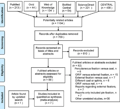
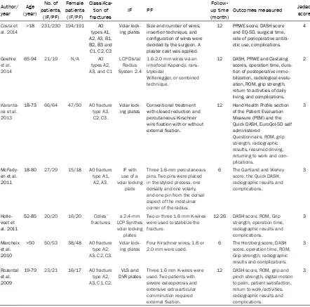
As shown in Figure 1, a total of 709 potentially relevant studies were identified and screened in the original search, and only six RCTs met the inclusion criteria. An additional study [17] was identified in the updated search, thus, seven publications were selected for the meta-sis. One RCT [18] was excluded from our analy-sis because it was restricted to patients with displaced distal radius fractures who present-ed within 3-6 weeks from injury. The basic char-acteristics of the included studies are shown in
Table 1. The quality of each article was graded from 2 to 4 according to the Jadad quality score [14]. A total of 874 patients were included in this analysis. The corresponding authors of six publications [17, 19-23], but not the other [24] responded to our email and original data were provided from five trials [19-23]. Because of the obvious nature of the intervention, no trials were double-blind.
Effects of volar locking plate versus percutane-ous fixation on functional outcome
[image:3.612.96.519.71.455.2]was no significant difference between volar locking plate and percutaneous fixation in the treatment of distal radius fractures at 3, 6 and 12 months, postoperatively (all P>0.05) (Table 2).
Independent analysis compared internal and percutaneous fixation in patients aged ≥50 years. For PRWE scores, data were only avail-able from ≥12 months postoperatively. Pooled analysis showed that there was no significant difference in PRWE scores between the two treatment groups (RR=-0.543, 95% CI -2.891
to 1.806, P=0.651). The pooled data of DASH scores showed that there were no significant differences between volar locking plate and percutaneous fixation groups at any time point postoperatively (Table 3).
Effects of volar locking plate versus percutane-ous fixation on operation time, wrist and fore-arm range of motion and grip strength
[image:4.612.84.526.87.524.2]Four of the seven included studies reported operation time data. Meta-analysis demon-strated that internal fixation with volar locking Table 1. Baseline characteristics of the included RCTs
Author/ year (year)Age
No. of patients,
(IF/PF)
Female patients (IF/PF)
Classifica-tion of
fractures IF PF
Follow-up time
(month) Outcomes measured
Jadad score
Costa et al. 2014
>18 231/230 194/191 AO types A1, A2, A3, B1, B2, B3 and C1, C2, C3
Volar lock-ing plates
Size and number of wires, insertion technique, and configuration of wires were decided by the surgeon. A plaster cast was applied.
12 PRWE score, DASH score and EQ-5D, surgical time, rate of perioperative antibi-otic use, complications.
4
Goehre et al. 2014
65-94 21/19 N/A AO
types A2, A3, and C1
LCP Distal Radius System 2.4
1.6-2.0 mm wires via an intrafocal Kapandji, rans-styloidal
Willenegger, or combined technique.
12 DASH, PRWE and Castaing scores, operation time, dura-tion of postoperative immo-bilization, radiological evalu-ation, ROM, grip strength, return to activities of daily living, and complications.
2
Karanta-na et al. 2013
18-73 66/64 47/50 AO fracture type A3, C2, C3.
Volar
lock-ing plates Conventional treatment with closed reduction and percutaneous Kirschner wire fixation with or without external fixation.
12 Hand Health Profile section of the Patient Evaluation Measure (PEM) and the Quick DASH, EuroQol-5D self administered
Questionnaire, ROM, grip strength, radiographic results, resumed driving, returning to work and com-plications.
3
McFady-en et al. 2011
18-80 27/29 15/18 AO fracture type A1,
A2, A3.
IF with use of a volar locking
plate
Three 1.6-mm percutaneous pins. Two pins were placed in the styloid process, one dorsally and one volarly, and one pin from the dorsal aspect of the mostulnar corner of the radius.
6 The Gartland and Werley score, the Quick DASH, radiographic results and complications.
3
Holle-voet et al. 2011
52-85 20/20 16/20 Colles’
fractures LCP Synthes a 2.4-mm volar locking
plates
Two or three 1.6 mm K-wires were used to stabilize the fracture.
12-26 DASH score, ROM, Grip strength, operation time, radiographic results and complications.
3
Marcheix et al. 2010
>50 50/53 38/48 AO fracture type A2, A3, C2, C3.
Volar
lock-ing plates Four Kirschner wires, 1.8 or 2.0 mm were used. 6 The Herzberg score, DASH score, operation time, ROM, Grip strength, radiographic results and complications.
3
Rozental et al. 2009
19-79 23/21 16/17 AO fracture type A2, A3, C1, C2.
VLS and DVR plates
Three 1.6 mm K-wires were used. Two patients with severe osteoporosis and extensive extra-articular comminution required external fixation.
12 DASH score, ROM, grip and pinch strength, digital motion to palm, patient satisfaction, return to work/activities, radiographic results and complications.
3
plates took significantly longer than percutane-ous fixation (RR=22.0, 95% CI: 18.648-25.352, P<0.001) (Figure 2).
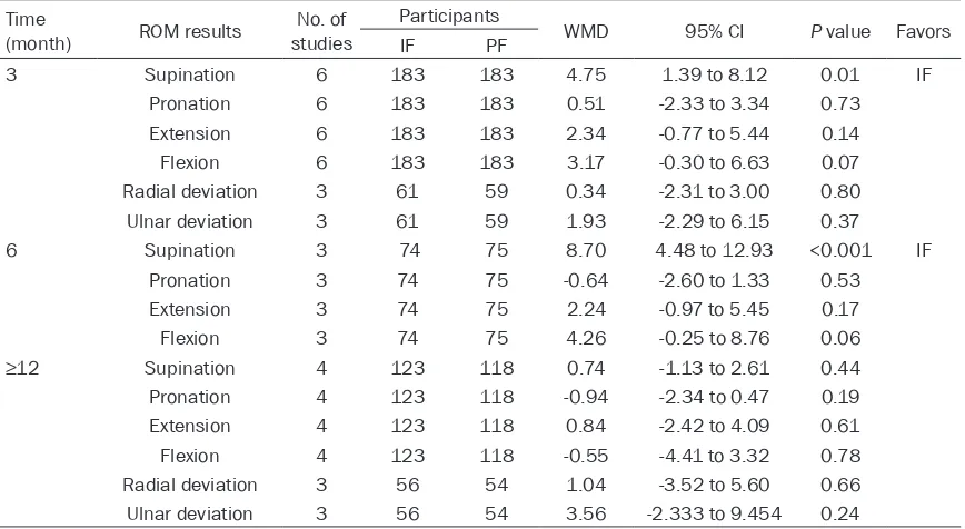
Pooled data of wrist and forearm ROM were available from 3-6 of the seven studies. Only Goehre et al. [24] reported postoperative radial and ulnar deviation data at 6 months, so we were not able to perform a pooled analysis on postoperative radial and ulnar deviation at that time. There was a significant difference in supi-nation favoring volar locking plate fixation at 3 and 6 months but not at ≥12 months postop-eratively. For any other ROM variables, there were no significant differences (all P>0.05) in treatment effect at 3, 6 and ≥12 months between these two fixation methods (Table 4). Independent comparative analysis of internal and percutaneous fixation in patients aged
The radiographic parameters including volar tilt, radial height and radial inclination were pooled and analyzed. Radiological outcomes comparing volar locking plate with percutane-ous fixation at different times postoperatively are shown in Table 6. Pooled data across 2 or 3 studies showed that there were no significant differences between the two treatment groups immediately post-operation, and at 6 and ≥12 months postoperatively (all P>0.05). For pa- tients aged ≥50 years, there were no radio-graphic data available, so we were not able to perform an independent analysis.
Effects of volar locking plate and percutane-ous fixation on final complications
The seven included trials reported a total of 874 patients and provided information on over-Table 2. Comparison of IF with volar locking plates and PF regarding
PRWE, DASH and grip strength
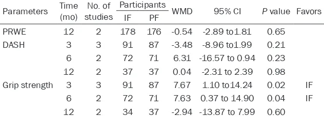
Parameters Time (mo) studiesNo. of Participants WMD 95% CI value FavorsP IF PF
PRWE 3 2 236 231 -2.69 -6.56 to 1.17 0.17 6 2 227 227 -1.82 -5.05 to 1.41 0.27 12 2 225 230 -0.54 -2.89 to 1.81 0.65 DASH 3 4 110 112 -6.19 -12.75 to 0.37 0.06 6 2 71 72 -6.31 -16.57 to 3.94 0.23 12 4 255 259 -1.53 -3.88 to 0.81 0.20 Grip strength 3 5 175 176 10.81 3.77 to 17.86 <0.01 IF
[image:5.612.91.405.98.241.2]6 3 137 136 12.05 3.05 to 21.05 <0.01 IF 12 4 126 119 3.03 -6.87 to 12.93 0.55 IF, internal fixation; PF, percutaneous fixation; WMD, Weighted mean difference; CI, confidence interval; PRWE, Patient-Rated Wrist Evaluation; DASH, Disabilities of the Arm, Shoulder and Hand.
Table 3. Comparison of IF with volar locking plates and PF regarding out-comes of PRWE, DASH and grip strength in patients aged ≥50 years Parameters Time (mo) studiesNo. of Participants WMD 95% CI P value Favors
IF PF
PRWE 12 2 178 176 -0.54 -2.89 to1.81 0.65
DASH 3 3 91 87 -3.48 -8.96 to1.99 0.21
6 2 72 71 6.31 -16.57 to 0.94 0.23 12 2 37 37 0.04 -2.31 to 2.39 0.98 Grip strength 3 3 91 87 7.67 1.10 to14.24 0.02 IF
6 2 72 71 7.63 0.37 to 14.90 0.04 IF 12 2 34 37 -2.94 -13.87 to 7.99 0.60 CI, confidence interval; DASH: Disabilities of the Arm, Shoulder, and Hand; IF, internal fixa-tion; PF, percutaneous fixafixa-tion; PRWE, Patient-Rated Wrist Evaluafixa-tion; WMD, Weighted mean difference.
≥50 years demonstra- ted the same results (Table 5).
Pooling of grip streng- th data was possible across 3 to 5 of the seven studies at diff- erent times. Meta-an- alysis of grip strength demonstrated that fix-ation with volar locking plates was superior to percutaneous fixation at 3 and 6 months postoperatively. How- ever, at ≥12 months, the grip strength was similar between the two treatment meth-ods (Table 2). For pa- tients aged ≥50 years, independent compara-tive analysis of volar locking plate and per-cutaneous fixation de- monstrated similar re- sults (Table 3).
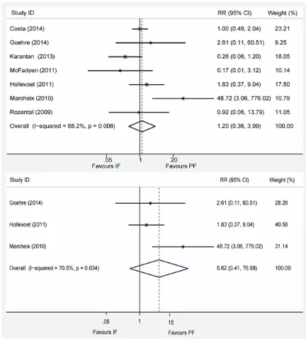
[image:5.612.90.406.325.440.2]all complications at final follow-up. Meta-analysis indicated that the final complications with volar locking plate did not differ significa- ntly from those with percutaneous fixation (RR=0.610, 95% CI: 0.364-1.022, P=0.06). Further analysis indicated that major compli- cations detected no significant difference be-
[image:6.612.90.529.73.295.2]tween the two groups (RR=0.708, 95% CI: 0.389-1.290, P=0.259). There were significant-ly fewer minor complications in the patients treated with volar locking plate than percutane-ous fixation (RR=0.561, 95% CI: 0.351-0.895 P=0.015) (Figure 3). We performed an indepen-dent meta-analysis regarding the incidence of Figure 2. Forest plot for estimation of operation time for internal fixation with volar locking plates versus percutane-ous fixation. IF, internal fixation; PF, percutanepercutane-ous fixation.
Table 4. Comparison of IF with volar locking plates and PF regarding ROM Time
(month) ROM results studiesNo. of
Participants
WMD 95% CI P value Favors
IF PF
3 Supination 6 183 183 4.75 1.39 to 8.12 0.01 IF
Pronation 6 183 183 0.51 -2.33 to 3.34 0.73 Extension 6 183 183 2.34 -0.77 to 5.44 0.14
Flexion 6 183 183 3.17 -0.30 to 6.63 0.07
Radial deviation 3 61 59 0.34 -2.31 to 3.00 0.80 Ulnar deviation 3 61 59 1.93 -2.29 to 6.15 0.37
6 Supination 3 74 75 8.70 4.48 to 12.93 <0.001 IF
Pronation 3 74 75 -0.64 -2.60 to 1.33 0.53
Extension 3 74 75 2.24 -0.97 to 5.45 0.17
Flexion 3 74 75 4.26 -0.25 to 8.76 0.06
≥12 Supination 4 123 118 0.74 -1.13 to 2.61 0.44
Pronation 4 123 118 -0.94 -2.34 to 0.47 0.19
Extension 4 123 118 0.84 -2.42 to 4.09 0.61
Flexion 4 123 118 -0.55 -4.41 to 3.32 0.78
Radial deviation 3 56 54 1.04 -3.52 to 5.60 0.66 Ulnar deviation 3 56 54 3.56 -2.333 to 9.454 0.24
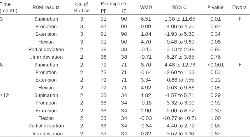
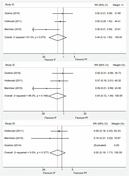
[image:6.612.90.524.361.599.2]second surgical intervention, and the need for further surgery was similar in both treatment groups (RR=1.201, 95% CI: 0.361-3.990, P= 0.765) (Figure 4). Independent analysis of patients aged ≥50 years demonstrated that there was no significant difference between internal and percutaneous fixation regarding final complications (RR=0.440, 95% CI: 0.121-1.601, P=0.075), minor and major complica-tions (RR=0.516, 95% CI: 0.155-1.713, P= 0.280; RR=0.427, 95% CI: 0.123-1.487, P= 0.181) (Figure 5), and further surgery (RR= 5.625, 95% CI: 0.411-76.976, P=0.196) (Figure 4).
Sensitivity and publication bias analysis
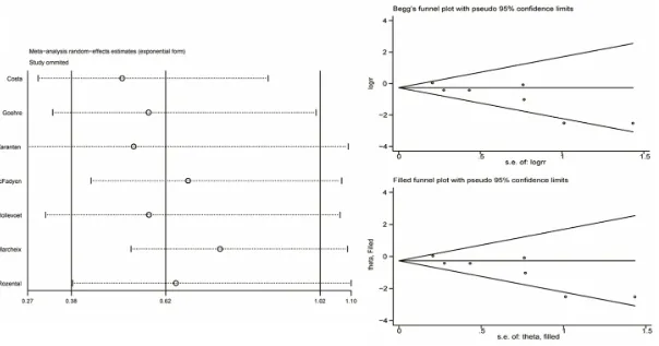
We selected complications data for sensitivity and publication bias analysis, since all of the studies had such data. Sensitivity analysis was evaluated by detecting the influence of any indi-vidual study on the overall RR. No indiindi-vidual study affected the overall RR dominance, because omission of any single study did not make a large difference (Figure 6).
[image:7.612.93.522.86.322.2]Begg’s funnel plot and Egger’s test evaluated the publication bias. The shape of the funnel plot showed symmetry (P=0.072), but the Table 5. Comparison of IF with volar locking plates and PF regarding ROM in patients aged ≥50 years Time
(month) ROM results studiesNo. of
Participants WMD 95% CI P value Favors
PF IF
3 Supination 3 91 90 6.51 1.38 to 11.65 0.01 IF
Pronation 3 91 90 0.09 -4.06 to 4.25 0.97
Extension 3 91 90 1.84 -1.93 to 5.60 0.34
Flexion 3 91 90 4.70 -0.48 to 9.89 0.08
Radial deviation 2 38 38 -0.13 -3.13 to 2.88 0.93 Ulnar deviation 2 38 38 -0.71 -5.27 to 3.85 0.76
6 Supination 2 72 71 8.70 4.48 to 12.93 <0.001 IF
Pronation 2 72 71 -0.64 -2.60 to 1.33 0.53
Extension 2 72 71 3.34 -0.86 to 7.55 0.12
Flexion 2 72 71 4.92 -0.03 to 9.86 0.05
≥12 Supination 2 33 34 1.82 -1.57 to 5.21 0.29
Pronation 2 33 34 -0.16 -3.32 to 3.00 0.92
Extension 2 33 34 2.96 -2.60 to 8.52 0.30
Flexion 2 33 34 -0.03 -10.77 to 10.71 1.00
Radial deviation 2 33 34 -0.84 -4.40 to 2.72 0.65
Ulnar deviation 2 33 34 0.32 -3.52 to 4.16 0.87
CI, confidence interval; IF, internal fixation; PF, percutaneous fixation; ROM, range of motion; WMD, weighted mean difference.
Table 6. Comparison of IF with volar locking plates and PF regarding radiographic results
Time Parameters studiesNo. of Participants WMD 95% CI P value Favors IF PF
Immediately post-operation Volar tilt 2 77 82 1.61 -9.37 to 12.58 0.77 Radial height 3 100 103 -0.17 -1.28 to 0.93 0.76 Radial inclination 3 100 103 0.26 -0.61 to 1.13 0.55
6 month Volar tilt 3 98 101 2.23 -9.84 to 14.30 0.72
Radial height 2 77 82 -0.68 -2.14 to 0.79 0.36 Radial inclination 2 77 82 1.01 -2.91 to 4.93 0.61
12 month Volar tilt 2 89 85 1.99 -5.85 to 9.83 0.62
[image:7.612.94.522.373.518.2]Egger’s test suggested a possible publication bias (P=0.023). We conducted a trim and fill method to investigate the publication bias fur-ther. The pooled analysis incorporating the hypothetical studies continued to show that the final complications with volar locking plate in
not significant (RR=0.621, 95% CI: 0.379-1.018, P=0.059) (Figure 6).
Discussion
[image:9.612.88.520.118.600.2]The studies included in our meta-analysis Figure 3. Forest plot for RR estimate for internal fixation with volar locking plates versus percutaneous fixation. Up-per graph, assessment of overall complication rate. Middle graph, assessment of major complication rate. Bottom graph, assessment of minor complication rate. IF, internal fixation; PF, percutaneous fixation.
uate the effects of volar locking plate and per-cutaneous fixation for treatment of distal radial fractures. Patient-reported, clinical and radio-graphic outcome measures were not performed in all of the RCTs and postoperative outcome was not measured at the same time points, such as 6 weeks, 6 months and 1 year. This variability suggested that, to make a precise and full evaluation of the outcomes of treating distal radius fractures, a well-designed proto-col with completed evaluation parameters needs to be established in advance.
Our meta-analysis showed that in treating dis-tal radial fractures, volar locking plate and per-cutaneous fixation yielded similar outcomes. Although volar locking plates have the advan-tage of supination and grip strength at 3 and 6 months postoperatively, this was not reflected in the functional outcome. In contrast, percuta-neous fixation is quicker to perform than volar locking plate fixation. Over the past decade, there has been a shift in the surgical approach for treatment of distal radial fractures in favor of open reduction and internal fixation, to achieve anatomical reconstruction of the frac-tured bone. By pooling and analyzing the origi-nal data provided by the corresponding author, our results suggested that if early grip strength and supination rehabilitation are important, volar locking plate fixation is an alternative method. Otherwise, percutaneous fixation is an ideal treatment. In contrast, the results of grip strength were not presented although the authors mentioned that the data on grip strength were collected in one recently pub-lished meta-analysis [11], and the grip strength data were analyzed with incorrect raw data in the other meta-analysis [10]. We believe that our results are correct and precise.
Our pooled data indicated that overall and major complication rates were similar and only the minor complication rate was lower in the volar locking plate fixation group compared with percutaneous fixation group. It could be postulated that most of the minor complica-tions were resolved without surgery, because the incidence of second surgical intervention was similar in both treatment groups.
Even though it was reported that anatomical function can be restored and maintained with volar locking plates in elderly patients with osteoporosis [25], our analysis indicated that
the PRWE and DASH scores did not differ between internal and percutaneous treatment groups in patients aged ≥50 years. Our results were similar to some aspects of Arora’s ran-domized controlled study [26], which compared volar locking plate fixation with another conven-tional method, closed reduction and cast immo-bilization for displaced and unstable distal radi-al fractures in patients aged ≥65 years. They found that there were no significant differences between these two treatment groups at 6 and 12 months.
stratified analysis regarding age and fracture type, paralleled with cost-effectiveness analy- sis.
Acknowledgements
Funding provided by the National Natural Science Foundation of China (grant nos. 81201440 and 81171704) http://www.nsfc. gov.cn/.
Disclosure of conflict of interest
None.
Address correspondence to: Dr. Gang Zhao, Depart- ment of Emergency and Trauma Center Shanghai Jiao Tong University Affiliated Sixth People’s Hospital, 600 Yishan Road, Shanghai 200233, PR China. Tel: 0086-189-3017-7631; Fax: 0086-21-6443-4556; E-mail: 18930177631@163.com
References
[1] Downing ND, Karantana A. A revolution in the management of fractures of the distal radius? J Bone Joint Surg Br 2008; 90: 1271-1275. [2] Yoon A, Grewal R. Management of distal radius
fractures from the North American perspec-tive. Hand Clin 2012; 28: 135-144.
[3] Drobetz H, Kutscha-Lissberg E. Osteosynthesis of distal radial fractures with a volar locking screw plate system. Int Orthop 2003; 27: 1-6. [4] Chung KC, Watt AJ, Kotsis SV, Margaliot Z,
Haase SC, Kim HM. Treatment of unstable dis-tal radial fractures with the volar locking plat-ing system. J Bone Joint Surg Am 2006; 88: 2687-2694.
[5] Rozental TD, Blazar PE. Functional outcome and complications after volar plating for dor-sally displaced, unstable fractures of the distal radius. J Hand Surg Am 2006; 31: 359-365. [6] Arora R, Lutz M, Hennerbichler A, Krappinger
D, Espen D, Gabl M. Complications following internal fixation of unstable distal radius frac-ture with a palmar locking-plate. J Orthop Trauma 2007; 21: 316-322.
[7] Ho AW, Ho ST, Koo SC, Wong KH. Hand numb-ness and carpal tunnel syndrome after volar plating of distal radius fracture. Hand (N Y) 2011; 6: 34-38.
[8] ohnson NA, Cutler L, Dias JJ, Ullah AS, Wildin CJ, Bhowal B. Complications after volar locking plate fixation of distal radius fractures. Injury 2014; 45: 528-533.
[9] Franceschi F, Franceschetti E, Paciotti M, Cancilleri F, Maffulli N, Denaro V. Volar locking plates versus K-wire/pin fixation for the
treat-ment of distal radial fractures: a systematic review and quantitative synthesis. Br Med Bull 2015; 115: 91-110.
[10] Zong SL, Kan SL, Su LX, Wang B. Meta-analysis for dorsally displaced distal radius fracture fix-ation: volar locking plate versus percutaneous Kirschner wires. J Orthop Surg Res 2015; 10: 108.
[11] Chaudhry H, Kleinlugtenbelt YV, Mundi R, Ristevski B, Goslings JC, Bhandari M. Are volar locking plates superior to percutaneous K-wires for distal radius fractures? A meta-analysis. Clin Orthop Relat Res 2015; 473: 3017-27.
[12] Berntsen GK, Fonnebo V, Tollan A, Sogaard AJ, Magnus JH. Forearm bone mineral density by age in 7,620 men and women: the Tromso study, a population-based study. Am J Epi- demiol 2001; 153: 465-473.
[13] Moher D, Liberati A, Tetzlaff J, Altman DG. Preferred reporting items for systematic re-views and meta-analyses: the PRISMA state-ment. PLoS Med 2009; 6: e1000097.
[14] Jadad AR, Moore RA, Carroll D, Jenkinson C, Reynolds DJ, Gavaghan DJ, McQuay HJ. Asse- ssing the quality of reports of randomized clin-ical trials: is blinding necessary? Control Clin Trials 1996; 17: 1-12.
[15] Higgins JPT GSe Cochrane Handbook for Systematic Reviews of Interventions Version 5.1.0 [updated March 2011]. The Cochrane Collaboration, 2011. Available from www.co-chrane-handbook.org.
[16] Hozo SP, Djulbegovic B, Hozo I. Estimating the mean and variance from the median, range, and the size of a sample. BMC Med Res Methodol 2005; 5: 13.
[17] Costa ML, Achten J, Parsons NR, Rangan A, Griffin D, Tubeuf S, Lamb SE; DRAFFT Study Group. Percutaneous fixation with Kirschner wires versus volar locking plate fixation in adults with dorsally displaced fracture of distal radius: randomised controlled trial. BMJ 2014; 349: g4807.
[18] Radwan M. Displaced distal radius fractures presented late: a randomized, prospective comparative study of two methods of treat-ment. The Internet J Orthop Surg 2008; 13. [19] Karantana A, Downing ND, Forward DP, Hatton
M, Taylor AM, Scammell BE, Moran CG, Davis TR. Surgical treatment of distal radial fractures with a volar locking plate versus conventional percutaneous methods: a randomized con-trolled trial. J Bone Joint Surg Am 2013; 95: 1737-1744.
wires? A prospective randomised controlled trial. Injury 2011; 42: 162-166.
[21] Hollevoet N, Vanhoutie T, Vanhove W, Verdonk R. Percutaneous K-wire fixation versus palmar plating with locking screws for Colles’ frac-tures. Acta Orthop Belg 2011; 77: 180-187. [22] Marcheix PS, Dotzis A, Benkö PE, Siegler J,
Arnaud JP, Charissoux JL. Extension fractures of the distal radius in patients older than 50: a prospective randomized study comparing fixa-tion using mixed pins or a palmar fixed-angle plate. J Hand Surg Eur Vol 2010; 35: 646-651. [23] Rozental TD, Blazar PE, Franko OI, Chacko AT,
Earp BE, Day CS. Functional outcomes for un-stable distal radial fractures treated with open reduction and internal fixation or closed reduc-tion and percutaneous fixareduc-tion. A prospective randomized trial. J Bone Joint Surg Am 2009; 91: 1837-1846.
[24] Goehre F, Otto W, Schwan S, Mendel T, Vergroesen PP, Lindemann-Sperfeld L. Com- parison of palmar fixed-angle plate fixation with K-wire fixation of distal radius fractures (AO A2, A3, C1) in elderly patients. J Hand Surg Eur Vol 2014; 39: 249-257.
[25] Figl M, Weninger P, Jurkowitsch J, Hofbauer M, Schauer J, Leixnering M. Unstable distal radius fractures in the elderly patient –volar fixed-an-gle plate osteosynthesis prevents secondary loss of reduction. J Trauma 2010; 68: 992-998.
[26] Arora R, Lutz M, Deml C, Krappinger D, Haug L, Gabl M. A prospective randomized trial com-paring nonoperative treatment with volar lock-ing plate fixation for displaced and unstable distal radial fractures in patients sixty-five years of age and older. J Bone Joint Surg Am 2011; 93: 2146-2153.
[27] Brogren E, Hofer M, Petranek M, Wagner P, Dahlin LB, Atroshi I. Relationship between dis-tal radius fracture malunion and arm-related disability: a prospective population-based co-hort study with 1-year follow-up. BMC Muscu- loskelet Disord 2011; 12: 9.
[28] Grewal R, MacDermid JC. The risk of adverse outcomes in extra-articular distal radius frac-tures is increased with malalignment in tients of all ages but mitigated in older pa-tients. J Hand Surg Am 2007; 32: 962-970. [29] Braziulis K, Rimdeika R, Kregzdyte R,
Tarasevicius S. Associations between the frac-ture type and functional outcomes after distal radial fractures treated with a volar locking plate. Medicina (Kaunas) 2013; 49: 399-402. [30] Tubeuf S, Yu G, Achten J, Parsons NR, Rangan