Received July 16, 2016; Accepted September 5, 2016; Epub April 15, 2017; Published April 30, 2017
Abstract: This study aimed to establish and validate a new volume estimation method for chronic subdural hema-toma (CSDH) based on the shape of CSDH similar to ellipsoid. Forty-six CT scans diagnosed as CSDH were reviewed. Based on the shape of CSDH more close to crescent or ellipsoid, we derivated a volume formula, and calculated the hematoma volume by using v (formula), v (2/3sh), v (1/2abc) and v (1/3abc), respectively, with computer-assisted volumetric analysis (CAVA) as gold standard. There are no statistical differences between v (formula) and v (Gold standard) groups (63.42 ± 9.92 vs. 63.42 ± 9.92, P > 0.05), whereas the hematoma volumes of v (2/3sh),
v (1/2abc) and v (1/3abc) groups significantly differed from v (formula) or v (Gold standard) group (71.69 ± 13.58,
81.40 ± 15.59, and 51.82 ± 9.92, P < 0.05). Besides, there was a significant correlation between v (formula) and v (Gold standard) groups using Pearson coefficient (r = 0.978, P < 0.001), suggesting that all of the hematoma volumes calculated by v (formula) is close to real volumes. In summary, the present study demonstrated the better validity and accuracy of our formula in estimating subdural hematoma volumes compared with 1/2ABC, 1/3ABC and 2/3Sh formula.
Keywords: Chronic subdural hematomas, hematoma volume, computer-assisted volumetric analysis, new volume estimation method
Introduction
Chronic subdural hematoma (CSDH) is a fre-quently encountered curable disease in neuro-surgical practice, especially in the elderly popu-lation [1, 2]. It is characterized by the persistent hematoma by encapsulation in the subdural
space which could evoke an inflammatory reac -tion [3, 4]. Recently, the increasing incidence has underlined the need for a simple and accu-rate method to determinate the volume of CSDH, as it is essential for the physicians’ deci-sions on a timely and appropriate treatment [5, 6].
Clinically, computer-assisted volumetric an- alysis (CAVA) has been used as the gold standard, but it is strenuous [7]. Currently, 1/2abc, the other most commonly used
meth-od for CSDH volumes, is confirmed to be a sim -ple and effective estimation of hematoma vol-ume [7]. However, the irregular shapes of CSDH makes its accuracy and validity for assessment
of CSDH volumes still questionable [7], as the source of the 1/2abc formula is derivative from the shape of hematoma similar to a sphere, but in fact the shape of CSDH is more close to cres-cent or ellipsoid [6]. Therefore, a more simple accurate approach for the calculation of CSDH volume is urgently needed. In this study, we aimed to establish and validate a new volume estimation method for CSDH based on the shape of CSDH similar to ellipsoid.
Materials and methods
Patient
The formula derivation of ellipsoid missing volume
With a plane to cut a section of ellipsoid and the part of the proceeds of the so-called ellip-soid missing, outer and inner circles are respec-tively the margins of two ellipsoid missing. Hence, the volume of crescent-shape is equal to the subtractive volume of two ellipsoids missing. The volume of fusiformate is equal to the sum of two ellipsoids missing volume. Here it is the focus on solving the volume of ellipsoid missing: Assuming arbitrary ellipsoid Equation
is: 1
ax b
y c z 2 2 2 2 2
+ + # , we take two planes on
the Z axis and make them perpendicular to the
Z axis, that is z = d (d∈[0, c]) and z = c, and then
calculate the volume of ellipsoid between that part of two planes.
Using slicing method, we obtain
dxdydz dz dxdy
D d Dz
c =
###
#
##
, where Dz isthe elliptical sections that parallel to the xoy
plane,
z ( ) 1 ,
D x, y ax2 by cz d z c
2 2 2
2 2
=
'
+ # - # #1
,that: ( )
1 1
1,
Dz x, y
a c z x b c z y
d z c
2 2 2 2 2 2 2 2 = -+ -# # #
*
c
m
`
j
4
Its area is:
1 b 1 1
S a cz2 cz ab cz
2
2 2
2 2
=r - - =r
c
-m
So the volume of ellipsoid missing is:
1 1
3 3
2 3
V sdz ab
c z dz ab c z dz ab z c z
ab c d c d d c d c d c d c 2 2 2 2 2 3 2 3 = = - = -= - = - + r r r r
c
c
c
c
m
m
m
m
#
#
#
If assuming the height of the ellipsoidal crown
is h, then d = c - h, into the Equation above that
volume of ellipsoid missing is:
1 3
V abhc hc
2
= r
`
-j
(1) The formula derivation of crescent-shaped he-matoma volumeAs the ellipsoid missing volume would be calcu-lated by the formula V abhc 1 3hc
2
= r
`
-j
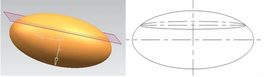
, the ellipsoid equation and the height of ellipsoidmissing must be known first, hence, the follow -ing purpose is to seek the value of a, b, and c. Any one of ellipsoid is being cut in a plane which is perpendicular to the major axis Z, as shown in Figure 1, leaving intercepted part to be the ellipsoid missing.
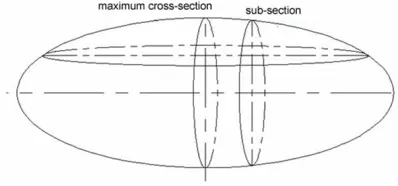
Then we intercept ellipsoid missing using equi-distantly parallel planes parallel to the long axis Z, which is similar to the process of CT scans, and then get different sections of ellipsoid missing, among which there must be a maxi-mum cross-section and sub-section of the ellip-soid missing, as shown in Figure 2.
Thereafter we select the maximum cross-sec-tion and separate it from the ellipsoid missing for further analysis (Figure 3). The maximum cross-section outer margin of the ellipsoid missing is must be an elliptic equations, by measuring then know the largest diameter and wide diameter is a1 and b1, respectively. Due to the outer margin of the maximum cross-section being elliptic equations, we set the Equation as bx2 cy 1
2 2 2
+ = , take (b-b1,
2
[image:2.612.91.525.75.203.2]a1) into
the elliptic Equation b bb2 4ac 1 1 2 2 1 2 -+ =
^
h
(in themodel assuming that b = c), and then obtain
8 4
b c b b a
1 1
2 1
2
= = + (2) Similarly, we select the sub-maximum cross-section intercepted from ellipsoid missing, and the outer margin of this section is bound to be an ellipse and on the projection line of point A which is on the outer margin of the sub-maxi-mum cross-section (Figure 4).
As shown in Figure 5, we take A coordina- te point (b-b1+b2, k) into the Equation
( )
1 a
b b b
b k 2 1 2 2 2
- + + = , where b
1 and b2 are the wide diameters of maximum cross-section and sub-maximum cross-section.
a
b b b b
bk 2
1 2
2 =
-
^
- +h
(3)Simultaneous Equation 2 and 3 to obtain:
4 4
a
b b b a b b
k b a
4 1 1 2 12
1 2 1 2 1 2 = - + +
^
^
h
^
h
h
(4) The formula ofellipsoid missing volume is1 3
V1 abhc hc
2
= r
`
-j
, where a is the axis of the ellipsoid, h is the height of ellipsoid missing that is b1, so, h = b1.The a, b and c are substituted into the volume formula of ellipsoid missing, get the ellipsoid missing volume of the outer margin:
1 3 4 4 3 V c abh c h
b b b a b b
kb b a
4 1
2
1 2 1
2 2 1 2 1 2 1 2 1 1 = - = - + + r r
^
^
h
^
h
h
`
j
Similarly, the ellipsoid missing volume of inner edge is:
1 3
4
4 3
V abhc hc
b b b a b b
kb b a
4 1
2
3 3 4 1
2 3 4 3 2 3 2 1 2 = -= - + + r r
^
^
h
^
h
h
`
j
the outer margin wide shaft of the sub-section,
b3 is the inner margin wide shaft of the
maxi-mum cross-section, b4is the inner margin wide
shaft of the sub-section, and k is thickness.
Data analysis
The cephalic hematoma imaging was per-formed by Japanese Toshiba aquilion 64 spiral CT with 5 mm slice thickness to determine the length and width of scheduled slice of hemato-ma as well as the thickness of hehemato-matohemato-ma by computer software. Subsequently, the hemato-ma volume was calculated by using v (formula), v (2/3sh), v (1/2abc) and v (1/3abc), respec-tively. We considered CAVA as gold standard, during which the hematoma margins were hand-traced by the radiologist on each axial slice. Then the hematoma volume (cm3) on that particular slice was calculated as the product of the area of the traced hematoma (cm2) mul-tiplying the corresponding slice thickness (cm). The sum of the volumes on each slice gave the total hematoma volume.
Statistical analyses
The discrepancy of all hematoma volume value and percent deviations from the gold standard were analyzed with one-way analysis of vari-ance (ANOVA; Welch and Brown-Forsythe meth-od), and post hoc pairwise comparisons were
performed with Fisher’s least significant differ
-ence (LSD) test. In addition, correlation coeffi -cients were in turn obtained using Pearson
coefficients; then, the effects of different esti -mated methods on hematoma volumes and percent deviations were respectively analyzed by error bars and box plots. Data were expressed as mean ± SD, and all statistical analyses were performed using the SPSS 19.0 statistical software (SPSS Inc., IL, USA). A
P-value of less than 0.05 was considered
[image:3.612.89.288.73.166.2]Results
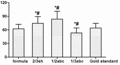
The volumes of CSDH were calculated respec-tively by the following methods: 1) v (formula), 2) v (2/3sh), 3) v (1/2abc), 4) v (1/3abc), 5) v (Gold standard). The one-way ANOVA results indicated that there are no statistical differenc-es between v (formula) and v (Gold standard) groups (63.42 ± 9.92 vs. 63.42 ± 9.92, P > 0.05, Figure 6), whereas the hematoma vol-umes of v (2/3sh), v (1/2abc) and v (1/3abc)
groups significantly differed from v (formula) or
v (Gold standard) group (71.69 ± 13.58, 81.40
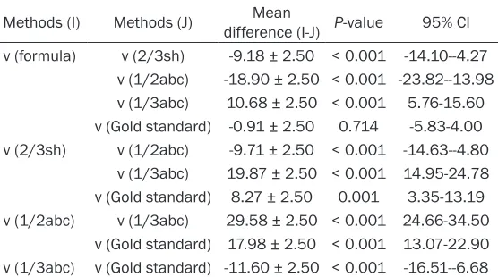
± 15.59, and 51.82 ± 9.92, P < 0.05, Table 1).
Besides, there was a significant correlation
between v (formula) and v (Gold standard)
groups using Pearson coefficient (r = 0.978, P < 0.001), suggesting that all of the hematoma volumes calculated by v (formula) was close to real volumes.
Discussion
Intracerebral hematoma volume has been vali-dated to be an independent predictor for poor outcome and mortality by early correlative research [8-10]. Precise determination of he- matoma volume is also one of the important
influence factors for an appropriate treatment
in clinical work [11, 12]. Thus, it is necessary for physicians to have a simple reliable method to measure the hematoma volume.
[image:4.612.92.524.71.209.2] [image:4.612.324.524.246.388.2]Considering the shape of subdural hemato- ma was closer to crescent, we found that the calculation method of ellipsoidal missing volume could assess the hematoma volume more precisely. For convenience in clinical Figure 3. The maximum cross-section image from ellipsoid missing.
Figure 4.The sub-maximum cross-section intercept-ed from ellipsoid missing.
Figure 5. The mean volumes of CSDH by each meth-od were analyzed by one-way analysis of variance. CSDH, chronic subdural hematomas; *, P < 0.05 vs. formula; #, P < 0.05 vs. Glod standard.
[image:4.612.90.287.254.352.2] [image:4.612.92.289.406.513.2]work, we derived the formula of crescent (
2
V
4
4 3
4
4 3
b b b a b b
kb b a
b b b a b b
kb b a
4 4
1 1
2 1 2 1
2 1
2 1
2
3 4 1 2
3 4 3
2 3
2 1
2
1 3
=
- +
+
-- +
+
r r
^
^
h
^
h
h
^
^
h
^
h
h
), the formula needs length and width of hema-toma on adjacent maximum slice. We also sim-ply made a programming to make the calcula-tion briefer that physicians can directly obtainthe result by inputting specific measured data
in clinical work.
In the present study, we considered CAVA as the gold standard, and calculated the hemato-ma volume by using v (formula), v (2/3sh), v (1/2abc) and v (1/3abc), respectively. The results showed a similar hematoma volume between v (formula) and v (Gold standard) groups (P > 0.05). Besides, a further significant correlation between the two groups (r = 0.978,
P < 0.001) also validated the accuracy of our formula most close to real volumes. This could be explained by the fact that estimation error
was more significantly associated with hema -toma shape compared with hema-toma size [13].
However, the 1/2abc method (81.40 ± 15.59) overestimated the subdural hematoma volume compared with gold standard (63.42 ± 9.92), which was consistent with Huttner et al who had also validated that the result obtained from the 1/2abc method measuring irregular shape of hematoma volume was overestimat-ed about 15% and 32% in comparison with actual volume [14]. Despite of its convenience, the method of 1/2abc was theoretically deduced with the assumption that hematoma
many situations [7], as evidenced by a calcu-lated of 51.82 ± 9.92 in comparison with gold standard (63.42 ± 9.92). Although some schol-ars maintain a viewpoint that the data assessed
by means of 2/3sh had no significant differ -ence with gold standard [7], this study did dem-onstrate an overestimated subdural hematoma volume (71.69 ± 13.58) compared with gold standard (63.42 ± 9.92).
Considering it was too intricate to use our for-mula for the calculation of subdural hematoma volume directly in clinical practice, it was nec-essary to develope a formula program so that physicians could obtain the result by only input-ting relevant data to the given program. Hence, further study were needed focusing on develop-ing and installed our grogram in mobile phones, computers or tablet PC etc. Therefore, the com-putational complexity should not be the obsta-cles for clinical hematoma volume estimation. In conclusion, the present study demonstrated the better validity and accuracy of our formula in estimating subdural hematoma volumes compared with 1/2ABC, 1/3ABC and 2/3Sh formula.
Acknowledgements
This study was funded by the Heilongjiang Postdoctoral Science-Research Foundation (NO. LBH-Q12042 to Yuehua Wang).
Disclosure of conflict of interest
None.
v (1/2abc) v (1/3abc) 29.58 ± 2.50 < 0.001 24.66-34.50 v (Gold standard) 17.98 ± 2.50 < 0.001 13.07-22.90 v (1/3abc) v (Gold standard) -11.60 ± 2.50 < 0.001 -16.51--6.68
Data were expressed as mean ± SD. A P-value < 0.05 was considered statistically
significant.
[image:5.612.91.369.97.252.2]Address correspondence to: Dr. Huaizhang Shi,
Department of Neurosurgery, First Affiliated Hospital
of Harbin Medical University, No. 23 Youzheng Street, Nangang District, Harbin 15001, Heilongjiang Province, China. Tel: 0451-8555090; Fax: +86-0451-85555090; E-mail: huaizhang_shi321@126. com
References
[1] Regan JM, Worley E, Shelburne C, Pullarkat R and Watson JC. Burr hole washout versus cra-niotomy for chronic subdural hematoma: pa-tient outcome and cost analysis. PLoS One 2015; 10: e0115085.
[2] Chan DY, Sun TF and Poon WS. Steroid for chronic subdural hematoma? A prospective phase IIB pilot randomized controlled trial on the use of dexamethasone with surgical drain-age for the reduction of recurrence with reop-eration. Chinese Neurosurgical Journal 2015; 1: 2.
[3] Tang J, Ai J and Macdonald RL. Developing a model of chronic subdural hematoma. Acta Neurochir Suppl 2011; 111: 25-9.
[4] Chon KH, Lee JM, Koh EJ and Choi HY. Independent predictors for recurrence of chronic subdural hematoma. Acta Neurochi- rurgica 2012; 154: 1541-1548.
[5] ur Rehman R, Noman MA, Ayoob S, Mewat S and Nabi A. Optimum management of chronic subdural hematoma: evaluation of various sur-gical options for the treatment of chronic sub-dural hematoma. Khyber Journal of Medical Sciences 2015; 7.
[6] Sucu HK, Gokmen M and Gelal F. The value of XYZ/2 technique compared with computer-as-sisted volumetric analysis to estimate the vol-ume of chronic subdural hematoma. Stroke 2005; 36: 998-1000.
[7] Zhao KJ, Liu Y, Zhang RY, Wang XQ, Gao C and Shen JK. A precise, simple, convenient and new method for estimation of intracranial he-matoma volume-the formula 2/3Sh. Neurol Res 2009; 31: 1031-1036.
[8] Zurasky J, Aiyagari V, Zazulia A, Shackelford A and Diringer M. Early mortality following spon-taneous intracerebral hemorrhage. Neurology 2005; 64: 725-727.
[9] Lim JK, Hwang HS, Cho BM, Lee HK, Ahn SK, Oh SM and Choi SK. Multivariate analysis of risk factors of hematoma expansion in sponta-neous intracerebral hemorrhage. Surg Neurol 2008; 69: 40-45.
[10] Juvela S. Risk factors for impaired outcome af-ter spontaneous intracerebral hemorrhage. Arch Neurol 1995; 52: 1193-1200.
[11] Becker K, Baxter A, Cohen W, Bybee H, Tirschwell D, Newell D, Winn H and Longstreth W. Withdrawal of support in intracerebral
hem-orrhage may lead to self-fulfilling prophecies.
Neurology 2001; 56: 766-772.
[12] Broderick JP, Brott TG, Duldner JE, Tomsick T and Huster G. Volume of intracerebral hemor-rhage. A powerful and easy-to-use predictor of 30-day mortality. Stroke 1993; 24: 987-993. [13] Xu X, Chen X, Zhang J, Zheng Y, Sun G, Yu X and
Xu B. Comparison of the Tada Formula With Software Slicer Precise and Low-Cost Method for Volume Assessment of Intracerebral Hematoma. Stroke 2014; 45: 3433-3435. [14] Huttner HB, Steiner T, Hartmann M, Köhrmann