Original Article
High-intensity focused ultrasound ablation:
an in vitro agarose gel model
Sen Yang1,2,4*, Yi-Jing Chen3*, Ran Cui1, Hua-Xin Zhao2, Yu Zhao1, Zhu-Qing Liu1, Yi Yu1, Xiao-Yan Shao1, Qing Xu1,2
1Department of Oncology, Shanghai Dermatology Hospital, Shanghai, China; 2Department of Medical Oncol-ogy, Shanghai Tenth People’s Hospital, School of Medicine, Tongji University, Shanghai, China; 3Department of Pathology, Traditional Chinese Medicine Hospital of Kun Shan, Suzhou, Jiangsu, China; 4Department of General Practice, Yangpu Hospital, Tongji University, Shanghai, China. *Equal contributors.
Received June 6, 2016; Accepted July 24, 2017; Epub November 15, 2017; Published November 30, 2017
Abstract: Purpose: There are many ultrasound studies involve the use of tissue-mimicking materials to research phenomena in vitro and predict in vivo biological effects. The goal of our study was to establish a typical agarose gel model of high-intensity focused ultrasound (HIFU) treatment for empirical study. Materials and methods: In this study, the model consists of human pancreatic tissues embedded in an agarose gel. The Sprague Dawley (SD) rat liver tissues were encapsulated in agarose gel and ablated by HIFU. The changes in vitro after ablation were assessed using ultrasonography, pathological examination, and histochemical staining. Results: After HIFU treat-ment, the results showed that the ultrasonic echo signal of the ablating region was strong, and the necrosis at the center of the ablation was monitored using hematoxylin and eosin (HE) staining. Shrinkage, chromatin condensa-tion, nuclear condensation and fragmentation of the mouse liver cancer cells were assessed during pathological examination. Acid phosphatase (ACP) and succinic dehydrogenase staining (SDH) were negative in the experimental group. Discussion: Our study demonstrated that agarose model was appropriate for HIFU ablation in in vitro human tissues. The in vitro model may provide a useful tool for studying ablation induced by HIFU and facilitate further investigation of HIFU in anticancer therapeutics.
Keywords: High-intensity focused ultrasound, in vitro model, agarose gel, ablation
Introduction
HIFU induces coagulative necrosis of tumors without any skin damage, and is a truly nonin-vasive modality [1]. In addition to thermal abla-tion, changes in cellular and molecular res- ponses occurred in the local environment of HIFU-ablated animal models [2].
Currently, in vitro animal model ablation is the most common modality for HIFU treatment. High-intensity focused ultrasound with
pulmo-nary flooding generated a thermal effect in vivo
in a porcine model of lung tumor [3]. Tumor-bearing mice were injected with micro-bubbles via tail vein and treated with HIFU to study the
in vivo effect of HIFU on breast cancer cell lines [4]. A number of models have been used to sim-ulate thermal lesion formation caused by ultra-sound-induced heating in high-intensity fo- cused ultrasound (HIFU) [5, 6]. While these can
accurately mimic the response of soft tissues to heating, they are unable to precisely visualize or quantify mechanical ablation caused by HIFU compared with human tissues.
A human tissue model for measurement of HIFU treatment would be valuable in both ultra-sound imaging and therapy. It could verify whether a given transducer and set of acoustic parameters are capable of causing therapeutic effect. However, the use of HIFU in in vitro
Materials and methods
Tissue preparation
Eighteen SD rats aged between 6 and 8 weeks were purchased from the Shanghai Institutes for Biological Sciences, the Chinese Academy of Sciences, and raised in the central laborato-ry of Shanghai Tenth People’s Hospital. After euthanasia, the rat liver tissues were divided randomly into three groups, six in each group, numbered 1, 2 and 3, respectively. Fresh human pancreatic cancer tissue was obtained from a department of pathology and placed in 0.9% saline solution at room temperature. All experiments were conducted within 6 hours of harvest. Experiments and animal care were carried out in compliance with the guidelines for the care and use of laboratory animals rec-ommended by the Ministry of Science and Technology of the People’s Republic of China [7].
HIFU
HIFU ablation was performed using a HIFU tumor therapy system (Model JC type, Chong- qing Haifu Tech Co., Ltd, Chongqing City, China)
comprising a firing system located in a
de-gassed water tank, an imaging system consist-ing of an ultrasound scanner coupled with a sterotaxic localizing arm, and a
computer-con-trolled system controlling the firing sequence and the three-dimensional movement of the fir -ing head {Martin, 2014 #81; Marinova, 2016 #98} [8, 9].
Gel acoustics
Ultrasonic attenuation coefficient (UAC) is a
parameter for acoustic propagation. The larger the value of UAC, the greater the sonic energy decrease [10, 11]. UAC was calculated using the insert-substitution method of GBT15261-2008 as follows (1) [12]:
d d20 log A /Ax
s w
1
1
=
- +
a
^
h
a (1)Where α represents the UAC of tested material,
and dl is the thickness of the thicker sample, and dx denotes the thickness of the thinner sample. As is the signal amplitude of thicker samples while A1 is the signal amplitude of
thin-ner samples. The αW is the corrected value, and
α and αW are indicated as dB/cm. The units of d1 and dx are cm, and the As and A1 are expressed in volts.
Preparation of in vitro agarose gel
Hydrogel was prepared by dissolving agarose (1% wt) in aqueous solvent at a temperature of 95°C. Gelation occurred when the temperature of this solution was lowered to room
tempera-ture. Briefly, rat liver tissues were encapsulated
in 1% agarose. The agarose molds were allowed to gel at 37°C for 10 min and loaded into aga-rose scaffolds.
HIFU ablation
Eighteen SD rats were euthanized after fasting for 24 h. Liver tissues were obtained and evenly divided into three groups (blank, control, and experimental), and were loaded immediately on the scaffolds. The HIFU ablation was compliant with the guidance of the National Standard of China and described previously [13, 14]. The blank group of liver tissues was left untreated, while the control and experimental groups were encapsulated in agarose gel. The experimental group was ablated with the HIFU system (Model JC type, Chongqing HaifuTech Co., Ltd, Chong- qing City, China) with a 10 cm diameter and 161 mm focal length was used as a therapy transducer. The transducer operates at 0.99
MHz, and is driven by a class D amplifier
with matching network developed in our lab. The transducer was positioned in a tank of
degassed, filtered water, and the focus was
aligned to the cortex of the rats liver sample in
plastic tubes filled with agarose gel submerged
in the tank. A real-time ultrasound imaging device was used to locate the liver tissue as the
predesigned target. Ultrasound was fired at a
3×3 grid of points sequentially, with 2 mm spacing between points. The total exposure time at each point was 5 seconds. The param-eters used were 1.03 MHz, 400 W. This pattern was chosen over single spot treatments so that lesions were more easily located in tissue for histological sectioning. Eventually, the entire target region was covered by HIFU, leading to coagulation necrosis of the whole target region. During the therapeutic process, real-time
eval-uation of the profile of tissue damage in the
model was conducted using the computer
sys-tem and graphic analysis of the target field and
Take a piece of human pancreatic cancer with a 4 cm diameter, then puts it into a plastic tube
filled with agarose gel submerged in the tank.
And then irradiated with HIFU, the process and parameters of HIFU ablation are the same as the rats liver tissue.
Hematoxylin and eosin (H&E) staining
The tissue samples were stained with 1% 2, 3, 5-triphenyltetrazolium chloride (TTC) solution for 5 to 7 min, and washed with water. Gross observations including the appearance, size and shape of the tissue were recorded. The
tis-sue samples were fixed in 40 g/L formaldehyde solution, embedded in paraffin and stained
with hematoxylin and eosin (H&E), respec- tively.
Histochemical staining
The liver samples were fixed in 40 g/L formal
-dehyde solution, embedded in paraffin and
respectively stained with ACP and SDH,
fol-lowed by light microscopy (Olympus BH2, Olympus Corporation, Tokyo, Japan). The sam-ples were deemed positive if brown granules were detected in the cytoplasm following ACP staining, and if they stained blue purple with SDH.
Statistical analysis
Sound speed and acoustic attenuation were
represented as mean ± S.D., and the signifi -cance level was set at P<0.05. The agarose concentration and acoustic attenuation were compared with linear analysis.
Results
Gel acoustics
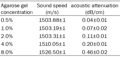
The acoustic properties of our agarose gel at various agarose concentrations are summa-rized in this study (Table 1). The gel concentra-tions ranged from 0.5% to 8%. Gels with higher concentrations of agarose showed a higher acoustic attenuation (Figure 1). A significant
correlation was found between the acoustic attenuation and agarose concentration, with a
correlation coefficient of 0.990 (P<0.01). The
magnitude of the attenuation coefficient was
controlled easily between 0.03 and 0.5 (dB/ cm), by varying the concentration of agarose.
In vitro agarose gel model and HIFU ablation
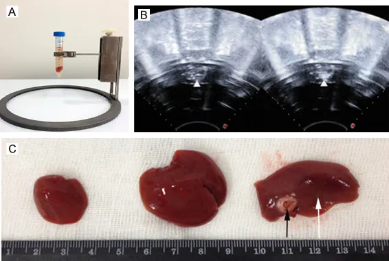
The agarose molds were allowed to gelate at 37°C for 10 min before loading on agarose scaffolds (Figure 2A). After HIFU treatment, changes in coagulation necrosis in the ultra-sonic echo of the ultrasound model were clear-ly found in the center of the ablating region, and the intensity of the echo grew stronger after ablation (Figure 2B).
Pathological manifestations
After HIFU treatment, pale coagulation necrosis
was easily identified in all the experimental liver
samples. Normal liver tissues were red, where-as tissues of coagulation necrosis were white after TTC staining. In the experimental group, there was a sharp boundary between HIFU necrosis and viable tissue (Figure 2C).
Light microscopy
Under a light microscope, the following necrotic features were observed in the livers of experi-mental group: karyopycnosis and nuclear
frag-Table 1. Acoustic properties of PAA gel at vari-ous agarose powder concentrations
Agarose gel
concentration Sound speed (m/s) acoustic attenuation (dB/cm)
0.5% 1503.88±1 0.04±0.01
1.0% 1503.19±1 0.07±0.02
2.0% 1503.31±1 0.11±0.01
4.0% 1510.05±1 0.20±0.01
8.0% 1526.50±1 0.46±0.02
Values are expressed as mean ± SD.
Figure 1. Gel acoustic characterization. Attenuation
coefficient and agarose gel concentration. x coor
[image:3.612.91.288.95.189.2] [image:3.612.92.287.216.350.2]mentation in most of the cells, and a sharp boundary between the normal tissue and tar-get zones. The tartar-get tissues contained necrot-ic cells and nuclear debris after ablation and HE staining. Further, the experimental group stained negative with ACP and SDH. However, the control and blank groups showed no differ-ences (Figure 3A-I).
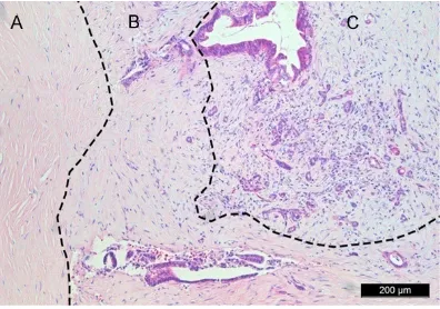
Human pancreatic cancer tissue in HE stain-ing
After HIFU treatment, the ablation area (A) showed homogeneous red dye and no cell structure, no ablation area (C) had clear cell structures and nucleolus, but the cells were arranged disorder. The junction between the two areas (B) could be seen the fuzzy structure, and nuclear pale staining (Figure 4).
Discussion
The ideal treatment for localized cancer should result in complete cell death without collateral damage [15, 16]. Previous researchers have directly observed the interaction of cavitation with cells, either with liquid and gel cell suspen-sions [17].
The novelty of our model as follows: we found that the tissue model described in this study
ducibly. It is user-friendly, and amenable to large-scale use [22].
Acoustic attenuation coefficient demonstrated
the ability of the media to transfer energy. A higher agarose gel concentration resulted in
greater acoustic attenuation coefficient [23,
24]. Therefore, to reduce the acoustic attenua-tion of agarose gels, low concentraattenua-tions of aga-rose gel are better than higher levels. However, if the agarose gel concentration is low, it is very soft and fragile, and the acoustic attenuation
coefficient of 1% agarose gel is 0.073±0.001
dB/cm [25]. The human tissue sound
attenua-tion coefficient is about 0.5 to 1.5 dB/cm/MHz
[26]. It is far less than the human tissue
attenu-ation coefficient and the ultrasonic attenuattenu-ation
is negligible. Furthermore, in preliminary experi-ments [27], a polypropylene model was used after HIFU treatment. Several bubbles were observed in the central rubber block, and the polypropylene plastic was destroyed, while 1% agarose gel model after HIFU treatment showed no reaction. These show similar appearance to HIFU treatment seen previously in agarose gels [28]. Therefore, 1% agarose gel was the optimal choice.
In this study, we selected the rat liver tissue as a sample. In vitro liver tissue wrapped with 1% agarose gel was exposed to HIFU. The
patho-Figure 2.HIFU ablation. A. The in vitro model was loaded on agarose scaf-folds. B. Changes in ultrasonic echo of the target regions after HIFU. Δdenotes
the view of the coagulated lesion. C. These models were: blank, control and experimental groups (from left to right), respectively; TTC staining of liver tissues after HIFU. Coagulation necrosis (black arrow) was obvious and the white arrow showed normal pancreas. The boundary was clear.
can be used to directly ob- serve cavitation and the re- sulting damage as it would occur in solid soft tissues [18]. Besides, the model provides a more controllable method to study the effects of different ultrasound parameters and transducers than in vivo tis- sue.
Agarose is a polysaccharide with alternating copolymers of
1,4-linked 3, 6-anhydro-α-l-galactose and 1,3-linked
[image:4.612.92.375.73.263.2]repro-logical section was observed under a micro-scope, which revealed that agarose gel had no
effect on the biological properties of the tissue and HIFU ablation. Furthermore, we used human pancreatic cancer tissue to conduct
experiments to confirm that this model was
suitable for HIFU therapeutics, and this result was consistent with previous studies of in vivo
tissue histotripsy lesions [29].
The model was simple and user-friendly facili-tating tissue location and control. Each experi-ment in this model was treated with HIFU irra-diation and ablative attenuation consistently, reducing the experimental error between the groups and any damage to the operator. Moreover, the gel model should be relatively simple and inexpensive to prepare, as one pur-pose is to avoid the costs associated with using experimental animals, and the experimental materials were wrapped in agrose gel, which prevented contamination of the tissue. Most
[image:5.612.91.524.70.400.2]importantly, this model was fixed by the brack -et, obviating the need for manual handling.
Figure 3. Light microscopy. Presentations of liver in the experimental group (C, F, I) under light microscope after HIFU (H&E, SDH, ACP×200). An apparent boundary was seen between normal and target tissues. However, no differences were found between the blank (A, D, G) and control groups (B, E, H). The cellular structures were normal.
[image:5.612.90.288.467.606.2]However, additional trials are underway to ascertain the utility of agarose gel model. Besides, it is essential to determine the histo-logical changes and molecular mechanisms of the agarose gel models. A number of prelimi-nary studies suggested the reliability of the agarose model, which may facilitate additional studies investigating the mechanisms of car- cinogenesis.
In conclusion, the novel agarose gel model is a safe and effective in vitro experimental tool for the investigation of HIFU anticancer thera- peutics.
Acknowledgements
This article is supported by the National Natural Science Foundation of China (NO 81372749). Disclosure of conflict of interest
None.
Address correspondence to: Dr. Qing Xu, Department of Medical Oncology, Shanghai Tenth People’s Hospital, School of Medicine, Tongji University, 301 Middle Yanchang Road, Zhabei District, Shanghai 200072, P. R. China. Tel: +86 188 1787 9518; E-mail: qingxumd@aliyun.com
References
[1] Hsiao YH, Kuo SJ, Tsai HD, Chou MC and Yeh GP. Clinical application of high-intensity fo-cused ultrasound in cancer therapy. J Cancer 2016; 7: 225-231.
[2] Kim YS, Bae DS, Park MJ, Viitala A, Keserci B, Rhim H and Lim HK. Techniques to expand pa-tient selection for MRI-guided high-intensity focused ultrasound ablation of uterine fibroids. Am J Roentgenol 2014; 202: 443-451. [3] Wolfram F, Reichenbach JR and Lesser TG. An
ex vivo human lung model for ultrasound-guid-ed high-intensity focusultrasound-guid-ed ultrasound therapy using lung flooding. Ultrasound Med Biol 2014; 40: 496-503.
[4] Wang J, Li P, Tian R, Hu W, Zhang Y, Yuan P, Tang Y, Jia Y and Zhang L. A novel microbub- ble capable of ultrasound-triggered release of drug-loaded nanoparticles. J Biomed Nano-technol 2016; 12: 516-524.
[5] Kyriakou Z, Corral-Baques MI, Amat A, Cous-sios CC. HIFU-induced cavitation and heating in ex vivo porcine subcutaneous fat. Ultra-sound 2011; 37: 568-579.
[6] Qian K, Li C, Ni Z, Tu J, Guo X and Zhang D. Uniform tissue lesion formation induced by
high-intensity focused ultrasound along a spi-ral pathway. Ultrasonics 2017; 77: 38-46. [7] Canney MS, Bailey MR, Crum LA, Khokhlova VA
and Sapozhnikov OA. Acoustic characterization of high intensity focused ultrasound fields: a combined measurement and modeling ap-proach. J Acoust Soc Am 2008; 124: 2406-2420.
[8] Merckel LG, Knuttel FM, Deckers R, Van DT, Schubert G, Peters NH, Weits T, van Diest PJ, Mali WP, Vaessen PH, van Gorp JM, Moonen CT, Bartels LW, van den Bosch MA. First clinical experience with a dedicated MRI-guided high-intensity focused ultrasound system for breast cancer ablation. Eur Radiol 2016; 26: 4037-4046.
[9] Möri N, Jud C, Salomir R and Cattin PC. Lever-aging respiratory organ motion for non-inva-sive tumor treatment devices: a feasibility study. Phys Med Biol 2016; 61: 4247-4267. [10] Martin JM, Handorf EA, Kutikov A, Uzzo RG,
Bekelman JE, Horwitz EM and Smaldone MC. The rise and fall of prostate brachytherapy: use of brachytherapy for the treatment of localized prostate cancer in the national cancer data base. Cancer 2014; 120: 2114-2121.
[11] Hoche S, Hussein MA and Becker T. Density, ultrasound velocity, acoustic impedance,
re-flection and absorption coefficient determina
-tion of liquids via multiple reflec-tion method. Ultrasonics 2015; 57: 65-71.
[12] Tang ZY, Zhao JN, Zhong WJ, Luo YD, Wu W, Chen WJ and Dai YB. The value of proton mag-netic resonance spectroscopy in high-intensity focused ultrasound treatment of experimental liver cancer. Transl Oncol 2015; 8: 163-168. [13] Poretti D, Lanza E, Sconfienza LM, Mauri G,
Pedicini V, Balzarini L and Sardanelli F. Simul-taneous bilateral magnetic resonance angiog-raphy to evaluate thoracic outlet syndrome. Radiol Med 2015; 120: 407-412.
[14] Li J, Krupka T, Yao J, Wang R, Jiang L, Zhou Y, Zuo G, Wang Z, Dai L and Ren J. Liquid-solid phase-inversion PLGA implant for the treat-ment of residual tumor tissue after HIFU abla-tion. PLoS One 2015; 10: e0117358.
[15] Zhou J, Zhou C, Zhan W, Jia X, Dong Y and Yang Z. Elastography ultrasound for breast lesions: fat-to-lesion strain ratio vs gland-to-lesion st- rain ratio. Eur Radiol 2014; 24: 3171-3177. [16] Uchida T, Tomonaga T, Kim H, Nakano M, Shoji
S, Nagata Y and Terachi T. Improved outcomes with advancements in high intensity focused ultrasound devices for the treatment of local-ized prostate cancer. J Urol 2015; 193: 103-110.
[18] Tako M, Kitajima S, Yogi T, Uechi K, Onaga M, Tamaki Y and Uechi S. Structure-function relationship of a gellan family of polysaccha-ride, S-198 gum, produced by alcaligenes ATCC31853. Advances in Biological Chemistry 2016; 6: 55-69.
[19] Maxwell AD, Cain CA, Duryea AP, Yuan L, Gurm HS and Xu Z. Noninvasive thrombolysis using pulsed ultrasound cavitation therapy-histot- ripsy. Ultrasound Med Biol 2009; 35: 1982-1994.
[20] Zucca P, Fernandez-Lafuente R and Sanjust E. Agarose and its derivatives as supports for enzyme immobilization. Molecules 2016; 21: 1577.
[21] Díazlópez A, Morenobueno G and Cano A. Role of microRNA in epithelial to mesenchymal tran-sition and metastasis and clinical perspec-tives. Cancer Manag Res 2014; 2014: 205-216.
[22] Batumalay M, Harun SW, Ahmad F, Nor RM, Zulkepely NR and Ahmad H. Study of a fiber optic humidity sensor based on agarose gel. J Mod Optic 2014; 61: 244-248.
[23] Daugschies M, Rohde K and Barkmann R. The preliminary evaluation of a 1 MHz ultrasound probe for measuring the elastic anisotropy of human cortical bone. Ultrasonics 2014; 54: 4-10.
[24] Mohd TMK, Ibrahim S, Yunus MAM and Fara-marzi M. Contact and non-contact ultrasonic measurement in the food industry: a review. Measurement Science & Technology 2016; 27: 012001.
[25] Feldmann A, Langlois C, Dewailly M, Martinez EF, Boulanger L, Kerdraon O and Faye N. Shear wave elastography (SWE): an analysis of breast lesion characterization in 83 breast lesions. Ultrasound Med Biol 2015; 41: 2594-2604. [26] Martin NE, Massey L, Stowell C, Bangma C,
Briganti A, Bill-Axelson A, Blute M, Catto J, Chen RC and D’Amico AV. Defining a standard set of patient-centered outcomes for men with localized prostate cancer. Eur Urol 2015; 67: 460-467.
[27] Zackrisson S, van de Ven SMWY and Gambhir SS. Light in and sound out: emerging transla-tional strategies for photoacoustic imaging. Cancer Res 2014; 74: 979-1004.
[28] Misono S, Marmor S, Yueh B, Virnig BA. Treat-ment and survival in 10,429 patients with lo-calized laryngeal cancer: a population-based analysis. Cancer 2014; 120: 1810-1817. [29] Egorov V, Tsyuryupa S, Kanilo S, Kogit M and