Original Article
The clinical value of real-time tissue
elastography in diagnosing thyroid malignancy
Juan Li, Mei Zhang, Li Sheng
Department of Ultrasound, The First People’s Hospital of Jingzhou, Jingzhou, Hubei Province, China
Received November 8, 2017; Accepted December 14, 2017; Epub February 15, 2018; Published February 28, 2018
Abstract: Objective: To investigate the diagnostic value of real-time tissue elastography (RTE) in defining the nature of thyroid nodules and its diagnostic efficiency in diagnosing malignant thyroid tumor. Methods: Sixty-eight patients who were diagnosed as having thyroid nodules and treated in our hospital were included as subjects (the total number of nodules is 102). All of them received both conventional B-mode ultrasound (BUS) and RTE tests prior to the operation. The diagnostic values and efficiencies of BUS and RTE in the differentiation of malignant and benign thyroid nodules were compared and analyzed by using receiver operating characteristic (ROC) curve and surgical pathological diagnosis as gold standards. Results: In the study, there were 61 benign thyroid nodules out of 38 cases and 41 malignant thyroid nodules out of 30 cases. By the analysis of ROC curve, the area under the curve and 95% of confidence interval of RTE and BUS were 0.923, 0.886-0.967, and 0.835, 0.761-0.896 respectively, with statistically significant difference (χ2=9.027, P=0.000). Moreover, there were intergroup differences in the sensitiv-ity, specificity and accuracy of BUS and RTE when they are used to define the nature of thyroid nodules, which were 67.21%, 46.34%, 58.82%, and 85.25%, 75.61% and 81.37% respectively (all P<0.05). Conclusion: Compared to BUS, the RTE method works more effectively in the differential diagnosis of thyroid nodules and demonstrates high diagnostic value in detecting thyroid malignancy.
Keywords: Real-time tissue elastography, thyroid ultrasonography, diagnostic value, thyroid malignancy
Introduction
In recent decades, incidence of thyroid nodules has been rising each year in China, and accord-ing to the clinical and epidemiological data, the incidence of thyroid malignancy accounts for around 5% of all these cases. Although there are similarities in the clinical manifestations of malignant and benign thyroid nodules, the treatment and the prognosis of these two kinds of nodules are extremely different [1, 2]. Therefore, it is of great clinical importance to choose a testing method that is highly accu-rate, easy to perform and non-invasive for diag-nosing and defining the nature of thyroid nod-ules during the early stage, so that physicians can decide on treatment options in a timely manner [3]. At present, the thyroid nodules are mainly examined by palpation and conventional two-dimensional B-mode ultrasound (BUS), but the specificity and accuracy of the examination are not very satisfactory [3]. In recent years,
been gaining clinical attention, as it has demon-strated good performance when applied in the differentiation of benign and malignant nodules in mammary gland, prostate and other organs. However, the reports of RTE being used for assessing the thyroid nodules are relatively few [4]. Therefore, this study was to investigate the clinical value of RTE in the differential diagnosis of thyroid nodules by analyzing the clinical data of the patients in our hospital, and to obtain evidence-based clinical information about using this method for the early detection of thy-roid malignancy.
Materials and methods
Participants/subjects
sisted of 40 males and 28 females with an average age of 45.8±9.2. Among them, 37 patients had single nodule and 31 patients had multiple nodules. Inclusion criteria were as fol-lows: 1) patients were diagnosed as having thy-roid nodules by palpation and general neck ultrasound; 2) patients received surgery and pathologic diagnosis; 3) patients signed the informed consent. Exclusion criteria were: 1) the nodules were completely cystic; 2) the nod-ules were too big or too close to the edge where there was no adequate thyroid tissue surround-ed for comparison; 3) patients receivsurround-ed head and neck radiotherapy in the past; 3) patients were obese with excessive fat in the larynx that could affect the results of BUS. Informed con-sents were obtained from all patients and the study was approved by the Ethics Committee of the hospital.
Examination equipment and methods
All patients in this study were given the conven-tional two-dimensional BUS and RTE tests prior to the operation. The instrument used for RTE examination was Philips Color Doppler Ultra- sound iU22, and the L12-5 convex array probe was applied. The detection frequency was set at 5-14 MHz. During the examination, patients were in supine position, with their heads tipped backward to fully expose the neck area. Then,
over twice the size of the lesion area. When the image quality became stable, the image was frozen and transmitted to the hard disk of the instrument. Images were scored by two physi-cians who were highly experienced in diagnos-tic sonography. Only the qualified images were taken for the measurement of the thyroid nod-ules, and the average value of three scores of the image was taken as the final value.
Determination of hardness by ultrasonic elas-tography
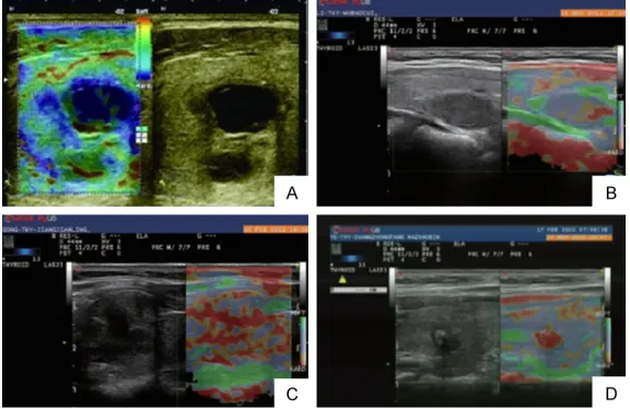
[image:2.612.89.377.72.259.2]The four-grade rating recommended by the Esaote Group was adopted as the elasticity scoring system. The ratings were as follows: grade 1, the whole lesion showed blue color (the blue color indicated an intermediate strain rate); grade 2, most of the lesion showed blue color, while few parts showed red color (the red color indicated no strain); grade 3, most of the lesion showed red color; grade 4, the whole lesion showed red color. By using this scoring system, the nodule in grade 1 or 2 was diag-nosed as benign, whereas the nodule in grade 3 or 4 was highly suspected of being malignant. Thus, grade 1 and 2 were used as diagnostic criteria for benign thyroid nodule, and grade 3 and 4 as criteria for malignant nodule [4]. See
Figure 1.
the volume of the thyroid lesions was measured by two-dimensional BUS, and the shape, quantity, border, blood supply, calcification, homoge-neity of the nodules, and pres-ence of any diffuse echo change in the surrounding tis-sues were carefully examined. The examination conditions were based on the size of the thyroids and the location and depth of the lesions. Patients were asked to refrain from swallowing, and the probe was placed onto the neck area. The coupling agent between the probe and neck skin could be increased if needed. The examiner vibrated the probe with a vibration frequency of twice per second and a pres-sure index of 3-4. The size of the target area was adjusted Figure 1. RTE images of the thyroid nodules with different grades in elasticity
Criteria for defining benign and malignant thy -roid nodules by conventional ultrasound The characteristics of malignant nodules inclu- ded: unclear boundary, irregular shape, internal hypoecho, posterior echo attenuation, micro-calcification, rich blood flow, anteroposterior/ transverse diameter ratio (A/T) ≥1, and vascu-lar resistance index (RI) ≥0.7 etc. The charac-teristics of benign nodules included: clear bo- undary, regular shape, internal isoecho or hy- perecho, no posterior echo attenuation, little blood flow or no blood flow, no presence of cal-cification or coarse calcal-cification, A/T ratio<1, and RI<0.7 etc. [5]. See Figure 2.
Pathological examination
The patients underwent pathological examina-tion of thyroid nodules during and after opera-tion, and surgical pathological diagnosis was used as gold standard for analyzing the clinical
value of two-dimensional BUS and RTE me- thods.
Statistical analysis
The SPSS software version 17.0 was used for statistical analysis. The count data was ex- pressed as percentage, and χ2 test was used
for intergroup comparison. Areas under the curve (AUC) of both methods were calculated by the receiver operating characteristic (ROC) curve and compared by χ2 test, for assessing
the diagnostic value of the two-dimensional BUS and RTE. Surgical pathological diagnosis was used as gold standard, and the sensitivity, specificity and accuracy in diagnosing thyroid nodules by two-dimensional BUS and RTE were calculated and compared; the diagnostic effi-ciencies of these two methods in the differen-tiation of benign and malignant thyroid no- dules were compared by paired χ2 test. The test
level was set as α=0.05.
Results
Basic information
In the study, there was no diffuse echo change detected in thyroids among all 68 patients and the diameter of the 102 thyroid nodules ranged from 0.4 to 5.2 cm with an average value of 2.8±1.1 cm. There were 58 nodules located in the right lobe, 34 nodules in the left lobe, and 10 nodules in the isthmus. All the 102 nodules were confirmed by surgical pathological diagno-sis. There was a total of 61 benign nodules out of 38 cases, which were identified as nodular goiter, 41 malignant nodules out of 30 cases, which were thyroid papillary carcinoma.
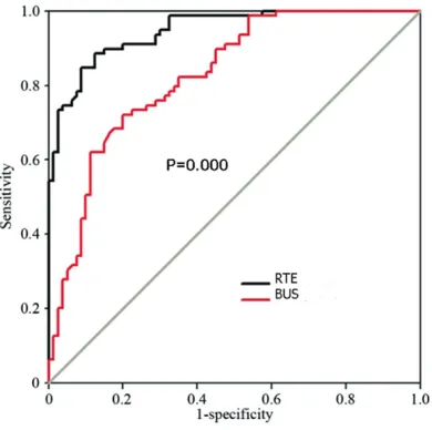
ROC curves for RTE and BUS
[image:3.612.87.524.72.172.2]The ROC curve was plotted using sensitivity as ordinate and 1-specificity as abscissa. Values Figure 2. Conventional ultrasound images of the thyroid nodules. A. Halos with different thickness can be seen around the nodules; B. Punctate hyperecho was detected in nodules; C. Signs of thyroid carcinoma are displayed, and halos can be seen in the surrounding area.
[image:3.612.92.287.233.427.2]of AUC were calculated accordingly. The AUC for RTE test in diagnosing the nature of thyroid nodules was 0.923, and the 95% confidence interval (CI) was 0.886-0.967. The RTE score 2.86 at the maximum of Youden index was cho-sen as the best critical point. If the RTE value ≥2.86, the nodule was considered malignant; if the RTE value <2.86, the nodule was consid-ered benign. The AUC for BUS was 0.835 and the 95% CI was 0.761-0.896. The difference in AUC value between two groups had statistical significance (χ2=9.027, P=0.000), indicating
that the RTE method had higher diagnostic value (Figure 3).
The sensitivity, specificity and accuracy of RTE and BUS
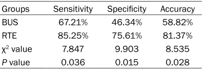
The sensitivity, specificity and accuracy of the conventional BUS and RTE were 67.21% (41/61), 46.34% (19/41), 58.82% (60/102) and 85.25% (52/61), 75.61% (31/41), 81.37% (83/102) respectively, which meant that all these three parameters were statistically differ-ent between the two testing methods (all P<0.05, Tables 1 and 2).
Discussion
Thyroid nodules are quite comon in clinical pratice and oftentimes discovered during physi-cal examination. Since there is a lack of speci-ficity in clinical manifestations of the malignant noules in early stage, and the conventional two-dimensional BUS can hardly differentiate the malignant from the benign nodules, missis could possibly occur [1, 5]. As early
diagno-sis and treatment are crucial for patients with thyroid malignancy, discovering an effective method that can diagnose and identify the nature of thyroid nodules during early stage has become one of the popular clinical topics [2, 6]. The tissue stiffness varies with the pathologi-cal types of the nodules. In the study, the tissue hardness of the thyroid papillary carcinoma was 2 to 3 times that of the nodular goiter. Clinicopathologic studies have shown that in malignant tumor, there would be abnormal pro-liferation of interstitial fibrous tissue, as well as an infiltrative growth of cancer cells in fibrous stroma, which can cause tissue to be denser and eventually increase its hardness [7]. Therefore, based on this pathological feature, a new type of ultrasonic imaging technique, i.e. ultrasonic elastography, has been applied clini-cally in recent years. Its principle is to form the image by evaluating the differences in the tis-sue stiffness, so that the hardness of the lesion can be reflected and the nature of the lesion can be determined [8].
One of the important ultrasonic elastography techniques is RTE, which can reflect the tissue strain through qualitative measurements of elastography [9, 10]. However, the results of RTE are obtained mainly by the subjective mea-surements of elastogram, and since in elastog-raphy, the image is mixed with multiple colors areas that are complex and diverse, it would be difficult to control the accuracy of the diagno-sis. Hence, the test results may have system-atic deviations, and can even cause misdiagno-sis or missed diagnomisdiagno-sis [11, 12]. Therefore, in view of this issue, clinical researchers have suggested the use of the elatography score to improve the accuracy in defining the nature of thyroid nodules. It is mainly based on the 5-point scoring system proposed by Itoh for rat-ing and diagnosis, in which the score was given according to the distribution of the color areas that was caused by different degrees of tissue displacement following compression. Tissues with high, medium and low elasticity
coeffi-Table 1. Diagnostic value of the two-dimensional BUS and RTE
BUS RTE
Benign Malignant Total Benign Malignant Total
Gold standard (surgical pathological diagnosis) Benign 41 20 61 52 9 61
Malignant 22 19 41 10 31 41
[image:4.612.95.523.84.152.2]Total 63 39 102 62 40 102
Table 2. Comparison of the sensitivity, speci-ficity and accuracy in two methods
Groups Sensitivity Specificity Accuracy
BUS 67.21% 46.34% 58.82%
RTE 85.25% 75.61% 81.37%
χ2 value 7.847 9.903 8.535
[image:4.612.91.289.197.266.2]cients are shown as red, green and blue respec-tively [13, 14]. At present, RTE is widely used in detecting the nature of lesions in breast, pros-tate and other areas, which has demonstrated good diagnostic performance. However, the clinical researches on the application of RTE in defining the nature of thyroid nodules and early detection of thyroid malignancy are still in prog-ress [15, 16]. Therefore, this study investigated and analyzed the diagnostic value of RTE, with a view to exploring an effective method for the early clinical detection of thyroid malignancy. In this study, the pathological examination was used as the gold standard; all benign thyroid nodules were identified as nodular goiter and malignant thyroid nodules were defined as thy-roid papillary carcinoma. This showed that the nature of the lesions in the subjects was not complex and the systematic error would be lit-tle. In ROC curves, it can be seen that the val-ues of AUC for RTE test were all greater than 0.9, which meant that RTE had good clinical value in terms of differentiating benign and malignant thyroid nodules. This result also aligned with the previous findings [17-20]. Besides, as a criterion for defining benign and malignant thyroid nodules, the maximum value of Youden index was chosen as the standard and the RTE score of 2.86 was set as the best critical point. Further research also showed that the sensitivity, specificity and accuracy of RTE method were all above 75% and higher than those in conventional two-dimensional BUS, based on the above-mentioned diagnos-tic criteria and with pathological diagnosis as gold standard. This presented that RTE method had good diagnostic value with a relatively low rate of misdiagnosing thyroid malignancy. In this study, we found that there were still some shortcomings in RTE, which were mainly due to the limitations of diagnosis in large or multiple thyroid nodules, for RTE requires ade-quate thyroid tissues in the surrounding area as controls. Thus, the target detection areas need to be at least twice the size of the nodule areas. Meanwhile, the large or multiple nod-ules could make neck lumpy, causing difficul-ties in examination as probes cannot be in fully contact with the neck. Since the overall sample size in this study was not large enough and the clinical study was single-centered, it would be necessary to conduct a multicentered clinical study with a larger-sample size for further ve- rification.
In conclusion, RTE test can perform more effec-tively in the differential diagnosis of benign and malignant thyroid nodules. The test has high diagnostic value in detecting thyroid malignan-cy, and could be recommended as the initial screening test of choice for patients who are highly suspected of having malignant thyroid tumors, with relatively small nodules and less than three unilateral nodules.
Disclosure of conflict of interest
None.
Address correspondence to: Li Sheng, Department of Ultrasound, The First People’s Hospital of Jing- zhou, No.8 Hangkong Road, Shashi District, Jing- zhou 434000, Hubei Province, China. Tel: +86-0716-8111888; E-mail: [email protected]
References
[1] Andrioli M and Persani L. Elastographic tech-niques of thyroid gland: current status. En- docrine 2014; 46: 455-461.
[2] Magri F, Chytiris S and Chiovato L. The role of elastography in thyroid ultrasonography. Curr Opin Endocrinol Diabetes Obes 2016; 23: 416-422.
[3] Liu BX, Liang JY, Xie XY, Huang GL, Zhou LY, Xu ZF and Lv MD. Preliminary comparative study of shear wave elastography versus quasi-sta- tic elastography on evaluation of thyroid nod-ules. Chinese Journal of Medical Ultrasound (Electronic Edition) 2014; 11: 57-62.
[4] Sui X, Liu HJ, Jia HL and Fang QM. Contrast-enhanced ultrasound and real-time elastogra-phy in the differential diagnosis of malignant and benign thyroid nodules. Exp Ther Med 2016; 12: 783-791.
[5] Zhang J, Xu HX, Zhang YF, Xu JM, Liu C, Guo LH and Liu LN. Value of acoustic radiation force impulse imaging in the differential diagnosis between benign and malignant solitary solid thyroid nodules. Chinese Journal of Medical Ultrasound (Electronic Edition) 2013; 10: 402-406.
[6] Yurttutan N, Gungor G, Bilal N, Kizildag B, Baykara M and Sarica MA. Interpretation of thyroid glands in a group of healthy children: real-time ultrasonography elastography study. J Pediatr Endocrinol Metab 2016; 29: 933-937.
[7] Sun J, Cai J and Wang X. Real-time ultrasound elastography for differentiation of benign and malignant thyroid nodules: a meta-analysis. J Ultrasound Med 2014; 33: 495-502.
elastog-raphy score system in differentiating malig-nant from benign thyroid nodules. Clin Imaging 2013; 37: 50-55.
[9] Zhang YZ, Xu T, Gong HY, Li CY, Ye XH, Lin HJ, Shen MP, Duan Y, Yang T and Wu XH. Applica-tion of high-resoluApplica-tion ultrasound, real-time elastography, and contrast-enhanced ultra-sound in differentiating solid thyroid nodules. Medicine (Baltimore) 2016; 95: e5329. [10] Kist JW, Nell S, de Keizer B, Valk GD, Borel
Rinkes IH and Vriens MR. The role of qualita-tive elastography in thyroid nodule evaluation: exploring its target populations. Endocrine 2015; 50: 265-267.
[11] Yoon JH, Yoo J, Kim EK, Moon HJ, Lee HS, Seo JY, Park HY, Park WJ and Kwak JY. Real-time elastography in the evaluation of diffuse thy-roid disease: a study based on elastography histogram parameters. Ultrasound Med Biol 2014; 40: 2012-2019.
[12] Rago T, Scutari M, Loiacono V, Santini F, Tonac-chera M, Torregrossa L, Giannini R, Borrelli N, Proietti A, Basolo F, Miccoli P, Piaggi P, Latrofa F and Vitti P. Low elasticity of thyroid nodules on ultrasound elastography is correlated with malignancy, degree of fibrosis, and high ex-pression of galectin-3 and fibronectin-1. Thy-roid 2017; 27: 103-110.
[13] Gungor G, Yurttutan N, Bilal N, Menzilcioglu MS, Duymus M, Avcu S and Citil S. Evaluation of parotid glands with real-time ultrasound elastography in children. J Ultrasound Med 2016; 35: 611-615.
[14] Ghajarzadeh M, Sodagari F and Shakiba M. Di-agnostic accuracy of sonoelastography in de-tecting malignant thyroid nodules: a systemat-ic review and meta-analysis. AJR Am J Roentgenol 2014; 202: W379-389.
[15] Menzilcioglu MS, Duymus M, Gungor G, Citil S, Sahin T, Boysan SN and Sarica A. The value of real-time ultrasound elastography in chronic autoimmune thyroiditis. Br J Radiol 2014; 87: 20140604.
[16] Nell S, Kist JW, Debray TP, de Keizer B, van Oostenbrugge TJ, Borel Rinkes IH, Valk GD and Vriens MR. Qualitative elastography can re-place thyroid nodule fine-needle aspiration in patients with soft thyroid nodules. A systemat-ic review and meta-analysis. Eur J Radiol 2015; 84: 652-661.
[17] Li W, Xiang FX, Li L, Yan TW, Zhang YK and Xie MX. The value of quantitative analysis in thy-roid nodules by real-time tissue elasticity imag-ing. Chinese Journal of Ultrasonography 2010; 19: 219-222.
[18] Wu Y, He RY, Duan HG, Wang LF, Dai WL and Li Q. Application value of ultrasonic elastography in benign and malignant thyroid nodules. Chi-na Modern Doctor 2015; 53: 115-117. [19] Liu BX, Liang JY, Xie XY, Huang GL, Zhou LY, Xu
ZF and Lv MD. Preliminary comparative study of shear wave elastography versus quasi-static elastography on evaluation of thyroid nodules. Chinese Journal of Medical Ultrasound (Elec-tronic Edition) 2014; 11: 57-62.