Original Article
Endotoxin-induced uveitis promotes retinal
endothelial cell injury and Muller cell proliferation
Lan Wu1, Yanshuang Wang1, Xuebing Xiao1, Yangyang Yu2
1Department of Ophthalmology, Daqing Oilfield Genaral Hospital, Daqing, China; 2Department of Ophthalmology,
Heilongjiang Provincial Hospital, Haerbin, China
Received June 19, 2017; Accepted January 7, 2018; Epub March 15, 2018; Published March 30, 2018
Abstract: Uveitis is one of the most common eye diseases that cause eye blind. It mainly occurs in young adults. The pathogenesis of uveitis has not been clarified, leading to great difficulties in the treatment. Rat endotoxin-induced uveitis (EIU) is an ideal animal model for the study of human endogenous uveitis. Retinal microvascular endothelial cells and Muller cells are responsible for the supply of retinal neurotrophic requirements. They play a key role in the visual protection through maintaining the blood-retinal barrier. However, the role and mechanism of EIU in retinal endothelial cell injury and Muller cell expression has not been reported. SD rats were randomly divided into two groups. The EIU animal model was established using Escherichia coli endotoxin (LPS). Retinal endothelial cells and Muller cells proliferation were detected by MTT assay. Cell apoptosis was evaluated by measurement of Caspase 3 expression. Real-time PCR and Western blot were used to test the vascular endothelial growth factor (VEGF) ex-pression. Enzyme-linked immunosorbent assay (ELISA) was applied to assess the level of interleukin-1β (IL-1β) and tumor necrosis factor-α (TNF-α). Our results showed that retinal endothelial cell and Muller cell proliferation was attenuated in EIU group. Caspase 3 activity was enhanced, with increased VEGF expression and IL-1β and TNF-α secretion in the EIU group compared with control group (P < 0.05). In conclusion, EIU causes retinal endothelial cell damage and Muller cell expression reduction by promoting inflammatory factors secretion, facilitating VEGF expres-sion, and inducing apoptosis.
Keywords: Endotoxin-induced uveitis, VEGF, inflammatory factors, retinal endothelial cell, Muller cell
Introduction
As one of the common diseases in ophthalmol-ogy, uveitis is a T cell-mediated autoimmune disease leading to eye blind [1, 2]. The inciden- ce of uveitis is relatively high as its incidence reaches 200/100,000 in developed countries and 700/100,000 in non-developed countries. Most patients are accompanied by conjunctivi-tis, visual impairment, and other serious eye complications. Among them, the number of blin- ding is up to a quarter [3, 4]. Uveitis is divided into anterior, intermediate, and posterior uve-itis, most of which occurs in young adults [5, 6]. The repeatability and persistent characteristics of uveitis makes its treatment extremely diffi-culty [7]. Due to the effect of inflammation on the posterior segment in the visually impaired uveitis patients, it often leads to retinal vasculi-tis, choroidal inflammation, and retinal inflam-mation [8]. Abnormal vascular function caused
by uveitis can induce retinal ischemia and neo-vascularization disorders [9]. Retinal blood ves-sels are complicated structures which are com-posed of a variety of cells, of which retinal mic- rovascular endothelial cells are responsible for the supply of retinal neurotrophic requirement. They play a crucial role in the visual protection through maintaining blood-retinal barrier, elimi-nating toxins, and alleviating inflammatory fac-tors [10, 11].
retinal neuron. Moreover, they maintain the sta-bility of the cell environment. Muller cells spe-cialized foot plate and retinal capillaries partici-pate in the composition of blood-retinal barrier [13, 14]. As the study of human uveitis is re- stricted by a number of factors, the current re- search is performed mainly using animal mod-els. Rat EIU is an ideal animal model for the study of human endogenous panuveitis [15, 16]. However, the impact and mechanism of EIU on retinal endothelial cell injury and Muller cell expression have not been reported. Materials and methods
Experimental animals
Specific pathogen free (SPF) grade male Spra- gue-Dawley (SD) rats aged 2 months with a body mass at 250 ± 20 g were purchased from the Experimental Animal Center of Heilongjiang. The feeding conditions contained constant temperature at 21 ± 1°C and relative humidity at 50-70%. The day/night cycle was 12 h/12 h. Rats were used for all experiments, and all pro-cedures were approved by the Animal Ethics Committee of Daqing Oilfield General Hospital.
Main materials and instruments
Escherichia coli endotoxin (LPS) was purchas- ed from Invitrogen (USA). Pentobarbital sodium and lidocaine were purchased from Zhpharma (Shanghai, China). IL-1α and TNF-α ELISA kits were purchased from R&D (USA). PVDF mem-brane was purchased from Pall Life Sciences. EDTA was purchased from Hyclone. Western blot related chemical reagents were from Beyo- time. ECL reagent was obtained from Amersham Biosciences. Rabbit anti-rats VEGF monoclonal and horseradish peroxidase (HRP) labeled IgG secondary antibodies were purchased from Ce- ll Signaling (USA). Caspase 3 activity detection kit was purchased from Nanjing Jiancheng bio-engineering research institute. Surgical micro-scopes were purchased from Suzhou Medical Equipment Factory. RNA extraction and reverse transcription kits were purchased from Axygen (USA). Labsystem Version1.3.1 Microplate rea- der was purchased from Bio-rad. ABI 7700 Fast Fluorescence Quantitative PCR was purchased from ABI (USA). Clean bench was purchased from Suzhou Sutai Purification Equipment Co., Ltd. Other common reagents were purchased from Sangon (Shanghai, China).
Grouping and treatment
The SD rats were equally and randomly divided into two groups, control and EIU group. Lipo- polysaccharides (LPS) were used to establish EIU rat model [15, 16]. LPS at 1 mg/ml were injected into the bilateral foot pad at 1 mg/kg.
Retinal endothelial cells and Muller cells isola-tion and cultivaisola-tion
The rats were anesthetized with 2% lidocaine and the bilateral eyeballs were aseptically re- moved. The eyeball was washed by gentamicin saline and transferred to the DMEM medium. Next, the eyeball was circuit cut at 3 mm poste-rior to the corneal limbus to remove the anteposte-rior segment and vitreous. Then the tissue was digested by 0.25% trypsin at room temperature for 10 min to separate the retinal endothelial cell layer and Muller cell layer. The tissue was further digested by 0.25% trypsin at 37°C for 30 min. Then the cell suspension was centri-fuged at 800 rpm for 10 min. At last, DMEM medium containing 100 U/ml penicillin and 100 μg/ml streptomycin was added into the cells and cultured at 37°C and 5% CO2.
MTT assay
20 μL MTT was added into retinal endothelial cells and Muller cells at logarithmic phase and incubated for 4 h. Then, 150 μL DMSO was add- ed into the plate for 10 min followed by measur-ing the absorbance value at 570 nm. Each experiment was repeated for three times.
Caspase 3 activity detection
Caspase 3 activity was tested according to the manual instructions. The cells were digested by trypsin and centrifuged at 600 g at 4°C for 5 min. Next, the cells were lysed on ice for 15 min and centrifuged at 20,000 g for 5 min at 4°C. At last, 2 mM Ac-DEVD-pNA was added into the cells and the absorbance value at 405 nm was measured to calculate Caspase 3 activity.
ELISA
was adopted to calculate the concentration of samples.
Real-time PCR
Total RNA was extracted from the retinal endo-thelial cells and Muller cells, and reversely tran-scribed into cDNA (Table 1). Real-time PCR was performed at 52°C for 1 min, followed by 35 cycles of 52°C for 1 min, 90°C for 30 s, and 72°C for 35 s. GAPDH was selected as an inter-nal reference. The relative expression of mRNA was calculated by 2-ΔCt method.
ultrasound at 5 s for 4 times and centrifuged at 10000 g for 15 min. The protein was trans-ferred to new tube and quantified by Bradford method. The protein was separated on 10% SDS-PAGE and transferred to PVDF membrane at 100 mA for 1.5 h. After blocked with 5% skim milk for 2 h, the membrane was incubated with VEGF antibody (1:1000) at 4°C overnight. Then the membrane was incubated with goat anti rabbit secondary antibody (1:2000) at room temperature for 30 min. Next, ECL substance was added into the membrane for 1 min fol-lowed by exposure to observe the result. The film was scanned by Quantity One software and analyzed by protein image processing system. Each experiment was repeated for four times.
Statistical analysis
All data analyses were performed on SPSS16.0 software presented as mean ± standard devia-tion and compared by one-way ANOVA. P < 0.05 was depicted as statistical significance. Results
Effect of EIU on retinal endothelial cell prolif-eration
MTT assay was adopted to test the impact of EIU on retinal endothelial cell proliferation. EIU significantly inhibited the retinal endothelial cell proliferation compared with control group (P < 0.05) (Figure 1).
Effect of EIU on Muller cell proliferation
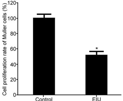
MTT assay was used to detect the effect of EIU on Muller cell proliferation. EIU obviously sup-pressed Muller cell proliferation compared with control group (P < 0.05) (Figure 2).
Influence of EIU on the activity of Caspase 3 in retinal endothelial cells
[image:3.612.91.286.138.322.2]The effect of EIU on the activity of Caspase 3 in retinal endothelial cells was analyzed using Caspase 3 activity detection kit. The results showed that EIU significantly promoted the ac- tivity of Caspase3 in retinal endothelial cells Table 1. Primer sequences
Gene Forward 5’-3’ Reverse 5’-3’
GADPH AGTGCCAGCCTCGTCTCATAG CGTTGAACTTGCCGTGGGTAG VEGF AGCGGCTCATCTAAACAATGG GGCGCACATTCTCTCCGTA
Western blot
[image:3.612.90.289.370.533.2]RIPA lysis buffer was added into the retinal endothelial cells and Muller cells and incubated on ice for 15-30 min. Next, the cells were treated with
Figure 1. The effect of EIU on retinal endothelial cell proliferation. *P < 0.05, compared with control.
compared with control group (P < 0.05) (Figure 3).
Influence of EIU on the activity of Caspase 3 in Muller cells
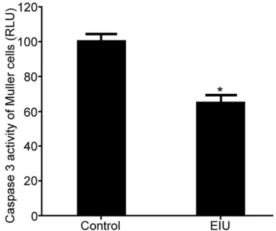
The effect of EIU on the activity of Caspase 3 in Muller cells was analyzed by Caspase 3 activity detection kit. The results demonstrated that EIU significantly facilitated the activity of Cas- pase 3 in Muller cells compared with control group (P < 0.05) (Figure 4).
Impacts of EIU on VEGF expression in retinal endothelial cells
The effect of EIU on the expression of VEGF in retinal endothelial cells was assessed by
Real-time PCR and Western blot. The results reveal- ed that the expression of VEGF mRNA and pro-tein in retinal endothelial cells was significantly higher in EIU group than that in control group (P < 0.05) (Figure 5).
Impacts of EIU on VEGF expression in Muller Cells
The effect of EIU on the expression of VEGF in EIU cells was assessed by Real-time PCR and Western blot. The results revealed that the expression of VEGF mRNA and protein in Muller cells was markedly higher in EIU group than that in control group (P < 0.05) (Figure 6).
Function of EIU on IL-1β and TNF-α contents in supernatant of retinal endothelial cells and Muller cells
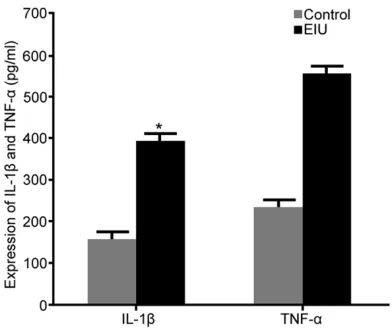
The contents of IL-1β and TNF-α in the superna-tant of retinal endothelial cells and Muller cells were analyzed by ELISA. The contents of IL-1β and TNF-α in the supernatant of retinal endo-thelial cells and Muller cells were apparently higher in EIU group than those in control group (P < 0.05) (Figures 7, 8).
Discussion
[image:4.612.91.290.71.247.2]Uveitis is a type of eye disease frequently occur-ring in young. It has a wide range of types with complicated pathogenesis. Improper treatment can lead to blindness, thus causing widespread concerns around the world. Since its pathogen-esis and recurrence mechanism is not entirely clear, the prevention has not been proceeded and the treatment effect is also unsatisfactory [17, 18]. Posterior EIU may cause retinal infiltra-tion, leading to disc damage, retinal tissue da- mage, and retinal vasculitis [19]. Retinal endo-thelial cells are one of the important cells that maintain retinal function, and endothelial cell damage and denudation can cause retinal damage [14]. As retinal-specific glial cells that play an important role in maintaining the mor-phology and physiological function of the reti-na, Muller cells account for more than 90% of all cells in the retina [20]. The morphological and physiological functions and metabolic chan- ges of Muller cells seriously affect the patho-genesis of the retinopathy [21]. This study con-firmed that retinal endothelial cells and Muller cell proliferation was decreased with enhanced Caspase 3 activity in EIU group, suggesting that uveitis damaged retinal endothelial cells and decreased Muller cell expression.
Figure 3. The influence of EIU on the activity of Cas-pase 3 in retinal endothelial cells. *P < 0.05, com-pared with control.
[image:4.612.90.288.306.472.2]In-depth investigation revealed that VEGF ex- pression was increased, while IL-1β and TNF-α secretion was enhanced in retinal endothelial cells and Muller cells from EIU group. VEGF has multiple isoforms that play a role through
[image:5.612.93.521.71.198.2]bind-ing to its correspondbind-ing receptor (VEGFR). Most VEGFRs are located on the surface of endothe-lial cells and can alter vascular permeability after ligand binding. Thus, in the formation of EIU, VEGF participates in regulation by altering
[image:5.612.95.523.252.383.2]Figure 5. The impacts of EIU on VEGF expression in retinal endothelial cells. A. Real-time PCR detection of VEGF mRNA expression; B. Western blot detection of VEGF protein expression; C. VEGF protein expression analysis. *P < 0.05, compared with control.
Figure 6. The impacts of EIU on VEGF expression in Muller cells. A. Real-time PCR detection of VEGF mRNA expres-sion; B. Western blot detection of VEGF protein expresexpres-sion; C. VEGF protein expression analysis. *P < 0.05, com-pared with control.
Figure 7. The function of EIU on IL-1β and TNF-α con-tents in supernatant of retinal endothelial cells. *P < 0.05, compared with control.
[image:5.612.91.287.442.607.2] [image:5.612.322.523.444.605.2]vascular permeability; and VEGF participates in the entire EIU pathology by promoting neovas-cularization [22]. IL-1β and TNF-α are potent regulators that inhibit the proliferation of retinal endothelial cells and Muller cells. They regulate the interaction between cells and the matrix, leading to the chemotaxis of macrophages and subsequent uveitis-induced retinopathy [23]. However, the specific mechanism of EIU on reti-nal endothelial cell injury and Muller cells still needs further investigation.
In conclusion, EIU causes retinal endothelial cell damage and Muller cell expression reduc-tion by promoting inflammatory factors secre-tion, facilitating VEGF expression, and inducing apoptosis.
Acknowledgements
This work was supported by the Scientific Re- search Project of Heilongjiang Provincial Health and Family Planning Commission (2016-520). Disclosure of conflict of interest
None.
Address correspondence to: Dr. Yangyang Yu, De- partment of Ophthalmology, Heilongjiang Provincial Hospital, 82 Zhongshan Road, Xiangfang District, Haerbin, Heilongjiang, China. Tel: +86-451-8802- 5079; Fax: +86-451-88025079; E-mail: [email protected]
References
[1] Chen CL, Chen JT, Liang CM, Tai MC, Lu DW, Chen YH. Silibinin treatment prevents endotox-in-induced uveitis in rats in vivo and in vitro. PLoS One 2017; 12: e0174971.
[2] Gutowski MB, Wilson L, Van Gelder RN, Pepple KL. In vivo bioluminescence imaging for longi-tudinal monitoring of inflammation in animal models of uveitis. Invest Ophthalmol Vis Sci 2017; 58: 1521-1528.
[3] Gu R, Lei B, Shu Q, Li G, Xu G. Glucocorticoid-induced leucine zipper overexpression inhibits lipopolysaccharide-induced retinal inflamma-tion in rats. Exp Eye Res 2017; 165: 151-163. [4] Ge Q, Wang S, Zheng Y. Systemic
administra-tion of an anti-tumor necrosis factor-alpha monoclonal antibody protects against endotox-in-induced uveitis in rats. Indian J Ophthalmol 2016; 64: 873-877.
[5] Sawada K, Hiraoka M, Abe A, Kelly R, Shayman JA, Ohguro H. Prolonged ocular inflammation in
endotoxin-induced uveitis in lysosomal phos-pholipase A2-deficient mice. Curr Eye Res 2017; 42: 611-616.
[6] Hsu YR, Chang SW, Yang CH, Lee YA, Kao TY. Expression profile of cationic amino acid trans-porters in rats with endotoxin-induced uveitis. Mediators Inflamm 2016; 2016: 6586857. [7] Bermudez MA, Sendon-Lago J, Seoane S, Eiro
N, Gonzalez F, Saa J, Vizoso F, Perez-Fernandez R. Anti-inflammatory effect of conditioned me-dium from human uterine cervical stem cells in uveitis. Exp Eye Res 2016; 149: 84-92. [8] Afarid M, Lashkarizadeh H, Ashraf MJ,
Now-roozzadeh MH, Shafiee SM. The efficacy of in-travitreal interferon alpha-2b for the treatment of experimental endotoxin-induced uveitis. In-dian J Ophthalmol 2016; 64: 376-381. [9] Sato K, Mihara Y, Kanai K, Yamashita Y,
Kimu-ra Y, Itoh N. Tyrosol amelioKimu-rates lipopolysac-charide-induced ocular inflammation in rats via inhibition of nuclear factor (NF)-kappaB activation. J Vet Med Sci 2016; 78: 1429-1438.
[10] Savage SR, McCollum GW, Yang R, Penn JS. RNA-seq identifies a role for the PPARbeta/ delta inverse agonist GSK0660 in the regula-tion of TNFalpha-induced cytokine signaling in retinal endothelial cells. Mol Vis 2015; 21: 568-576.
[11] Loukovaara S, Gucciardo E, Repo P, Vihinen H, Lohi J, Jokitalo E, Salven P, Lehti K. Indications of lymphatic endothelial differentiation and en-dothelial progenitor cell activation in the pa-thology of proliferative diabetic retinopathy. Acta Ophthalmol 2015; 93: 512-523.
[12] Matsuda M, Krempel PG, Marquezini MV, Sholl-Franco A, Lameu A, Monteiro MLR, Miguel NCO. Cellular stress response in hu-man Muller cells (MIO-M1) after bevacizumab treatment. Exp Eye Res 2017; 160: 1-10. [13] Ajayi AM, Martins DTO, Balogun SO, Oliveira
RG, Ascencio SD, Soares IM, Barbosa RDS, Ademowo OG. Ocimum gratissimum L. leaf fla-vonoid-rich fraction suppress LPS-induced in-flammatory response in RAW 264.7 macro-phages and peritonitis in mice. J Ethnophar- macol 2017; 204: 169-178.
[14] Kanai K, Hatta T, Nagata S, Sugiura Y, Sato K, Yamashita Y, Kimura Y, Itoh N. Luteolin attenu-ates endotoxin-induced uveitis in Lewis rats. J Vet Med Sci 2016; 78: 1229-1235.
[15] Erturkuner SP, Yaprak Sarac E, Gocmez SS, Ek-mekci H, Ozturk ZB, Seckin I, Sever O, Keskin-bora K. Anti-inflammatory and ultrastructural effects of Turkish propolis in a rat model of endotoxin-induced uveitis. Folia Histochem Cy-tobiol 2016; 54: 49-57.
Silva-Cunha A, Bousquet E, Naud MC, Ayres E, Orefice RL, Tekaya M, Kowalczuk L, Zhao M, Behar-Cohen F. Anti-inflammatory effect of dexamethasone controlled released from an-terior suprachoroidal polyurethane implants on endotoxin-induced uveitis in rats. Invest Ophthalmol Vis Sci 2016; 57: 1671-1679. [17] Okamoto T, Ozawa Y, Kamoshita M, Osada H,
Toda E, Kurihara T, Nagai N, Umezawa K, Tsub-ota K. The neuroprotective effect of rapamycin as a modulator of the mTOR-NF-kappaB axis during retinal inflammation. PLoS One 2016; 11: e0146517.
[18] Impellizzeri D, Ahmad A, Bruschetta G, Di Paola R, Crupi R, Paterniti I, Esposito E, Cuzzo-crea S. The anti-inflammatory effects of palmi-toylethanolamide (PEA) on endotoxin-induced uveitis in rats. Eur J Pharmacol 2015; 761: 28-35.
[19] Qin YJ, Chan SO, Chong KK, Li BF, Ng TK, Yip YW, Chen H, Zhang M, Block NL, Cheung HS, Schally AV, Pang CP. Antagonist of GH-releas-ing hormone receptors alleviates experimental ocular inflammation. Proc Natl Acad Sci U S A 2014; 111: 18303-18308.
[20] Ji HP, Xiong Y, Zhang ED, Song WT, Gao ZL, Yao F, Sun H, Zhou RR, Xia XB. Which has more stem-cell characteristics: Muller cells or Muller cells derived from in vivo culture in neuro-spheres? Am J Transl Res 2017; 9: 611-619. [21] Dong E, Bachleda A, Xiong Y, Osawa S, Weiss
ER. Reduced phosphoCREB in Muller glia dur-ing retinal degeneration in rd10 mice. Mol Vis 2017; 23: 90-102.
[22] Di Pierdomenico J, Garcia-Ayuso D, Pinilla I, Cuenca N, Vidal-Sanz M, Agudo-Barriuso M, Villegas-Perez MP. Early events in retinal de-generation caused by rhodopsin mutation or pigment epithelium malfunction: differences and similarities. Front Neuroanat 2017; 11: 14.