Original Article
Evaluating the expression of MACC1 and c-myc
in cervical cancer and their correlation
Ying Zhang, Xiaolin He
Department of Obstetrics and Gynecology, The People’s Hospital of Mianyang City, Mianyang, Sichuan, China Received January 15, 2018; Accepted March 15, 2018; Epub June 15, 2018; Published June 30, 2018
Abstract: Objective: The association of metastasis-associated in colon cancer (MACC1) and c-myc with the clini-copathological features of cervical cancer patients is unknown. This study aimed to investigate the expression of MACC1 and c-myc in cervical cancer and the correlation of these proteins with clinicopathological features. Method: In total, 503 cervical specimens from patients undergoing gynecological surgery and out-patient biopsies were collected, including 260 cervical cancer tissue specimens (cervical cancer group), 137 out-patient biopsy cervical intraepithelial neoplasia (CIN) II-III tissue specimens (CIN II-III group), and 106 normal cervical tissue specimens. RT-PCR was used to examine the expression of MACC1 and c-myc in the tissues of the three groups, and the as-sociation between the expressions and each clinicopathological feature was analyzed. Results: MACC1 and c-myc mRNA expression in cervical cancer tissues was related to the degree of tumor pathological differentiation, pelvic lymph node metastasis, and invasion depth, and the difference was statistically significant (P < 0.01). The expres-sion of both MACC1 and c-myc mRNA in tissues of the cervical cancer group was higher than that in the CIN II-III and normal control groups (P < 0.01), and the pairwise comparisons were statistically significant (P < 0.01). The expression of MACC1 and c-myc in cervical cancer tissues was positively related (r = 0.537, P < 0.05). Conclusions: Both MACC1 and c-myc were highly expressed in cervical cancer tissues. MACC1 and c-myc may facilitate the oc-currence of cervical cancer.
Keywords: Cervical cancer, MACC1, c-myc, pathological feature, lymphatic metastasis
Introduction
Cervical cancer is a serious disease threaten-ing women’s health globally and ranks third in morbidity among cancers affecting women. Most patients with intraepithelial neoplasia are aged between 30 and 35 years whereas those with invasive cervical carcinoma are aged between 45 and 55 years. In recent years, cer-vical cancer has shown a youth-oriented ten-dency. About 528,000 new cases of cervical cancer occur each year worldwide, and about 266,000 patients die of cervical cancer annu-ally [1, 2]. Lifestyle and habits change with the development of the society, thus, resulting in increasing human papillomavirus (HPV) infec-tion in females. Genital HPV infecinfec-tion has become a common sexually transmitted dis-ease (STD) in females, and most cervical can-cer cases are caused due to high-risk HPV infections [3].
Presently, the expression of MACC1 and c-myc in cervical cancer and their association with the clinicopathological features of patients is not known, and examining the expression of both genes simultaneously for monitoring cervi-cal cancer is also not reported. The association of MACC1 and c-myc expression with the occur-rence and development of cervical cancer is investigated in this study by examining the expression of MACC1 and c-myc in cervical can-cer tissues, thus, providing a reference for the clinical diagnosis and therapy of cervical cancer.
Materials and methods
General information
In total, 503 cervical specimens from patients undergoing gynecological surgery admitted to Department of Obstetrics and Gynecology, The People’s Hospital of Mianyang City, as well as out-patient biopsies were collected, including 260 cervical cancer tissue specimens (cervical cancer group), 137 out-patient biopsies of cer-vical intraepithelial neoplasia (CIN) II-III tissue specimens (CIN II-III group), and 106 normal cervical tissue specimens from hospitalized patients undergoing hysteromyomectomy (nor-mal control group). The patients were 24-68 years old with an average age of 42.36 ± 7.26 years. According to the WHO female genital organ cervical cancer TNM 2014 and FIGO classification standard [8], the patients in the cervical cancer group were classified as fol -lows: on the basis of (i) tissue typing: 63 cases of adenocarcinoma and 197 cases of squa-mous carcinoma; (ii) clinical stages: 118 cases in phase I and 142 cases in phase II; (iii) degree of pathological differentiation: 112 cases of high differentiation, 83 cases of intermediate differentiation, and 65 cases of low differentia-tion; 81 cases of lymphatic metastasis and 179 cases of no lymphatic metastasis; and 236 cases of positive high-risk HPV and 24 cases of negative high-risk HPV infection. The inclusion
data of the patients were complete. All cervical tissues after extraction were stored at -80°C. The study was approved by the Ethics Committee of our hospital. All the subjects included in this study provided signed informed consent.
Main instruments and reagents
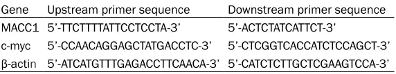
The Agilent Mx3000P/3005P real-time fluor-escence quantitative PCR system was pur-chased from Beijing Keyu Xingye Science and Technology Development Co., Ltd. GelDoc-It- TS3 automatic gel-imaging system was pur-chased from Shanghai Yuansheng Instrument and Equipment Co., Ltd. Total RNA extraction kit (Trizol method) was purchased from Applied Biosystems, USA. M-MLV reverse transcription kit was purchased from Promega (Beijing) Biotechnology Co., Ltd. MACC1 and c-myc PCR kits were purchased from Biomiga Company. Primers for the real-time fluorescence quantita -tive PCR for MACC1, c-myc, and the β-actin internal reference gene were synthesized by Dalian Kakara Company. The primer sequences are listed in Table 1.
Test method
[image:2.612.92.378.98.152.2]From cervical tissue specimens stored at -80°C, 100 mg tissues were sliced, placed in liquid nitrogen, and completely grinded. The prepared tissues were mixed with Trizol reagent, placed at room temperature, and allowed to stand for 30 min to ensure complete lysis. Total RNA was extracted in strict accordance with the manufacturer’s instructions, and the con-centration and purity of extracted RNA was determined by ultraviolet spectrophotometry and protein electrophoresis. The extracted RNA was reverse transcribed, according to the man-ufacturer’s instructions. The extracted cDNA samples were stored at -20°C. The PCR sam-ples were prepared, according to the manufac-turer’s instructions in a 12.62-µL volume made up to 20 µL with DEPC-treated water. The PCR Table 1. Primer sequences used for MACC1, c-myc, and β-actin
genes
Gene Upstream primer sequence Downstream primer sequence MACC1 5’-TTCTTTTATTCCTCCTA-3’ 5’-ACTCTATCATTCT-3’
c-myc 5’-CCAACAGGAGCTATGACCTC-3’ 5’-CTCGGTCACCATCTCCAGCT-3’ β-actin 5’-ATCATGTTTGAGACCTTCAACA-3’ 5’-CATCTCTTGCTCGAAGTCCA-3’
conditions were as follows: Initial denaturation at 94°C for 10 min followed by 40 cycles of 94°C for 45 s, 60°C for 45 s and 72°C for 45 s. The experiment was repeated three times and the mean results were considered. The results were analyzed using the 2-ΔΔCT method.
Statistical analyses
SPSS19.0 (SPSS China) was used for statistical analysis. All measurement data are presented as mean ± standard deviation (_x±s). ANOVA was used for comparison among multiple groups and t-test was used for two groups’ comparison. Pearson correlation analysis was performed to determine the correlation be-
tween MACC1 and c-myc expression in cervical cancer tissues. P < 0.05 was considered as statistically significant.
Results
Association between MACC1 and c-myc mRNA in cervical cancer tissues and clinicopathologi-cal features
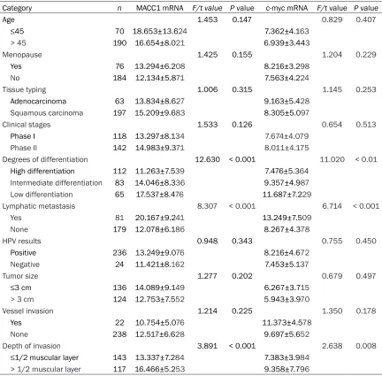
MACC1 and c-myc mRNA expression in cervical cancer tissues was related to the degree of tumor pathological differentiation, pelvic lymph node metastasis, and invasion depth, with a statistically significant difference (P < 0.01). The expression was unrelated to age, meno-Table 2. Relationship between relative expression of MACC1 and c-myc mRNA in cervical cancer tis-sues and clinicopathological parameters (_x±s)
Category n MACC1 mRNA F/t value P value c-myc mRNA F/t value P value
Age 1.453 0.147 0.829 0.407
≤45 70 18.653±13.624 7.362±4.163
> 45 190 16.654±8.021 6.939±3.443
Menopause 1.425 0.155 1.204 0.229
Yes 76 13.294±6.208 8.216±3.298
No 184 12.134±5.871 7.563±4.224
Tissue typing 1.006 0.315 1.145 0.253
Adenocarcinoma 63 13.834±8.627 9.163±5.428
Squamous carcinoma 197 15.209±9.683 8.305±5.097
Clinical stages 1.533 0.126 0.654 0.513
Phase I 118 13.297±8.134 7.674±4.079
Phase II 142 14.983±9.371 8.011±4.175
Degrees of differentiation 12.630 < 0.001 11.020 < 0.01
High differentiation 112 11.263±7.539 7.476±5.364 Intermediate differentiation 83 14.046±8.336 9.357±4.987
Low differentiation 65 17.537±8.476 11.687±7.229
Lymphatic metastasis 8.307 < 0.001 6.714 < 0.001
Yes 81 20.167±9.241 13.249±7.509
None 179 12.078±6.186 8.267±4.378
HPV results 0.948 0.343 0.755 0.450
Positive 236 13.249±9.076 8.216±4.672
Negative 24 11.421±8.162 7.453±5.137
Tumor size 1.277 0.202 0.679 0.497
≤3 cm 136 14.089±9.149 6.267±3.715
> 3 cm 124 12.753±7.552 5.943±3.970
Vessel invasion 1.214 0.225 1.350 0.178
Yes 22 10.754±5.076 11.373±4.578
None 238 12.517±6.628 9.697±5.652
Depth of invasion 3.891 < 0.001 2.638 0.008
≤1/2 muscular layer 143 13.337±7.284 7.383±3.984
[image:3.612.90.522.95.520.2]pause, tumor tissue typing, clinical stages, high-risk HPV infection test results, tumor size, and vessel invasion (P > 0.05). The details are summarized in Table 2.
Expression of MACC1 mRNA in tissues of the cervical cancer group, CIN II-III group, and the normal control group
The expression levels of MACC1 mRNA in tis-sues of the cervical cancer group, CIN II-III
group, and the normal control group were 14.079 ± 8.297, 3.046 ± 1.531, and 1.186 ± 0.671, respectively. Compared with those in the normal control group, the expression levels of MACC1 mRNA in tissues of the cervical can-cer group and CIN II-III group were significantly higher (P < 0.01); compared with that in the CIN II-III group, the expression level of MACC1 mRNA in tissues of the cervical cancer group was significantly higher (P < 0.01). The trend of MACC1 mRNA expression among the three groups was as follows: cervical cancer group > CIN II-III group > normal control group, and the pairwise comparisons among the three groups were statistically significant (P < 0.01, Figure 1).
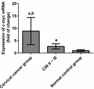
Expression of c-myc mRNA in tissues of the cervical cancer group, CIN II-III group, and the normal control group
The expression levels of c-myc mRNA in tissues of the cervical cancer group, CIN II-III group, and the normal control group were 8.857 ± 5.479, 2.673 ± 1.047, and 0.951 ± 0.376, respectively. Compared with those in the nor-mal control group, the expression levels of c-myc mRNA in tissues of the cervical cancer group and CIN II-III group were significantly higher (P < 0.01); compared with that in CIN II-III group, the expression level of c-myc mRNA in tissues of the cervical cancer group was sig-nificantly higher (P < 0.01). The trend of c-myc mRNA expression among the three groups was as follows: cervical cancer group > CIN II-III group > normal control group, and the pairwise comparisons among the three groups were sta-tistically significant (P < 0.01, Figure 2). Correlation between MACC1 mRNA and c-myc mRNA expression in cervical cancer tissues
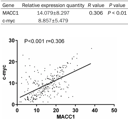
The expression of MACC1 and c-myc in cervical cancer tissue was positively related (r = 0.306, P < 0.05), as indicated by Pearson linear regres-sion analysis. See Table 3 and Figure 3. Discussion
Globally, cervical cancer is the most common malignancy among women’s cancers, besides breast cancer and colorectal cancer. Cervical cancer is a unique female malignancy with an identified pathogen that is closely related to high-risk HPV infection [9]. HPV infection plays an important role in the occurrence and
devel-Figure 1. Comparison of MACC1 mRNA expression results in tissues of the cervical cancer group, CIN II-III group, and the normal control group. Note: aP
< 0.01 compared with the normal control group;
[image:4.612.93.285.74.257.2]bP<0.01 compared with CIN II-III.
Figure 2. Comparison of c-myc mRNA expression re-sults in tissues of the cervical cancer group, CIN II-III group, and the normal control group. Note: aP < 0.01
compared with the normal control group; bP < 0.01
[image:4.612.93.286.339.523.2]opment of cervical cancer. At present, an HPV vaccine has been popularized and used to reduce the occurrence of cervical cancer to some extent. However, with the changing social lifestyle, the rate of HPV infection has remained high, and the prevalence of cervical cancer also tends to be greater in the younger popula-tion [10]. The main clinical therapy for cervical cancer is surgery assisted by chemotherapy or radiotherapy. Although the mortality rate of patients has been reduced greatly, metastasis, invasion, and local recurrence of cervical can-cer are still the major factors resulting in patient death [11]. Therefore, to investigate the occur-rence and development of cervical cancer, latent genes that inhibit tumor growth and reduce tumor metastasis and invasion are of great importance clinically.
In this study, the expression levels of MACC1 and c-myc mRNA in normal cervical tissues, CIN II-III, and cervical cancer tissues were found to be gradually elevated and were relat-ed to the degree of tumor pathological differen-tiation, pelvic lymph node metastasis, and invasion depth, indicating that MACC1 and c-myc may participate in the occurrence and development of cervical cancer and play an important role in its invasion and metastasis.
MACC1 is a recently identified gene that is closely related to colon cancer metastasis and can regulate tumor growth and metastasis with a key role in the process of cell proliferation and differentiation [12]. MACC1 can be expressed independently of c-Met, and is a regulator of c-Met. High c-Met expression can induce autocrine expression of HGF, resulting in invasion and malignant transformation of cells. Increased expression of MACC1 can induce c-Met expression and HGF overexpres-sion, which can cause c-Met activation in the cell nucleus by MACC1, thus, forming a feed-back pathway involving the three proteins and facilitating invasion, metastasis, and malignant transformation of tumor cells [13, 14]. MACC1 may also be involved in the occurrence and development of cervical cancer through this feedback pathway. These observations are sim-ilar to the conclusions of the study conducted by Chandrasinghe et al. [15], which were that high expression of MACC1 in colon cancer can promote the invasion and metastasis of tumor cells; MACC1 can also be used as an indepen-dent test index of prognosis and life expectancy in patients with colon cancer.
c-myc is a common cancer-promoting gene involved in cell proliferation, differentiation, apoptosis, and metastasis, and many other bio-logical functions are mediated by the transcrip-tion factor encoded by this gene [16]. High expression of c-myc can induce the evolution of cells from normal cells to malignant tumor cells. Studies indicate that when body cells are stimulated, the stability of c-myc is altered, which results in c-myc activation, leading to over-proliferation of cells that are, thus, immor-talized and form tumor cells [17]. c-myc is acti-vated in the evolution process in the normal cervix uteri and participates in the develop-ment of cervical cancer [18]. This observation is similar to that reported by Cui et al. [19]. In this study, MACC1 and c-myc expression in cer-vical cancer tissues was found to be positively related, indicating that the joint detection of both has better clinical predictive value for the occurrence and development of cervical ca- ncer.
[image:5.612.87.305.108.317.2]This study includes a large number of speci-mens, which can suitably indicate the expres-sion of MACC1 and c-myc in different cervical tissues and ensure the reliability of the study. Table 3. Correlation of expression quantity
be-tween MACC1 and c-myc mRNA in cervical cancer tissues
Gene Relative expression quantity R value P value MACC1 14.079±8.297 0.306 P < 0.01 c-myc 8.857±5.479
The limitations of this study are that patients in phase III-IV were not included in this study, the analyses for prognosis and life expectancy were not performed. Tumor occurrence, devel-opment, invasion, and metastasis constitute a complex process regulated by multiple factors. The proliferation and apoptosis of tumor cells can be promoted by cytokines secreted from tumor cells [20]. MACC1 and c-myc are also highly expressed in other tumors. The mecha-nism of action of both in female malignancies is not yet known. We, therefore, hope that the prognosis and survival of patients with cervical cancer can be analyzed in the next study and that the mechanism of action of the proteins encoded by both genes in cervical cancer can be elucidated.
In conclusion, both MACC1 and c-myc are high-ly expressed in cervical cancer tissues and may facilitate its occurrence, development, inva-sion, and metastasis; thus, MAAC1 and c-myc expression may have an important predictive value clinically.
Disclosure of conflict of interest
None.
Address correspondence to: Ying Zhang, Depart- ment of Obstetrics and Gynecology, The People’s Hospital of Mianyang City, No. 10-12, the West of Jiannan Road, Fucheng District, Mianyang, Sichuan 621000, China. Tel: +86-0816-2342105; E-mail: [email protected]
References
[1] Cancer Genome Atlas Research Network; Al-bert Einstein College of Medicine; Analytical Biological Services; Barretos Cancer Hospital; Baylor College of Medicine; Beckman Re-search Institute of City of Hope; Buck Institute for Research on Aging; Canada’s Michael Smith Genome Sciences Centre; Harvard Med-ical School; Helen F. Graham Cancer Center &Research Institute at Christiana Care Health Services; HudsonAlpha Institute for Biotech-nology; ILSbio, LLC; Indiana University School of Medicine; Institute of Human Virology; Insti-tute for Systems Biology; International Genom-ics Consortium; Leidos Biomedical; Massachu-setts General Hospital; McDonnell Genome Institute at Washington University; Medical College of Wisconsin; Medical University of South Carolina; Memorial Sloan Kettering
Can-cer Center; Montefiore Medical Center; Nan -tOmics; National Cancer Institute; National Hospital, Abuja, Nigeria; National Human Ge-nome Research Institute; National Institute of Environmental Health Sciences; National Insti-tute on Deafness &Other Communication Dis-orders; Ontario Tumour Bank, London Health Sciences Centre; Ontario Tumour Bank, Ontar-io Institute for Cancer Research; OntarOntar-io Tu-mour Bank, The Ottawa Hospital; Oregon Health &Science University; Samuel Oschin Comprehensive Cancer Institute, Cedars-Sinai Medical Center; SRA International; St Joseph’s Candler Health System; Eli &Edythe L. Broad Institute of Massachusetts Institute of Technol-ogy &Harvard University; Research Institute at Nationwide Children’s Hospital; Sidney Kim-mel Comprehensive Cancer Center at Johns Hopkins University; University of Bergen; Uni-versity of Texas MD Anderson Cancer Center; University of Abuja Teaching Hospital; Univer-sity of Alabama at Birmingham; UniverUniver-sity of California, Irvine; University of California Santa Cruz; University of Kansas Medical Center; Uni-versity of Lausanne; UniUni-versity of New Mexico Health Sciences Center; University of North Carolina at Chapel Hill; University of Oklahoma Health Sciences Center; University of Pitts-burgh; University of São Paulo, Ribeir ão Preto Medical School; University of Southern Califor-nia; University of Washington; University of Wisconsin School of Medicine &Public Health; Van Andel Research Institute; Washington Uni-versity in St Louis. Integrated genomic and mo-lecular characterization of cervical cancer. Na-ture 2017; 543: 378-384.
[2] Vaccarella S, Laversanne M, Ferlay J and Bray F. Cervical cancer in Africa, Latin America and the Caribbean and Asia: regional inequalities and changing trends. Int J Cancer 2017; 141: 1997-2001.
[3] Rodriguez AC, Avila C, Herrero R, Hildesheim A, Sherman ME, Burk RD, Morales J, Alfaro M, Guillen D, Trejos ME, Vargas RM, Torres G and Schiffman M. Cervical cancer incidence after screening with HPV, cytology, and visual meth-ods: 18-year follow-up of the Guanacaste co-hort. Int J Cancer 2017; 140: 1926-1934. [4] Wieringa HW, van der Zee AG, de Vries EG and
van Vugt MA. Breaking the DNA damage re-sponse to improve cervical cancer treatment. Cancer Treat Rev 2016; 42: 30-40.
-match repair status for recurrence risk predic-tion in stage II colon cancer patients: the BI-OGRID studies. Ann Oncol 2017; 28: 1869-1875.
[6] Zhao Y, Dai C, Wang M, Kang H, Lin S, Yang P, Liu X, Liu K, Xu P, Zheng Y, Li S and Dai Z. Clini-copathological and prognostic significance of metastasis-associated in colon cancer-1 (MACC1) overexpression in colorectal cancer: a meta-analysis. Oncotarget 2016; 7: 62966-62975.
[7] Raman D and Pervaiz S. 133-tumor pro-oxi-dant enviroment stabilizes onco-protein c-myc via sustained phosphorylation at serine 62 that promotes its oncogenic activity. Free Radi-cal Biology and Medicine 2017; 112: 98. [8] Marth C, Landoni F, Mahner S, McCormack M,
Gonzalez-Martin A, Colombo N and Committee EG. Cervical cancer: ESMO clinical practice guidelines for diagnosis, treatment and follow-up. Ann Oncol 2017; 28: iv72-iv83.
[9] Clifford GM, Tully S and Franceschi S. Carcino-genicity of human papillomavirus (HPV) types in HIV-positive women: a meta-analysis from HPV infection to cervical cancer. Clin Infect Dis 2017; 64: 1228-1235.
[10] Chetty R. 70 years of the JCP-highly cited pa-pers: the causal relation between human pap-illomavirus and cervical cancer. J Clin Pathol 2017; 70: 997.
[11] Kim SW, Chun M, Ryu HS, Chang SJ, Kong TW, Oh YT and Kang SH. Long-term results of early adjuvant concurrent chemoradiotherapy for high-risk, early stage uterine cervical cancer patients after radical hysterectomy. BMC Can-cer 2017; 17: 297.
[12] Hua FF, Liu SS, Zhu LH, Wang YH, Liang X, Ma N and Shi HR. MiRNA-338-3p regulates cervi-cal cancer cells proliferation by targeting MACC1 through MAPK signaling pathway. Eur Rev Med Pharmacol Sci 2017; 21: 5342-5352.
[13] Lemos C, Hardt MS, Juneja M, Voss C, Förster S, Jerchow B, Haider W, Bläker H and Stein US. Abstract 2689: first MACC1 transgenic mice demonstrate tumor progression via the newly discovered MACC1/Nanog/Oct4 axis. Cancer Research 2016; 76: 2689-2689.
[14] Mudduluru G, Ilm K, Dahlmann M and Stein U. MACC1, a novel player in solid cancer carcino-genesis. 2017.
[15] Chandrasinghe P, Stebbing J and Warusavi-tarne J. The MACC1-SPON2 axis: a new bio-marker and therapeutic target in colorectal cancer. Oncogene 2017; 36: 1474-1475. [16] Liu J, Zhu M, Xia X, Huang Y, Zhang Q and Wang
X. Jumonji domain-containing protein 1A pro-motes cell growth and progression via transac-tivation of c-Myc expression and predicts a poor prognosis in cervical cancer. Oncotarget 2016; 7: 85151-85162.
[17] Sun L, Liu M, Sun GC, Yang X, Qian Q, Feng S, Mackey LV and Coy DH. Notch signaling activa-tion in cervical cancer cells induces cell growth arrest with the involvement of the nuclear re-ceptor NR4A2. J Cancer 2016; 7: 1388-1395. [18] Zhao WH, Hao M, Cheng XT, Yang X, Wang ZL,
Cheng KY, Liu FL and Bai YX. c-myc gene copy number variation in cervical exfoliated cells de-tected on fluorescence in situ hybridization for cervical cancer screening. Gynecol Obstet In-vest 2016; 81: 416-423.
[19] Cui F, Hou J, Huang C, Sun X, Zeng Y, Cheng H, Wang H and Li C. C-Myc regulates radiation-in-duced G2/M cell cycle arrest and cell death in human cervical cancer cells. J Obstet Gynaecol Res 2017; 43: 729-735.