Original Article
Identification of candidate genes associated with
Xiang pig estrus by genome-wide transcriptome analysis
Fuping Zhang1, Xi Niu1, Liangting Tang1, Xueqin Ran1, Sheng Li1, Jiafu Wang1,2, Shihui Huang1
1College of Animal Science/Key Laboratory of Plant Resource Conservation and Germplasm Innovation in
Moun-tainous Region (Ministry of Education), Collaborative Innovation Center for Mountain Ecology & Agro-Bioengineer-ing (CICMEAB), Institute of Agro-BioengineerAgro-Bioengineer-ing, Guizhou University, Guiyang 550025, Guizhou Province, China;
2Tongren University, Tongren, China
Received December 3, 2017; Accepted June 30, 2018;Epub November 15, 2018; Published November 30, 2018
Abstract: Oestrus is one of the most important physiological processes especially in pig reproduction and breed-ing. Oestrus is affected by interactions between multiple genes and the environment. Although recent studies have
identified some genes associated with prolificacy in pigs, transcriptomic pattern of specific genes affecting estrus in
porcine is unclear. In order to identify candidate genes associated with estrus in swine, we assessed gene expres-sion changes of the ovaries from Xiang pigs within estrus or no-estrus stage using the RNA-Seq method. A total of
432 differentially expressed genes were identified: 204 genes were upregulated and 228 genes were downregu -lated in oestrous ovary samples when compared with non-oestrous samples. A large number of these genes re-lated to steroid hormone regulation in animal ovaries, including 51 Gene Ontology terms and top 20 Kyoto Encyclopedia of Genes and Genomes pathways involved in steroid biosynthesis and ovarian steroidogenesis. From these
differ-entially expressed genes, we identified a total of 14 genes using a bioinformatics screen that may be associated
with oestrus in Xiang pigs, which were CYP51, EBP, TM7SF2, MSMO1, SQLE, LSS, DHCR24, FDFT1, HMGCS1, FDPS, MVK, IDI1, ACAT2, and ACAT1. These results provide a list of new candidate genes for porcine prolificacy to
be further investigated.
Keywords: Oestrus, production, Xiang pig, genome-wide transcriptome analysis
Introduction
As an important part of the national economy, the pig industry is crucial to China’s livestock industry. And oestrus is one of the most impor-tant physiological processes in pig production and is affected by interactions between multi-ple genes and the environment. Oestrus is a behavioral performance, as well as an external and visible sign of ovulation. During each oes-trus cycle, the ovary undergoes proliferation, differentiation and apoptosis. And these nor-mal physiological changes directly affect and/ or determine the ovulation, fertilization rate, the litter size and production of female animals. Detection and regulation of oestrus help us to
better understand animal reproduction effi -ciency, human reproductive medicine and bio-chemical research. As the rapid development of swine industry, oestrus and ovulation play a
crucial role in improving production efficiency.
Therefore, the objective of this current study was to investigate the candidate genes related
to swine oestrus, with the hope to increase the reproductive ability and create greater econom-ic value.
There has been some recent progress in char-acterizing the major genes involved in the
pro-lificacy of swine, such as the estrogen receptor
(ESR) [1], follicle-stimulating hormone beta sub-unit (FSH-β) [2], retinol-binding protein 4 (RBP4) [3]. With the recent publication of the pig, more candidate genes or quantitative trait loci (QTLs) have been extensively investigated for their involvement in porcine production [4-6]; but genomic location, function and interaction of these genes requires further research. RNA sequencing (RNA-Seq) can measure genes, both quantitatively and functionally, at the tran-scriptome level [7]. Up to now, RNA-Seq has
been used to study specific ovarian genes of
cattle [8, 9], goat [10, 11] and pig [12], but
explain heredity of estrus, and be used to iden-tify key genes relating to oestrus.
In this study, to identify the candidate genes
that influence pig oestrous, we performed a
comparative analysis of the whole transcrip-tomes of ovary between estrus group and
non-estrus group. We identified a series of differen -tially expressed genes (DEGs) between these two groups, which represent potential candi-date genes affecting pig oestrus traits. Using
these data, we identified several important GO
terms and metabolic pathways associated with pig oestrus. In conclusion, our data provides a solid foundation for identifying the critical genes whose functions affect pig oestrus, and can facilitate studies on the molecular regula-tory mechanisms underlying animal oestrus, hoping to increase the yield of domesticated animals.
Materials and methods
Ethics statement
All experimental procedures and sample collec-tion were performed according to the Re- gulations for the Administration of Affairs Con- cerning Experimental Animals (Ministry of Science and Technology, China; revised in June 2004) and approved by the Biological Studies Animal Care and Use Committee of Guizhou Province, PR China. This report fully adhered to the ARRIVE Guidelines for the reporting of
ani-the non-oestrous group was 152.0±3 days, which had no statistical difference and been comparable (P>0.05). Their intact ovaries were rapidly harvested from their carcasses and immediately frozen in liquid nitrogen. All tissue samples were stored at -80°C until the total RNA extraction was performed.
Oestrus detection
Oestrus detection was performed once a day. Standing oestrus was determined by the back-pressure test and the degrees of swelling and reddening of the vulva, according to Eliasson method (1989) [14]. The same oestrous detec-tion procedure was performed twice daily in front of a boar [15]. A sow not showing signs of oestrus within 10 days after last ovulation was considered non-oestrous.
mRNA library preparation and sequencing
Total RNA was extracted from ovaries using TRIzol (Invitrogen, Carlsbad, CA, USA) and was
purified using DNase I [16, 17]. The quality of
the total RNA was checked using the Agilent 2100 Size Bioanalyzer system (Santa Clara, CA,
USA). A total amount of 3 μg RNA per sample
was used as input material for the RNA sample preparations. Sequencing libraries were gener-ated using NEBNext® UltraTM RNA Library Prep Kit for Illumina® (NEB, USA) according to
manu-facturer’s instructions. Briefly, mRNA was
ex-tracted from total RNA using oligo (dT) magnet-Table 1. Primer sequences
Gene Name Forward (5’-3’) Reverse (5’-3’)
β-actin TGCGTGACATTAAGGAGAAG GCTCGTAGCTCTTCTCCA CYP51 GGCTCTTACCAGGCTGGCTT GTCTGCGTTTCTGGATTGCC TM7SF2 CAACCCACGCATCTGTTCCT GTTCTGCCTCCTGCATCAGC EBP ATCCTGGCTGGCCTCTTCTC AGTCTCCGCCAAGTGCTCAG MSMO1 GCACACCCTCTGGAAACCCT ATCAAACGAACGGTCACCCA SQLE ACCTTTCTCGGCATTGCCAC AGGAATACCAGCACGCCCAG LSS AAAGCCCTAGCCGAGAGCAG ACGTCATCCCATTCAGAGCG DHCR24 CTGCCTGTGTTGCCAGAGCT CAGATGTGCTGGAACAGGCC FDFT1 GGAATTGGCCTTTCCCGTCT TTTTCTGCAGGAACAGGCCC HMGCS1 TACGGCTGCCTTGCATCTGT CAGAGTGGCAGCCAAACCAG FDPS GTTGCCCGACTCAAGGAGGT CAATACACCAGCCCACGGTC MVK ATGCGAGGAGATCCCAAACC CATGAATCACCCTCTCCCCC IDI1 GCTTGTTGCAGCCATCCACT GGAATGCCCAGTTCAGCCTT ACAT2 AAACATGAGCAAGGCCCCTC AACGCATCTGTCAGCCCATC ACAT1 CCGTGCACCACGATAAACAA ATGCTCTCCATTCCACCTGC
mal research [13]. The animals were fed in the same environment and given the same diet ad libitum during the experimental period. Food was not given to the animals on the night before they were slaughtered.
Animals and ovary collection
[image:2.612.92.358.82.297.2]ic beads and sheared into short fragments of about 200 bases. These fragmented mRNAs were then used as templates for cDNA
synthe-sis. The cDNAs were amplified by PCR method
to complete the library. The cDNA library was sequenced using the Illumina HiSeqTM 2000 platform.
Analysis of RNA-Seq data
Raw RNA-Seq reads were processed through in-house perl scripts. Clean reads were ob- tained by removing reads containing low quality reads and/or adaptor sequences from raw reads [18], and mapped to the pig genome (Sus scrofa 11.1) using TopHat software [19] (Electronic, allowing up to two base mismatch-es. The exon expression level was then calcu-lated using RSEM [20]. Differences in expres-sion were determined using NOISeq. The results showed that genes with a probility value
of ≥8, and a difference of 2 in FPKM value was
designated as differently expressed gene to reduce the occurrence of false positives [21]. DEGs lists were submitted to the databases of Gene Ontology (GO) and Kyoto Encyclopedia of Genes and Genomes (KEGG) for enrichment
analysis of the significant over representation
of GO terms and KEGG-pathway categories [22-24]. In all tests, P values were calculated using
the Benjamini-corrected modified Fisher’s
exact test and <0.05 was taken as the
differ-ence significantly.
Quantitative PCR analysis
The total RNA from pig tissues was extracted with an RNApure Tissue Kit (CWBIO, China), according to the manufacturer’s instructions,
and reverse-transcribed into first strand cDNA
using the HiFiScript 1st Strand cDNA Synthesis Kit (CWBIO, China). Quantitative PCR was per-formed with the TransStart Green qPCR SuperMix (Transgen, China) and tested by the
DA7600 Real-time Nucleic Acid Amplification
Fluorescence Detection System (Bio-Rad) in a
20 µL reaction system. β-actin was tested at the same time to normalize mRNA levels. The 2-ΔΔCt method was used to calculate the relative
[image:3.612.91.527.96.208.2]mRNA expression changes. The thermal cycling conditions were 95°C for 10 min, followed by 39 cycles of 95°C for 15 s and 59°C for 1 min. The primers were synthesized by the Beijing Genomics Institute and are listed in Table 1. Comparison of gene expression levels related to estrus was performed by using the student’s Table 2. RNA sequencing results of mRNA from the ovaries of oestrous or non-oestrous groups of Xiang pigs
Sample ID1 N1 N2 N3 O1 O2 O3
Clean Reads 52218318 50004320 52994446 49981708 50375286 50368172
Total Base Pairs 7832747700 7500648000 7949166900 4498353720 4533775740 4533135480
Unique Match 26660588 24588262 23679778 32974828 32714413 33256352
Unique Match Rate 51.06% 49.17% 44.68% 65.97% 64.94% 66.03%
Total Mapped Reads 28480570 26308398 25665310 36502081 36259741 36774725
Total Mapped Reads Rate 54.54% 52.61% 48.43% 73.03% 71.98% 73.01%
Expressed Gene 15881 15453 16340 17504 17116 17151
Expressed Transcripts 17579 17134 18163 19669 19326 19295
1. N1, N2, N3 and O1, O2, O3 are replicate from the oestrous and non-oestrous groups.
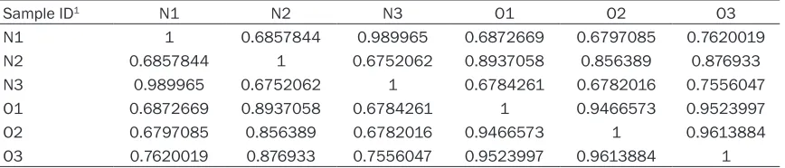
Table 3. Correlations value between each two samples
Sample ID1 N1 N2 N3 O1 O2 O3
N1 1 0.6857844 0.989965 0.6872669 0.6797085 0.7620019
N2 0.6857844 1 0.6752062 0.8937058 0.856389 0.876933
N3 0.989965 0.6752062 1 0.6784261 0.6782016 0.7556047
O1 0.6872669 0.8937058 0.6784261 1 0.9466573 0.9523997
O2 0.6797085 0.856389 0.6782016 0.9466573 1 0.9613884
O3 0.7620019 0.876933 0.7556047 0.9523997 0.9613884 1
[image:3.612.91.525.255.348.2]t test, and correlations between qPCR and RNA-Seq measures were calculated.
Results
Overview of sequencing data
After removing the low quality and adaptor sequences, we obtained approximately 49 to 52 million clean reads for six RNA-Seq libraries, and the percentages of mapped reads ranging from 48.43% to 73.03%, the unique match rate ranging from 44.68% to 66.03% (Table 2). These results indicated that our six libraries were of high quality, and had high coverage of the pig genome. This allowed us to compare the ovary transcriptomes from pigs with oestrous or not.
Differentially expressed genes between estrus and no-estrus groups
Subsequently, the present study detected the
gene expression levels and identified the differ -entially expressed genes between oetrous and non-oetrous samples using the RSEM software package. RSEM maximum likelihood abun-dance was estimated by using the
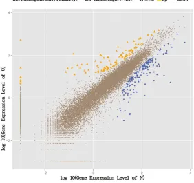
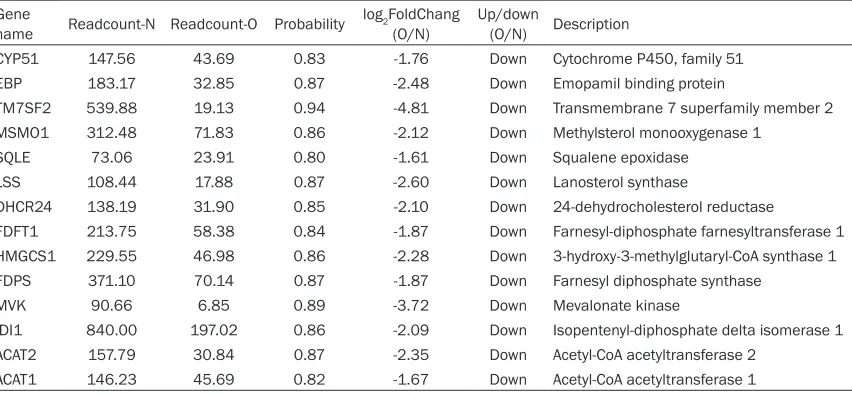
Expectation-expressed between the two groups, in which 204 genes were upregulated and 228 genes were downregulated in the oestrous group (Figure 1). The 14 most differentially down expressed genes related to the pigs’
produc-tion from the total of 432 DEGs identified
between the non-oestruos and oestrous sam-ples were: cytochrome P450, family 51 (CYP- 51), emopamil binding protein (EBP), trans-membrane 7 superfamily member 2 (TM7S- F2), methylsterol monooxygenase 1 (MSMO1), squalene epoxidase (SQLE), lanosterol syn-thase (LSS), 24-dehydrocholesterol reductase (DHCR24), farnesyl-diphosphate farnesyltrans-ferase 1 (FDFT1), 3-hydroxy-3-methylglutaryl-CoA synthase 1 (HMGCS1), farnesyl diphos-phate synthase (FDPS), mevalonate kinase (MVK), isopentenyl-diphosphate delta isomer-ase 1 (IDI1), acetyl-CoA acetyltransferase 2 (ACAT2), acetyl-CoA acetyltransferase 1 (AC- AT1) (Table 4).
Functional enrichment analysis of differentially expressed genes
To define the biological functions of the 432
DEGs, GO and KEGG analysis were carried out.
Fifty-one significantly enriched GO terms (cor
-Figure 1. Genes expression level in oestrous and non-ostrous samples (O: oestrous group; N: non-ostrous group).
Maximization (EM) algorithm of its statistical model, includ-ing the modelinclud-ing of PE and variable-length reads, frag-ment length distributions and quality scores, to determine which transcripts were iso-forms of the same gene. The FPKM method was used to determine the gene expres-sion levels. The analysis con-tained the majority of the annotated pig genes. The cor-relation of the gene expres-sion between two samples was evaluated. Results reve- aled that gene expression lev-els among two groups were highly correlated, suggesting that the experiments were reli-able and the samples selec-tion were reasonable (Table 3).
[image:4.612.92.374.92.358.2]rected P<0.05) were identified, including bio -logical regulation, cellular component organiza-tion or biogenesis, cellular process, develop- mental process, regulation of biological pro-cess, metabolic propro-cess, negative regulation of biological process, positive regulation of bio-logical process, reproduction, reproductive pro-cess (Figure 2). Meanwhile, 20 significantly enriched KEGG pathways were identified,
including metabolic pathways, terpeniod back-bone biosynthesis, PPAR signaling pathway, steroid biosynthesis, starch and sucrose me- tabolism, ribosome, fat digestion and absorp-tion (Figure 3). Among these GO terms and KEGG pathways, the steroid biosynthesis, reproduction and reproductive process were the ones related to steroid hormone regulation in animal ovaries and therefore likely to be con-tributing to osterous. However, as most GO and KEGG assignments and distributions are relat-ed to reproduction, growth and development, and metabolism, our results indicate that the DEGs are involved in a wide range of regulatory functions in Xiang pig ovaries.
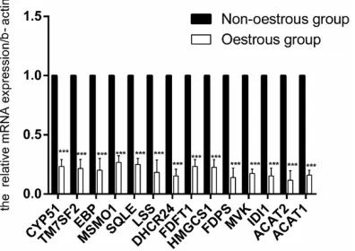
qRT-PCR validation
The transcript levels of 14 genes (CYP51, EBP, TM7SF2, MSMO1, SQLE, LSS, DHCR24, FDFT1, HMGCS1, FDPS, MVK, IDI1, ACAT2, and ACAT1) were re-evaluated using qRT-PCR technology. RNA from the same animals assigned to the RNA-Seq analysis was used for the qRT-PCR validation experiment (Figure 4). Overall, the
results obtained from qRT-PCR were in agree-ment with the results of RNA-seq.
Discussion
Ovaries are one of the most important animal reproductive organs, which directly regulate ovulation and female hormone secretion and
have a significant impact on the fecundity of
mammals [25, 26]. Also, animals showing clear visible oestrous signs have high yield [27, 28]. But, until now, rare study investigated the oes-trous candidate genes related to the produc-tion of Xiang pig. To the best of our knowledge,
this is the first report focused on pig oestrous
using RNA-seq.
It is well known that there is gene expression
specificity in different tissues and cells. The
ovaries contain a mixture of different tissue, and the expression of candidate genes may dif-fer between them. We strove to ensure that we obtained intact ovaries and ground them com-pletely for the purpose of RNA extraction, to ensure that the RNA-Seq results were repre-sentative of the complete Xiang pig ovarian transcriptome. In order to minimize the effect of age, we selected six pigs with similar age for RNA-Seq. Two sequencing libraries were con-structed from the non-oestrous and oestrous samples. High quality transcriptome data was generated (about 52 million clean reads for
each sample), which was sufficient for the
[image:5.612.95.521.85.282.2]quantitative analysis of gene expression. Table 4. Detailed information on the 14 differentially expressed genes related to oestrus
Gene
name Readcount-N Readcount-O Probability log2FoldChang (O/N) Up/down (O/N) Description
CYP51 147.56 43.69 0.83 -1.76 Down Cytochrome P450, family 51
EBP 183.17 32.85 0.87 -2.48 Down Emopamil binding protein
TM7SF2 539.88 19.13 0.94 -4.81 Down Transmembrane 7 superfamily member 2
MSMO1 312.48 71.83 0.86 -2.12 Down Methylsterol monooxygenase 1
SQLE 73.06 23.91 0.80 -1.61 Down Squalene epoxidase
LSS 108.44 17.88 0.87 -2.60 Down Lanosterol synthase
DHCR24 138.19 31.90 0.85 -2.10 Down 24-dehydrocholesterol reductase
FDFT1 213.75 58.38 0.84 -1.87 Down Farnesyl-diphosphate farnesyltransferase 1
HMGCS1 229.55 46.98 0.86 -2.28 Down 3-hydroxy-3-methylglutaryl-CoA synthase 1
FDPS 371.10 70.14 0.87 -1.87 Down Farnesyl diphosphate synthase
MVK 90.66 6.85 0.89 -3.72 Down Mevalonate kinase
IDI1 840.00 197.02 0.86 -2.09 Down Isopentenyl-diphosphate delta isomerase 1
ACAT2 157.79 30.84 0.87 -2.35 Down Acetyl-CoA acetyltransferase 2
ACAT1 146.23 45.69 0.82 -1.67 Down Acetyl-CoA acetyltransferase 1
The increased plasma concentrations of oes-trogen and luteinizing hormone (LH), and the decreased concentration of progesterone be- fore ovulation can initiate the occurrence of oestrous behavior [29]. Besides, enzymes in ovarian tissue, such as the cytochrome P450 (CYP) family, hydroxy steroid dehydrogenase and catechol-O-methyl-transferaseenzyme, di- rectly catalyze the synthesis and metabolism of oestrogen from cholesterol [30]. The expres-sion of key steroidogenic enzymes responsible for the production of oestrogens - oestradiol (E2) and oestrone (E1) as well as testosterone
(T), namely CYP17A1 and CYP19A3 was noted in the porcine uterus during early pregnancy and the oestrous cycle [31, 32]. And in the study, we found 14 candidate genes related to the oestrus of swine: CYP51, EBP, TM7SF2, MSMO1, SQLE, LSS, DHCR24, FDFT1, HMGC- S1, FDPS, MVK, IDI1, ACAT2, and ACAT1. CYP51
is a member of CYP family, just as descripted, can catalyze the synthesis and metabolism of oestrogen. TM7SF2 gene has been reported to be involved in cholesterol biosynthesis by
encoding the protein 3β-hydroxysterol
[image:6.612.89.522.72.513.2]Δ14-reductase [33], indicating it may involve in the
process of oestrus. Besides, SQLE is also reported to regulate cholesterol synthesis [34],
LSS has relationship with oestrogen [35] and so on.
To experimentally validate the DEGs identified
from the sequencing data, we analyzed 14
DEGs using qPCR. The results confirmed that
cess, metabolic pathways, terpeniod backbone biosynthesis, PPAR signaling pathway, steroid biosynthesis, starch and sucrose metabolism, ribosome, fat digestion and absorption. It is accepted that the molecular regulation of ani-mal traits is very complex and the relationship between genes and traits is often that of “one-to-many” or “many-to-one” [36]. The DEGs were not only enriched in reproduction-related path-ways but also in those involved with steroid bio-synthesis and fatty metabolism. This suggests that the genes may be associated with both reproduction and fat metabolism. The synthe-sis of steroid hormones is closely related to fat metabolism. It has been demonstrated that all of steroid hormones are synthesized from cho-lesterol [37]. The pathways mentioned above are, to a greater or lesser degree, involved in the development of follicular cells and oocytes. Consequently, functional studies should be performed with these DGEs in order to identify
key candidate genes influencing reproductive
traits in swine.
In conclusion, this study screened for DEGs in the pigs’ ovarian tissues with oestrous or non-oestrous character using RNA-Seq. We
identi-Figure 3. The most enriched 20 pathways. (The size of black circle repre-sents the DEGs number).
the expression patterns of these DEGs were consistent with those obtained from tran-scriptome sequencing data (Figure 4). These results
indi-cate that the DEGs identified
in the genome-wide transcrip-tome sequencing data are reliable.
[image:7.612.89.371.74.364.2]We also performed GO enrich-ment analysis of DEGs from the DEGseq comparisons N (non-oestroud group) vs. O (oestrous group). Notably, from the GO and KEGG analy-sis, we found that the func-tions of DEGs between the two groups were mainly in- cluding biological regulation, cellular component organiza-tion or biogenesis, cellular process, developmental pro-cess, regulation of biological process, metabolic process, negative regulation of biologi-cal process, positive regula-tion of biological process, re- production, reproductive pro-
Figure 4. The expression levels of genes related to
oestrus were quantified using qRT-PCR. The data are expressed as means ± SD. Statistical significance
[image:7.612.92.290.417.557.2]fied 204 genes that were upregulated and 228
genes that were downregulated in the oestrous samples compared with the non-oestrous group. After analyzing the function of these genes, we found 14 DEGs that maybe relevant
to the prolificacy of pigs and verified these
genes by qPCR technology. This new informa-tion provides a solid foundainforma-tion for further studies of the molecular mechanisms
underly-ing porcine prolificacy. In the future, biochemi -cal and physiologi-cal analyses of these candi-date genes will be conducted.
Acknowledgements
This work was supported by National High Te- chnology Research and Development Program of China (863 Program) (grant 2013AA102503) (http://www.most.gov.cn/bszn/new/863/jbxx/), Guizhou Province Science and Technology Innovation Team Building Special (Guizhou Branch of talent team 2009-4006) (http://kjt. gzst.gov.cn), National Natural Science Foun- dation of China (31672390) (http://kjt.gzst. gov.cn), the Guizhou Province “Hundreds” inno-vative talents project (QKHRC[2016]-4012) (http://kjt.gzst.gov.cn), and the Guizhou Agri- culture Research Program ([2013] 3073, [20- 17] 2585, [2017] 2587) (http://kjt.gzst.gov.cn). Disclosure of conflict of interest
None.
Address correspondence to: Xueqin Ran, College of Animal Science, Guizhou University, Guiyang 5500- 25, China. Tel: +86-85188298005; E-mail: xqran@ gzu.edu.cn; Jiafu Wang, College of Animal Science/ Key Laboratory of Plant Resource Conservation and Germplasm Innovation in Mountainous Region (Ministry of Education), Collaborative Innovation Center for Mountain Ecology & Agro-Bioengineering (CICMEAB), Institute of Agro-Bioengineering, Gui- zhou University, Guiyang, China; Tongren University, Tongren, China. Tel: +86-085188298070; E-mail: [email protected]
References
[1] Munoz G, Ovilo C, Estelle J, Silio L, Fernandez A and Rodriguez C. Association with litter size of new polymorphisms on ESR1 and ESR2 genes in a Chinese-European pig line. Genet Sel Evol 2007; 39: 195-206.
[2] Zhao Y, Li N, Xiao L, Cao G, Chen Y, Zhang S, Wu C, Zhang J, Sun S and Xu X. FSHB subunit gene is associated with major gene controlling litter size in commercial pig breeds. Sci China
[3] Sun YX, Zeng YQ, Tang H, Fan XZ, Chen QM, Li H, Qian Y and Song YP. [Relationship of genetic polymorphism of PRLR and RBP4 genes with litter size traits in pig]. Yi Chuan 2009; 31: 63-68.
[4] Martinez-Montes AM, Fernandez A, Perez-Montarelo D, Alves E, Benitez RM, Nunez Y, Ovilo C, Ibanez-Escriche N, Folch JM and Fer-nandez AI. Using RNA-Seq SNP data to reveal potential causal mutations related to pig pro-duction traits and RNA editing. Anim Genet 2017; 48: 151-165.
[5] Rohrer GA, Wise TH and Ford JJ. Deciphering the pig genome to understand gamete produc-tion. Soc Reprod Fertil Suppl 2006; 62: 293-301.
[6] Reiner G. [Product quality and genome analy-sis in pig production-a review]. Dtsch Tierarztl Wochenschr 2006; 113: 65-69.
[7] Marioni JC, Mason CE, Mane SM, Stephens M and Gilad Y. RNA-seq: an assessment of tech-nical reproducibility and comparison with gene expression arrays. Genome Res 2008; 18: 1509-1517.
[8] Gonella-Diaza AM, Andrade SC, Sponchiado M, Pugliesi G, Mesquita FS, Van Hoeck V, Strefezzi Rde F, Gasparin GR, Coutinho LL and Binelli M. Size of the ovulatory follicle dictates spatial dif-ferences in the oviductal transcriptome in cat-tle. PLoS One 2015; 10: e0145321.
[9] McGettigan PA, Browne JA, Carrington SD, Crowe MA, Fair T, Forde N, Loftus BJ, Lohan A, Lonergan P, Pluta K, Mamo S, Murphy A, Roche J, Walsh SW, Creevey CJ, Earley B, Keady S, Kenny DA, Matthews D, McCabe M, Morris D, O’Loughlin A, Waters S, Diskin MG and Evans AC. Fertility and genomics: comparison of gene expression in contrasting reproductive tissues of female cattle. Reprod Fertil Dev 2016; 28: 11-24.
[10] Zi XD, Lu JY and Ma L. Identification and com -parative analysis of the ovarian microRNAs of
prolific and non-prolific goats during the follicu -lar phase using high-throughput sequencing. Sci Rep 2017; 7: 1921.
[11] Ling YH, Quan Q, Xiang H, Zhu L, Chu MX,
Zhang XR and Han CY. Expression profiles of
differentially expressed genes affecting fecun-dity in goat ovarian tissues. Genet Mol Res 2015; 14: 18743-18752.
[12] Zhang X, Huang L, Wu T, Feng Y, Ding Y, Ye P and Yin Z. Transcriptomic analysis of ovaries from pigs with high and low litter size. PLoS One 2015; 10: e0139514.
[13] Kilkenny C, Browne WJ, Cuthi I, Emerson M and Altman DG. Improving bioscience research reporting: the arrive guidelines for reporting animal research. Vet Clin Pathol 2012; 41: 27-31.
46-[15] Hulten F, Wallenbeck A and Rydhmer L. Ovari-an activity Ovari-and oestrous signs among
group-housed, lactating sows: influence of behaviour,
environment and production. Reprod Domest Anim 2006; 41: 448-454.
[16] Li X, Ye Y, Zhou X, Huang C and Wu M. Atg7 enhances host defense against infection via downregulation of superoxide but upregulation of nitric oxide. J Immunol 2015; 194: 1112-1121.
[17] Li X, Zhou X, Ye Y, Li Y, Li J, Privratsky B, Wu E, Gao H, Huang C and Wu M. Lyn regulates
in-flammatory responses in Klebsiella pneumoni -ae infection via the p38/NF-kappaB pathway. Eur J Immunol 2014; 44: 763-773.
[18] Kerpedjiev P, Frellsen J, Lindgreen S and Krogh A. Adaptable probabilistic mapping of short
reads using position specific scoring matrices.
BMC Bioinformatics 2014; 15: 100.
[19] Kim D and Salzberg SL. TopHat-Fusion: an al-gorithm for discovery of novel fusion tran-scripts. Genome Biol 2011; 12: R72.
[20] Mortazavi A, Williams BA, McCue K, Schaeffer L and Wold B. Mapping and quantifying mam-malian transcriptomes by RNA-Seq. Nat Meth-ods 2008; 5: 621-628.
[21] Wang Z, Gerstein M and Snyder M. RNA-Seq: a revolutionary tool for transcriptomics. Nat Rev Genet 2009; 10: 57-63.
[22] Ashburner M, Ball CA, Blake JA, Botstein D, Butler H, Cherry JM, Davis AP, Dolinski K, Dwight SS, Eppig JT, Harris MA, Hill DP, Issel-Tarver L, Kasarskis A, Lewis S, Matese JC, Richardson JE, Ringwald M, Rubin GM and
Sherlock G. Gene ontology: tool for the unifica -tion of biology. The Gene Ontology Consortium. Nat Genet 2000; 25: 25-29.
[23] Kanehisa M, Araki M, Goto S, Hattori M, Hi-rakawa M, Itoh M, Katayama T, Kawashima S, Okuda S, Tokimatsu T and Yamanishi Y. KEGG for linking genomes to life and the environ-ment. Nucleic Acids Res 2008; 36: D480-484. [24] Mao X, Cai T, Olyarchuk JG and Wei L.
Automat-ed genome annotation and pathway identifica -tion using the KEGG Orthology (KO) as a con-trolled vocabulary. Bioinformatics 2005; 21: 3787-3793.
[25] Weiner S, Wright KH and Wallach EE. Studies on the function of the denervated rabbit ovary: human chorionic gonadotropin-induced ovula-tion. Fertil Steril 1975; 26: 363-368.
[26] Balen A. The effects of ovulation induction with gonadotrophins on the ovary and uterus and implications for assisted reproduction. Hum Reprod 1995; 10: 2233-2237.
[27] Gerritsen R, Soede NM, Hazeleger W, Lan-gendijk P, Dieleman SJ, Taverne MA and Kemp B. Intermittent suckling enables estrus and pregnancy during lactation in sows: effects of stage of lactation and lactation during early pregnancy. Theriogenology 2009; 71: 432-440.
[28] Patterson J, Wellen A, Hahn M, Pasternak A, Lowe J, DeHaas S, Kraus D, Williams N and Foxcroft G. Responses to delayed estrus after weaning in sows using oral progestagen treat-ment. J Anim Sci 2008; 86: 1996-2004. [29] Soede NM, Langendijk P and Kemp B.
Repro-ductive cycles in pigs. Anim Reprod Sci 2011; 124: 251-258.
[30] Low YL, Li Y, Humphreys K, Thalamuthu A, Da-rabi H, Wedren S, Bonnard C, Czene K, Iles MM, Heikkinen T, Aittomaki K, Blomqvist C, Ne-vanlinna H, Hall P, Liu ET and Liu J. Multi-vari-ant pathway association analysis reveals the importance of genetic determinants of estro-gen metabolism in breast and endometrial cancer susceptibility. PLoS Genet 2010; 6: e1001012.
[31] Chen X, Li A, Chen W, Wei J, Fu J and Wang A. Differential gene expression in uterine endo-metrium during implantation in pigs. Biol Re-prod 2015; 92: 52.
[32] Kiezun M, Smolinska N, Dobrzyn K, Szeszko K, Rytelewska E and Kaminski T. The effect of orexin A on CYP17A1 and CYP19A3 expression and on oestradiol, oestrone and testosterone secretion in the porcine uterus during early pregnancy and the oestrous cycle. Therio-genology 2017; 90: 129-140.
[33] Zuleger N, Boyle S, Kelly DA, de las Heras JI, Lazou V, Korfali N, Batrakou DG, Randles KN, Morris GE, Harrison DJ, Bickmore WA and
Schirmer EC. Specific nuclear envelope trans -membrane proteins can promote the location of chromosomes to and from the nuclear pe-riphery. Genome Biol 2013; 14: R14.
[34] Howe V, Sharpe LJ, Prabhu AV and Brown AJ. New insights into cellular cholesterol acquisi-tion: promoter analysis of human HMGCR and SQLE, two key control enzymes in cholesterol synthesis. Biochim Biophys Acta 2017; 1862: 647-657.
[35] Muth-Kohne E, Westphal-Settele K, Bruckner J, Konradi S, Schiller V, Schafers C, Teigeler M and Fenske M. Linking the response of endo-crine regulated genes to adverse effects on sex differentiation improves comprehension of
aromatase inhibition in a fish sexual develop -ment test. Aquat Toxicol 2016; 176: 116-127. [36] Guo ZL, Zhu Y, Su XT, Liu J, Yang QX, Nan JY,
Zhao BC, Zhang YY, Yu YN, Li B, Xiao HB and Wang Z. DanHong injection dose-dependently varies amino acid metabolites and metabolic pathways in the treatment of rats with cerebral ischemia. Acta Pharmacol Sin 2015; 36: 748-757.