Original Article
Effects of changes in the intestinal function of
rats with intestinal dysfunction induced
by new intestinal obstructions
Nan Zhang1*, Jun-Hong Ma1*, Man-Li Qi2, Zhen-Li Zhou1
1Department of Gastrointestinal Surgery, Tianjin Nankai Hospital, Tianjin, China; 2Department of Dermatology,
Tianjin General Hospital of Medical University, Tianjin, China. *Equal contributors and co-first authors.
Received March 27, 2018; Accepted July 14, 2018; Epub November 15, 2018; Published November 30, 2018
Abstract: The aim of this study was to establish an animal model of reversible mechanical intestinal obstruction to observe alterations of intestinal function at different time points. A reversible mechanical intestinal obstruction model was developed in Wister rats, in which a homemade atraumatic intestinal clamp was used to externally op-press the intestines. This model was used to study changes in intestinal histopathology, damage indexes of ileum mucosa, serum carbamyl ornithine levels, D-lactic acid levels, biochemical markers, and cytokine levels at different time points (6, 12, 24, 48, 72 and 96 hours after obstruction and 48, 72 and 96 hours after recovery in rats that had been obstructed for 48 hours). The model was successfully developed and showed good stability. Using this model, prolonged obstruction duration led to aggravated pathological changes of the intestines, increased damage indexes of ileum mucosa, and decreased serum carbamyl ornithine, Cr, and IL-2 levels, along with increased D-lactic
acid, ALT, LDHF, IL-6 and TNF-α levels. Mortality gradually increased with increasing obstruction time. Signs of sys
-temic inflammatory responsible syndrome (SIRS) and multiple organ dysfunction syndrome (MODS) appeared after
48 hours of obstruction. After clamps were removed, recovery of intestines was observed. This method can be used to successfully establish a rat model of reversible complete mechanical intestinal obstruction. The animals
devel-oped signs of MODS after 48 hours of obstruction, making this model appropriate for studying intestinal dysfunction
associated with intestinal obstruction.
Keywords: Intestinal obstruction, animal model, intestinal function, multiple organ dysfunction syndrome
Introduction
Intestinal obstruction remains one of the most common abdominal diseases in general sur-gery, leading to high morbidity and mortality [1]. Intestinal obstruction can alter the anatomy and function of the intestines and can also induce systemic physiological disorders, lead-ing to intestinal dysfunction and even progress-ing to multiple organ dysfunction syndrome (MODS) [2]. Therefore, developing a simple and convenient animal model that reflects the physiological and pathological alterations of intestinal obstruction is of significance for studying the mechanisms and treatments of intestinal obstruction and secondary intestinal dysfunction.
The pathogenesis of mechanical intestinal obstruction can be summarized into three
cat-egories: intraluminal obstruction, extraluminal oppression, and disease of the intestinal wall [3]. Intraluminal obstruction is due to the ob- struction by stools or intestinal stones and is usually incomplete. Local intestinal stenosis or external compression caused by intestinal ad- hesion, tumors, or hernia, accounts for 90% of mechanical intestinal obstruction [4]. Previ- ously, most large animal models of intestinal obstruction were developed using intraluminal obstruction [4-7], characterized by complicated procedures for modeling, many factors affect-ing obstruction, inaccurate obstruction efficien-cy, and high costs.
the silk thread is very thin and ligature strength cannot be controlled uniformly. In addition, a tight ligature is prone to cause intestinal necro-sis and even incise and perforate the intes-tines, whereas loose ligatures do not achieve complete intestinal obstruction.
Therefore, in this study, a new mechanical intestinal obstruction model was developed, in which a homemade atraumatic intestinal clamp was used. The clamp was made of a double flat iron-core binding-wire with a width of 3.0 mm, not likely to incise the intestines. Since it has a circumference of 10 mm, it was appropriate for the diameter of the small intestines of rats. Furthermore, its two ends were bent for 3 mm to avoid damaging the intestines. This model was used to investigate alterations of intestinal function at different time points, to study MODS in the process of intestinal obstruction and pro-vide a basis for further investigating intestinal dysfunction.
Materials and methods
Animals and grouping
A total of 200 healthy and pathogen-free Wister rats, half male and half female, weighing 250-270 g, were provided by the Laboratory Animal Center of the Institute of Hygiene Environment- al Medicine of the Academy of Military Medical Science (Certificate # SCXK-(Military)
2009-003). This study was approved by the Animal Ethics Committee of Tianjin Nankai Hospital. Before the experiment, the animals were raised adaptively at the Standard Laboratory Animal Center of the Institute of Acute Abdominalgia of Tianjin Nankai Hospital for 1 week. They had free access to standard feed and water in ven-tilated and specific pathogen-free conditions, at a temperature of 20-25°C, relative humidity 50±10%, and light/dark cycles of 12/12 hours. The animals were randomized to seven groups using a random number table (n = 20 in each group): I0 (sham operation group), I6 (intestinal obstruction for 6 hours), I12 (intestinal obstruc-tion for 12 hours), I24 (intestinal obstrucobstruc-tion for 24 hours), I48 (intestinal obstruction for 48 hours), I72 (intestinal obstruction for 72 hours), and I96 (intestinal obstruction for 96 hours). In addition, 60 rats underwent intestinal obstruc-tion for 48 hours. Clamps were then removed and rats were randomized into three recovery groups (each containing 20 rats): R48 (recovery for 48 hours), R72 (recovery for 72 hours), and R96 (recovery for 96 hours).
Development of the atraumatic intestinal clamp
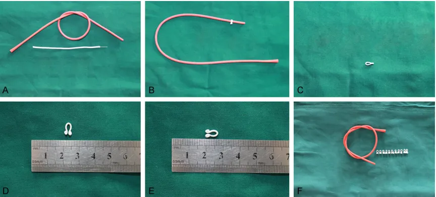
[image:2.612.91.523.71.266.2]The innovative and key point of this model was the homemade atraumatic intestinal clamp. A disposable 10 Fr bladder catheter was used to Figure 1. Preparation of the atraumatic intestinal clamp. A: A 10 Fr bladder catheter was used for simulating the
simulate the distal ileum intestine. A double- flat iron-core binding wire (width of 3.0 mm, diameter of the iron core of 0.3 mm, referred to as binding wire) was cut into pieces of 16 mm, which were wrapped around the cathet- er to make a “Ω” shape. These pieces were then referred to as “atraumatic intestinal clamps”. The clamp’s circumference was 10 mm and its two ends were curved for 3 mm to prevent puncturing the intestines. The clamps were treated with silicone oil and autoclaved (Figure 1).
Based on pilot results, the mean external pr- essure on the intestines when using the at- raumatic clamp to clip the distal ileum intes- tine (disposable 10 Fr bladder catheter) was 12.81±3.95 N. This tended to cause comple- te intestinal obstruction, but was unlikely to cause intestinal necrosis, as tested by the Tianjin Medical Device Quality Supervision and Inspection Center.
Modeling
Before surgery, the animals were fasted for 12 hours, with free access to water. Rats in each group received an intraperitoneal injection of 10% chloral hydrate (0.3 mL/100 g). They were placed in the supine position and fixed on the operating table to expose the chest and
abdo-men. The limbs were symmetrically fixed using elastic cloth. Abdominal hair was removed, fol-lowed by disinfection with iodine and alcohol and sterile draping. All models were prepared by the same operator.
A skin incision of about 2 cm was made at one third below the ventral median line, isolated layer-by-layer to cut open the abdomen to expose the ileocecal part. This was taken out with notched forceps and placed on sterile gauze. A relatively isolated intestine part with a long mesenterium was selected in the ileoce-cal part, 1 cm towards the distal ileum, where a hole was poked in the avascular zone of the mesenteric small intestine with ophthalmic hemostatic forceps. The atraumatic clamp was covered outside the intestine and clipped th- rough the mesenterium, thereby causing com-plete obstruction of the lumen. Next, foreign bodies on the intestinal surface were rinsed with warm saline and the intestine was restored in the abdomen. About 1 mL of 5% levofloxacin glucose injection was dripped into the abdomi-nal cavity, followed by abdomiabdomi-nal closure layer by layer. In the sham group, only opening and closure of the abdomen was conducted. Releasing the obstruction, rats were anesthe-tized, the abdomen was opened to find the obstruction point, and the atraumatic clamp was removed. This model was developed very conveniently and took only about 3 minutes to complete.
Post-operative management and sampling
[image:3.612.89.523.84.179.2]After modeling, the animals were transferred to the animal room (temperature of 20-25°C and relative humidity of 50±10%). Mental attitudes and activities of the rats were observed and their general status at 6, 12, 24, 48, 72 and 96 hours after modeling was recorded. At the same time points, the abdomen was opened to Table 1. Criteria for Chiu’s intestinal mucosal damage
Score Grading Criteria 1 Grade 0 Normal villi
2 Grade 1 Submucosal space on top of villi, capillary congestion
3 Grade 2 Expanded Submucosal space, isolation of intestinal mucosa and submucosa 4 Grade 3 Isolation of intestinal mucosa and submucosa was extended to bilateral villi 5 Grade 4 Blunted villi, exposure of lamina propria and vessels, inflammatory infiltration 6 Grade 5 Digestion and disintegration of lamina propria, with bleeding and ulceration
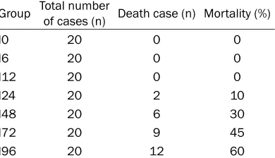
Table 2. Mortalities of obstruction groups Group Total number of cases (n) Death case (n) Mortality (%)
I0 20 0 0
I6 20 0 0
I12 20 0 0
I24 20 2 10
I48 20 6 30
I72 20 9 45
[image:3.612.94.289.216.328.2]expose the visceral organs to observe injuries to the intestine. Complications and mortality were recorded. In cases of death, explorative laparotomies were performed to investigate causes.
Pathological analyses of ileum after modeling
Color, activity, and obstruction of the small intestines were observed visually. A segment of 2 cm of small intestine at the obstruction point and 1 cm at the proximal end were selected. After removing the foreign bodies from the lumen, segments were rinsed repeatedly with cool saline, fixed with 10% neutral formalin solution, followed by paraffin-embedding, sec-tioning, and H&E staining. Damage to the intes-tinal mucosa was assessed using Chiu’s crite-ria by a pathologist blinded to grouping [9] (Table 1).
Diagnoses of SIRS and MODS
SIRS was diagnosed, as previously described [10], based on abnormal white cell counts with
at least 10% of immature neutrophils and abnormal blood pH. MODS was diagnosed, as previously described [11], based on a compre-hensive review of systems, symptoms, and bio-chemistry. Blood pH, lactic acid, urea, creati-nine (Cr), carbamyl ornithine, and glucose were monitored as signs of SIRS or MODS.
Serum carbamyl ornithine levels
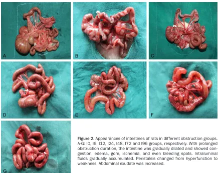
[image:4.612.88.523.68.414.2]Blood (10 mL) was collected from the abdomi-nal aorta in two different 5-mL tubes contain-ing anticoagulant. Blood was centrifuged at 3000 rpm for 15 minutes. Serum was collected and kept at -20°C. Serum carbamyl ornithine levels were determined by high performance liquid chromatography (HPLC), using an AC- QUITY Ultra HPLC workstation (Waters, Milford, MA, USA). The chromatographic column was an ACQUITY UPLC BEH C18 (1.7 µm, 100×4.6 mm; Waters, Milford, MA, USA). Mobile phase A was 0.1% formic acid-water solution. Mobile phase B was methanol. Column temperature was 40°C, flow velocity was 0.4 mL/min, and the Figure 2. Appearances of intestines of rats in different obstruction groups. A-G: I0, I6, I12, I24, I48, I72 and I96 groups, respectively. With prolonged obstruction duration, the intestine was gradually dilated and showed con-gestion, edema, gore, ischemia, and even bleeding spots. Intraluminal
fluids gradually accumulated. Peristalsis changed from hyperfunction to
wavelength was sampled in time series at λ = 254 nm.
Serum D-lactic acid levels
Serum D-lactic acid levels were tested using a rat D-lactate dehydrogenase (D-LDH) ELISA Kit (E02D0015, BlueGene Biotech, Shanghai, Ch- ina), according to manufacturer instructions.
Biochemical indicators
Serum alanine transaminase (ALT), lactate dehydrogenase (LDH), and Cr were tested using a MEGA biochemical analyzer (Toshiba Corp., Tokyo, Japan). Reagents for ALT, LDH, and Cr were purchased from Shanghai Huachen Biological Reagent Co., Ltd. (Shanghai, China).
Serum inflammatory cytokines levels
Tumor necrosis factor-α (TNF-α), interleukin (IL)-6, and IL-2 were tested using rat ELISA kits for TNF-α (E02T0008), IL-6 (E02I0006), and IL-2 (E02I0308) from Shanghai BlueGene Bio- tech Co., Ltd. (Shanghai, China), according to manufacturer instructions.
Statistical analysis
Equality of variance was tested using the Levene test and normality was tested using the Shapiro-Wilk test. All data are expressed as mean ± standard deviation and were analyzed using one-way ANOVA with LSD-t post hoc test. For non-normally distributed data or heteroge-neity of variance, comparisons were conducted using Kruskal-Wallis rank sum test and pair-wise comparisons were conducted using Wilcoxon rank sum test. Correlation was tested using Pearson’s analysis (normal distribution) or Spearman’s rank correlation (non-normal distribution). All statistical analyses were per-formed using SPSS 17.0 (IBM, Armonk, NY, USA). Two-sided P-values < 0.05 are consid-ered statistically significant.
Results
Model success
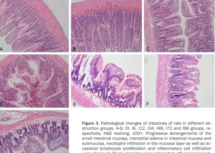
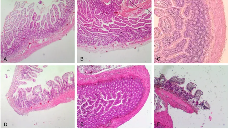
[image:5.612.94.523.74.378.2]Rats in the I0 group were in good condition, without mortality. After modeling, all rats were awake after about 3 hours. With prolonged Figure 3. Pathological changes of intestines of rats in different ob-struction groups. A-G: I0, I6, I12, I24, I48, I72 and I96 groups, re-spectively. H&E staining, 100×. Progressive derangements of the small intestinal mucosa, interstitial edema in intestinal mucosa and
submucosa, neutrophil infiltration in the mucosal layer as well as oc
-casional lymphocyte proliferation and inflammatory cell infiltration
obstruction duration, rats gradually showed fatigue, reduced activity, slow response, ab- dominal tic, and cage adhesion. Furthermore, with prolonged obstruction duration, intestinal obstruction symptoms became more signifi-cant and mortality gradually increased. The complete intestinal obstruction model was con-sidered successful if rats did not produce fecal pellets. There was no mortality in the I6 and I12 groups, while the mortality rates were 10% (2/20), 30% (6/20), 45% (9/20), and 60% (12/20) in the I24, I48, I72 and I96 groups, respectively (Table 2).
Intestines of rats after obstruction
In the I0 group, intestines appeared normal in diameter and color (Figure 2A). In the I6 group, small intestines at the proximal obstruction point was mildly dilated and the intestinal wall had a normal color and mobility (Figure 2B). In the I12 group, intestines were gradually dilat- ed, intestinal wall color was darker, with local congestion and edema, and the gas-liquid accumulation was more significant than in the I6 group (Figure 2C). In the I24 group, small
intestines were significantly dilated and the gas-liquid accumulation was also significant. The high jejunum was not significantly dilated and the intestine was mildly congested with sig-nificant edema and intestinal hyperperistalsis (Figure 2D).
[image:6.612.89.524.72.318.2]In the I48 group, small intestines showed aggravated congestion and edema, with signifi-cant congestion, edema, and dilation, along with more significant gas-liquid accumulation. In addition, there was mild congestion of the intestinal wall and a clear yellowish exudate in the abdominal cavity, with relatively active peristalsis (Figure 2E). In the I72 group, small intestines were highly dilated, the ileum lu- men was significantly dilated, and there was a large amount of gas-liquid accumulation. There was a purulent or bloody turbid exudate in the abdominal cavity, with a foul smell (Figure 2F). In the I96 group, severe edema and con-gestion was observed in the small intestin- es, with hemorrhage and necrosis. Further- more, there was a manure-like exudate, with a foul smell and disappearance of peristalsis (Figure 2G).
Figure 4. Pathological sections of intestines at the obstruction sites. A-F: I6, I12, I24, I48, I72 and I96 groups, re-spectively. H&E staining, 100×. There was no necrosis at any time point after obstruction. In I6 to I72 groups, the mucosal epithelium was intact and with a normal structure, while mucosa and submucosa showed mild edema, vessels had mild congestion, the myometrium structure was normal, and the vessels in the serosa showed mild congestion and dilation. In the I96 group, part of the epithelium mucosa was denaturized with intact structure, while
the mucosa and submucosa showed mild edema, vascular congestion and dilation, with a small amount of inflam
Pathological changes of intestines in obstruct-ed rats
In the I0 group, intact ileum mucosal layers were observed, with orderly intestinal epitheli-um, regularly arranged intestinal villi, along with absence of lymphocyte proliferation and inflam-matory cell infiltration. In the I6 group, mild derangement of the small intestinal mucosa,
in intestinal mucosa and submucosa, capillary congestion in the villi, reduced number of intes-tinal epithelial cells, frangible villi, and inflam-matory exudate in the serous layer (Figure 3A-D).
[image:7.612.91.523.73.277.2]In the I48 group, there was edema, congestion and inflammatory cell infiltration, shedding of the intestinal mucosal epithelium, derange-Figure 5. Pathological changes of the intestines of rats after recovery. A-C: Intestine conditions were continuously observed for 48, 72 and 96 h after removal of the obstruction in rats from the I48 group. D-F: Pathological changes of the intestines were continuously observed for 48, 72, and 96 h after the obstruction was removed from rats of the I48 group. H&E staining, 100×. In the R72 group, edema in the intestinal wall was alleviated, but the intestine was still dilated, with poor peristalsis, a small amount of intra-abdominal exudate and mild adhesion. However, in the R96 group, intestinal appearance was improved.
Figure 6. Chiu’s intestinal mucosal injury scores in rats after obstruction. All
data are expressed as mean ± SD, n = 20/group. *P < 0.05 vs. the I0 group.
[image:7.612.91.373.372.568.2]ment of the villi, as well as ulceration of partial villus tip, shedding of a large amount of epithe-lium, and increased mast cells in the intestinal mucosal laminae propria. In the I72 group, there was significant injury, congestion, and epithelium shedding of the intestinal mucosa, as well as a large amount of mast cells in the
[image:8.612.91.521.72.227.2]intestinal mucosal laminae propria and su- bmucosa. In the I96 group, severely damag- ed intestinal mucosa, shedding of mucosa, infusion of villi, ulceration of villus tip, shedd- ing of a large amount of mucosal epithelium, reduced number of villus epithelial cells, shed-ding of epithelium, interstitial bleeshed-ding, isolated Figure 7. Serum carbamyl ornithine and D-lactic acid levels in rats after intestinal obstruction. A: Serum carbamyl ornithine level (μmol/L). B: Serum D-lactic acid (μg/mL). All data are expressed as mean ± SD, n = 20/group. *P <
0.05 vs. the I0 group. +P < 0.05 vs. the I48 group. #P < 0.05 vs. the I96 group.
[image:8.612.90.521.291.691.2]laminae propria, and a significantly increased number of mast cells in laminae propria and submucosa were observed (Figure 3E-G). There was no necrosis in the obstructed sec-tions of intestines. In the I6 to I72 groups, the mucosal epithelium was intact and with a nor-mal structure, while mucosa and submucosa showed mild edema. Vessels had mild conges-tion, the myometrium structure was normal, and vessels in the serosa showed mild con- gestion and dilation. In the I96 group, part of the epithelium mucosa was denaturized but with intact structure, while the mucosa and submucosa showed mild edema, vascular con-gestion, and dilation, with a small amount of inflammatory cell infiltration. There was mild dilation and congestion in the vessels of the serosa (Figure 4).
Recovery test of the rat obstruction model
Among rats that were re-operated to remove obstructions, there were six deaths within 48 hours and four afterwards. In the R48 group, there was significant congestion and edema in the intestines, with a thickened intestinal wall,
poor peristalsis, and intra-abdominal exudate. In the R72 group, edema in the intestinal wall was alleviated, but the intestines were still dilated, with poor peristalsis, a small amount of intra-abdominal exudate, and mild adhesion. However, in the R96 group, intestinal appear-ance was improved (Figure 5A-C).
Under the microscope, the R48 group showed interstitial edema and vascular congestion in the intestinal mucosa and submucosa, neutro-phil infiltration in the mucosa, capillary hyper-emia in villi, and significant inflammatory exu-date in the serosa. Additionally, epithelial cell mitochondria were damaged, most of the villi were injured, and part of the laminae propria showed increased mast cells. In the R72 group, the small intestinal mucosa was arranged irreg-ularly and there was shedding of villus epitheli-al cells. In the R96 group, epitheli-all of the above disor-ders were improved (Figure 5D-F).
Intestinal mucosal injury scores
[image:9.612.95.520.70.439.2]Compared to the I0 group, Chiu’s mucosal inju-ry scores of the I6 group did not show signifi-cant differences (P > 0.05). With prolonged
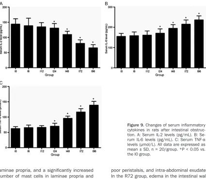
Figure 9. Changes of serum inflammatory
cytokines in rats after intestinal obstruc-tion. A: Serum IL-2 levels (pg/mL). B: Se-rum IL-6 levels (pg/mL). C: SeSe-rum TNF-α
in the I96 group (P < 0.05) (Figure 7A).
Changes in D-lactic acid levels
There were no differences in D-lactic acid le- vels between I0 and I6 groups (P > 0.05). Serum D-lactic acid levels began to slowly increase from 12 to 24 hours (P < 0.05) and began to elevate sharply from 48 hours (P < 0.05), elevating almost 6-folds by 96 hours (Figure 7B).
Biomedical indicators
ALT levels were similar in I0, I6, and I12 groups (P > 0.05), but were significantly increased from 24 hours (P < 0.05). Serum LDH started to sig-nificantly elevate from 24 hours (P < 0.05). Serum Cr levels did not change significantly at 6 and 12 hours but started to decrease from 24 hours (P < 0.05). These findings suggest that the rats began to suffer from liver dysfunc-tion and tissue injuries, as well as decreas- ed nutrition levels, after 24 hours of obstruc-tion (Figure 8). Results suggest that signs of MODS started to appear from 48 hours after obstruction.
Serum inflammatory cytokines
Serum IL-6 and TNF-α levels were gradually increased with prolonged obstruction duration, starting from 24 hours (P < 0.05). On the other hand, IL-2 levels were stably decreased after 24 hours (P < 0.05) (Figure 9).
Discussion
There is a lack of proper rat models of intestinal obstruction. Therefore, the present study aimed to establish an animal model of reversible mechanical intestinal obstruction to observe alterations of intestinal function at different
atraumatic intestinal clamp was the key for ensuring the success rate of the model. Tra- ditionally, simple complete intestinal obstruc-tion models have used silk threads to block the intestines [8], but this method is difficult to con-trol and may injure the intestine wall. In the present study, the model was minimally trau-matic to the intestine wall.
In the present study, intestinal pathology after 48 hours of obstruction showed significant damage and the number of activated mast cells in the lamina propria, as well as submu-cosa, was significantly increased. This was in agreement with previous studies [12, 13]. This was likely caused by the increased permeabi- lity of intestinal mucosa after intestinal obs- truction. Histopathological examination reveal- ed a large amount of activated mast cells in the lamina propria and submucosa. Indeed, the intestinal flora can then migrate from its origi-nal location to the deep intestiorigi-nal mucosa, perhaps migrating through lymphatic and vas-cular pathways [13, 14]. Increased intestinal permeability might lead to increased systemic inflammation.
Serum carbamyl ornithine mainly originates from the epithelial cells of the small intestinal mucosa and reflects intestinal function [15]. Some authors have used carbamyl ornithine levels to assess quantitatively chemotherapy-induced intestinal mucosal damage [16, 17]. In the present study, serum carbamyl ornithi- ne levels decreased with increasing duration of intestinal obstruction, reflecting intestinal dysfunction.
shock, intestinal ischemia, intestinal obstruc-tion, and other intestinal injuries, this may lead to intestinal mucosal injury and increased intestinal mucosal permeability, leading to increasing serum D-lactic acid levels [18, 19]. These levels may reflect injury to intestinal mucosa and change of intestinal permeability. In this study, serum D-lactic acid levels started to increase after 12 hours of obstruction, sup-porting the appearance of intestinal mucosa injuries.
Signs of SIRS and MODS began to appear about 48 hours after intestinal obstruction, as mani-fested by significantly decreased serum creati-nine levels, elevated ALT levels, and increased LDH levels. These changes are direct conse-quences of sustained intestinal ischemic injury and systemic inflammation, as revealed by decreased IL-2 levels and increased IL-6 and TNF-α levels. These changes are suggestive of SIRS and MODS [20].
This method can be used to successfully es- tablish an animal model of reversible comple- te mechanical intestinal obstruction. Animals developed MODS after 48 hours of obstruction, making this model appropriate for studying intestinal dysfunction associated with intesti-nal obstruction.
Acknowledgements
This work was supported by the Tianjin Tra- ditional Chinese Medicine Administration of Traditional Chinese Medicine Integrated Tra- ditional and Western Medicine Research Fund (13041).
Disclosure of conflict of interest
None.
Address correspondence to: Nan Zhang, Depart- ment of Gastrointestinal Surgery, Tianjin Nankai Hospital, Tianjin 300100, China. Tel: +86-22-2743- 5252; Fax: +86-22-27435252; E-mail: znndyx021@ 163.com
References
[1] Zinner MJ and Ashley SW. Maingot’s abdomi -nal operations. 12th edition. New York: Mc-Graw Hill Medical; 2013.
[2] Zhou ZL. Chinese west medicine journal of gastroenterology. 1st edition. Wuhan: Hua-
zhong University of Science and Technology Press; 2009.
[3] Tito WA and Sarr MG. Intestinal obstruction. In: Zuidema GD, editor. Surgery of the alimentary tract. Philadelphia: WB Saunders; 1996. pp. 375-416.
[4] Yuan ML, Yang Z, Li YC, Shi LL, Guo JL, Huang
YQ, Kang X, Cheng JJ, Chen Y, Yu T, Cao DQ, Pang H and Zhang X. Comparison of different
methods of intestinal obstruction in a rat mod-el. World J Gastroenterol 2013; 19: 692-705. [5] Chang IY, Glasgow NJ, Takayama I, Horiguchi K,
Sanders KM and Ward SM. Loss of interstitial
cells of Cajal and development of electrical dysfunction in murine small bowel obstruction. J Physiol 2001; 536: 555-568.
[6] Mulvihill SJ, Pappas TN, Fonkalsrud EW and Debas HT. The effect of somatostatin on ex-perimental intestinal obstruction. Ann Surg 1988; 207: 169-173.
[7] Collins J 3rd, Vicente Y, Georgeson K and Kelly
D. Partial intestinal obstruction induces sub-stantial mucosal proliferation in the pig. J Pedi-atr Surg 1996; 31: 415-419.
[8] Moore BA, Manthey CL, Johnson DL and Bauer AJ. Matrix metalloproteinase-9 inhibition
re-duces inflammation and improves motility in
murine models of postoperative ileus. Gastro-enterology 2011; 141: 1283-92, 1292, e1-4. [9] Chiu CJ, McArdle AH, Brown R, Scott HJ and
Gurd FN. Intestinal mucosal lesion in low-flow
states. I. A morphological, hemodynamic, and metabolic reappraisal. Arch Surg 1970; 101: 478-483.
[10] Sprung CL and Dellinger RP. Systemic inflam -matory response syndrome criteria for severe sepsis. N Engl J Med 2015; 373: 880.
[11] Dellinger RP, Levy MM, Rhodes A, Annane D,
Gerlach H, Opal SM, Sevransky JE, Sprung CL, Douglas IS, Jaeschke R, Osborn TM, Nunnally ME, Townsend SR, Reinhart K, Kleinpell RM,
Angus DC, Deutschman CS, Machado FR,
Ru-benfeld GD, Webb S, Beale RJ, Vincent JL and
Moreno R. Surviving sepsis campaign: interna-tional guidelines for management of severe sepsis and septic shock, 2012. Intensive Care Med 2013; 39: 165-228.
[12] Ghoshal UC. Is diarrhea-predominant irritable
bowel syndrome a low-grade gut inflammatory
disorder? Trop Gastroenterol 2012; 33: 239-241.
[13] Rana SV, Sharma S, Sinha SK, Parsad KK, Ma
-lik A and Singh K. Pro-inflammatory and anti-inflammatory cytokine response in
diarrhoea-predominant irritable bowel syndrome pa- tients. Trop Gastroenterol 2012; 33: 251-256. [14] Liu D, Guo Y, Wang Z and Yuan J. Exogenous
lysozyme influences clostridium perfringens
Kim YS and Park YK. Inhibitory effects of the