UNIVERSITY OF SOUIHAMPIW
Studies on the relationship between mitochondrial structure and functioning in Amoeba proteus.
Thesis submitted for the degree of Doctor of Philosophy by Robert A. Smith.
X X
TABLE OF CDNTEMTS Page.
Title page i
Table of contents ii
Abstract v
Acknowledgements vi
Abreviations and nomenclature vii
Diagram of mitochondrial characters viii
1. GHMERAL INTRODUCTim
1.1. Basic structure and chemical composition 1 1.2. Mitochondrial size and numbers 2
1^3. Mitochondrial autonomy 4
1.4. Energy-production by mitochondria 5 1.5. Structural changes correlated to function
'in vitro'. 7
1.6. Mitochondrial changes observed 'in vivo' 9 1.7. Different structural forms existing
simultan-eously within cells and tissues. 11
2. MATm^IALS AND METHODS
2.1. Culturing 13
2.2. Cell treatments 14
2.3. Cell synchronisation 15
2.4. Anaerobic culturing 16
2.5. Micrurgical operations 19
2.6. Preparation of cells for the Electron
micro-scope 20
2.7. Cytochemical method for demonstrating
cyto-chrome oxidase 23
2.8. E.M. autoradiography 25
2.9. Collection of data from the E.M. 27
STRUCTURAL CHARACTERISTICS OF MITOCHCNDRIAL TYPES IN A. PROTEUS AND THEIR PRESENCE THROUGHOUT THE CELL CYCLE
3.1. Introduction 29
X X X
3.2.1. Gross shape 33
3.2.2. y^pearance of cristae 35
3.3. Relative proportions throughout the cell cycle 37
3.4. Discussion 38
4. INFLUENCE OF GROWTH TEMPERATURE AND STARVATIOM CM THE MITOCHONDRIAL TYPES
4.1. Introduction 46
4.2ol. Effect of temperature on the cell cycle 49 4.2.2.1. Ultrastructural changes associated
with growth temperature 52 4.2.2.2. Reversion of temperatures to 20°C 59 4.3.1. Effect of starvation on ultrastructure 60 4.3.2. Observations on refed cells 66
Discussion 68
5. ACTION OF SUBSTANCES WHICH UNCOUPLE OXIDATIVE PHOSPHORYLATION
5.1. Introduction 74
5.2. Effects of uncouplers on cell morphology and
viability 77
5.2.1. Effects of DNP 80
5.2.2. Effects of PCP 88
5.2.3. Effects of CCCP 88
5.3. Ultrastructural changes associated with
un-couplers 89
5.3.1. DNP treatments 89
5.3.2. PCP treatments 96
5.3.3. CCCP treatments 97
5.3.3.2. Reversibility of structural changes 101
5.4. Discussion 101
6. EFFECTS OF FACTORS ACTING CN ELECTRONf TRANSPORT
6.1. Introduction 109
6.1.1. Anaerobiosis 109
6.1.2. Metabolic inhibitors 111
6.2.1. Effects of anaerobiosis on the whole cell 112 6.2.2. Effects of anaerobiosis on mitochondria 115 6.3. Exposure to KCN
6.3.1. Viability studies 123
IV
6.4. Exposure to Antimycin A 129
6.5. Discussion 129
7 . THE USE OF CYTOQjBVIISTRY, AUTORADIOGRAPHY AND MICRURGY IN STUDYING MITOCHONDRIAL TYPES
7.1. DAB cytochemistry to demonstrate CO activity 134
7.I0I. Introduction 134
7.1.2.1. DAB staining of untreated cells 137 7.1.2.2. DAB staining of anaerobic cells 141 7.1.2.3. DAB staining of MNU-treated cells 141 7.2. Studies involving mitochondrial DNA
7.2.1. Introduction 141
7.2.2. Incorporation of labelled thymidine
by A. proteus 143
7.3. Micrurgical investigations 149
7.3.1. Introduction 149
7.3.2. Observations on cells injected with
nucleotides, phosphate and succinate 150 7.4. Discussion of the results of the three es^eri 154
-mental approaches.
8. GENERAL DISCUSSION AND C m C L U S i m S 160
UNIVERSITY- OF Abstract
FACULTY OF SCimCE-BIOLOGY
Doctor of Philosophy
'Studies on the relationship between mitochondrial structure and functioning in Amoeba proteus.'
By Robert Anthony Smith.
The mitochondrial polymorphism existing within A. proteus upon aldehyde fixation was investigated using electron microscopy, related cytochemical techniques and autoradiography. Its significance was ascertained in relation to whole cell activity.
Type I, II and Int configurations were quantified by analysis of overall shape, cristal form and matrix density. Structural changes occurred following treatments which affected mitochondrial functiining and the work aimed to relate these with altered function. The relative proportions of the different mitochondrial types varied in different aged cells and could be experimentally changed when cells were cultured under stress conditions of lowered and raised growth temperatures and prolonged starvation. Control levels were again approached once the adverse culturing was terminated.
Hydrogen or nitrogen anaerobic culturing caused the elimination of Type I forms, with the generation of vacuolated Type II—Int organelles. Identical cristal vacuolation was observed in potassium cyanide-treated amoebae. On re-aeration Type I forms returned; the reappearance correlated with a reversion to normal whole cell activities.
longed anaerobiosis induced cristal degradation and a concommitont
appearance of matrical filamentous inclusions. Similar inclusion)resulted after exposure to metabolic inhibitors (KCN) emd uncoupling agents (DNP
and PCP); these were considered indicative of impaired respiratory functionir The rapidity with which uncouplers and metabolic inhibitors induced
structural changes supported the conclusion that mitochondrial structure was associated with organelle metabolic functioning. For CCCP treatment,
structural changes were shown to be completely reversible.
Initial results from microinjection experiments using nucleotides and substrate reinforced the conclusions that structural forms represented different organelle functional states.
VI
Acknowledgement s
I would like to express my gratitude to Dr. M.J. Ord and Dr. L.G,E. Bell for all help, guidance and time so readily given to me throughout this project.
I also appreciate the assistance of the other members of the Biology Department, especially Dr. I. Minassian and Mr. T.H. Courtenay for teaching me the intricacies of electron microscopy.
Thanks are extended to the staff of the M.R.C. Toxicology Unit, Carshalton; particularly Mr. V.H. Parker for the gift of recrystal— ised pentachorophenol and chloro-carbonyl cyanide phenylhydrazone and, with Dr. W.N. Aldridge and Mr. B.W. Street, for all helpful assistance. Financial support from a Medical Research Council Trainee Studentship is gratefully acknowledged.
The sympathy and comfort of many friends, particularly Agatha, Martyn, Ross and Eric, has been invaluable during the long months of writing this manuscript; as has Julie's efficient typing.
V I Z
ABBREVIATIONS and NOVIENCLATURE 1. Cellular Structures
CV - Contractile vacuole ch - cytoplasmic helix DB - DNA—containing body ER - Endoplasmic reticulum
f - cytoplasmic filamentous bundle GB — Golgi body
HSB — Heavy spheroid body - Type I mitochondrion - Type Int mitochondrion - Type II mitochondrion
mf - matrical filaments within the mitochondrion N — Nucleus
nh - Nuclear helix V - Vacuole
2.
ChemicalsADP Adenosine diphosphate ATP Adenosine triphosphate Ant A - Antimycin A
CCCP mChloro carbonyl cyanide phenylhydrazone DNP 2, 4 dinitrophenol
DAB Diaminobenzidine KCN Potassium cyanide PCP Pentachlorophenol
mit DNA - Mitochondrial deoxyribose nucleic acid
V X X l
Diagramatic representation to show the mitochondrial forms of Amoeba proteus and to demonstrate the characters used in their classification.
A
Type I. Type II.
(a.) The length:width (L:W) ratio or index, (b.) The width and complexity of the cristae, (c.) The density of the matrix.
- 1
-Chapter One General Introduction
Over recent years there has been a growing interest in relating the structure of the various cellular components to the different functions which they perform. Too often in the past isolated mitochondria have been studied by biochemists from a functional viewpoint with little regard to their morphology, so that it was difficult to perceive the reaction of the organelle within the living cell. Similarly the early ultrastructural
studies tended to be divorced from any functional approach, changes in mitochondrial morphology being recorded but with no corresponding functional data,. Three years ago, however, two important reviews of the knowledge of mitochondria were published bringing together the
literature
extensive on mitochondrial structure with the equally voluminous reports of functional properties (Lloyd, 1974; Munn, 1974).
In considering together the relationship between mitochondrial structure and function 'in situ' a system conducive to experimental manipulation is sought. Such a cell model is found with Amoeba proteus; the advantages of which have already been well argued
(Ord, 1970). Amoebae offer a means of studying cell components at particular regions of the cell cycle, with controlled culture
conditions and under various chemical and physical treatments. The relatively large size of the cell permits micrurgical operations. In addition modern fixatives provide good preservation of the cell, allowing BVI investigations to ascertain any changes at the ultra-structural level. With particular reference to mitochondria, A, proteus seemed worthy of further study as at least two distinct mitochondrial forms have been observed within the control cells
(Flickinger, 1968a). It was the intention of this project therefore to extend the amoeba model in discerning what such mitochondrial differences were reflecting in the cell, so that similar changes form resulting from chemical treatments might be correlated to any alterations in cell activity and functioning.
!• Basic Structure and Chemical Composition
- 2
-membrane separated by the peripheral space. The internal -membrane is thrown into a series of foldings constituting the cristae, and in general the mitochondria of cells requiring the greatest sustained demands for energy transduction tend to have the largest cristal membrane surface. The inner membrane enclose^ the matrix, a semi-fluid state, where the enzymes of the tricarboxylic acid cycle; fatty acid oxidation; mitochondrial protein and nucleic acid syn-thesising enzymes are restricted.
Water constitutes the major component of mitochondria, while the bulk of the dry mass is represented by protein and lipid. Nucleo-tides and nucleic acids are also inportant factors. Most of the mitochondrial protein is enzymic in nature, or functions in asso-ciated carrier molecules such as the cytochromes, with the possible exception of a structural or core protein. Over 900 enzymes have been listed; the oxidoreductases, transferases and hydrolases accounting for approximately 75%; with lysases, isomerases and ligases also of inportance. Some enzymes, such as cytochrome oxidase, have their sole cell localisation in the mitochondria while others e.g. isocit-rate dehydrogenase, may exist as isoenzymes with one form mitochondrial whereas the other is present in the cytosol. Many of the enzymes
are restricted in functional complexes i.e. succinic dehydrogenase. These coup 1exes, involved in oxidative phosphorylation, are found bound to the inner membrane (Schi^aitman and Greenwalt, 1968), and it is considered that the position of these with respect to each other is of importance in energy transduction (Racker, 1970).
In beef heart mitochondria 26% of the dry mass is made up of lipid uniformly distributed throughout the membranes. Over 90% of the total lipid content is of phospholipids. Lipids serve as a permeability barrier preventing the unrestricted flow of solutes and aiding the movement of substrates across the membrane. Lipids are further required to maintain a suitable orientation of the vari-ous haem proteins functional in oxidative phosphorylation (Green and
3
-.lengths of up to 3|J-. Reports of larger mitochondria were known:
mitochondria in the large amoeba Chaos chaos reached 8[i (Torch, 1955); in cells of ctenophore comb-plates profiles up to 8|i were seen
(Horridge, 1964); 7p- organelles were present in human uterus mucosa cells (Merker et al., 1968); while in HeLa cells organelles of up to SOjJ, with diameters of 15jJ- have been recorded (Posakony et al., 1975). Giant profiles were generated in hepatocytes by the copper chelating agents Cuprizone and diethylthiocarbamate (Susuki, 1969; Asano and Wakabayashi, 1974), from which respiratory measurements have been made on single organelles (Succhy and Cooper, 1974). Alter-ations in nutritional and culture conditions may also produce
enlarged profiles (Vartapetian et al., 1977).
Interest in recent years has focused on the construction of 3-dimensional models of mitochondria from serial sections. Such techniques have suggested that the numbers of organelles in certain cells is most probably lower than had previously been assumed from thin sectioning. In one strain of yeast a single mitochondrion was reported (Hoffman and Avers, 1973), although others suggested that diploid yeasts may contain over a hundred organelles (Grimes et al., 1974). A single organelle was constructed from serial thick sections of trypanosomes (Paulin, 1975), and below ten mitochondria/cell
were estimated in the flagellate Polytoma agilis (Burton and Moore, 1974). The related species, P. papillatum, possessed one large chondriome prior to division but the two new daughter cells had a content of many small mitochondria (Gaffal and Kreutzer, 1977). Similar findings have been reported in Euqlena gracilis where up to forty mitochondria are present during division but as the cell neared the end of its cell cycle less than ten giant forms were seen, indicative of organelle fusion (Calvayrac et al., 1972; 1974; Osafune, 1973).
4
-demonstrated (Posakony et al., 1975). With the alga Gonyostomum semen numerous separate organelles were indicated and it was proposed that the number of mitochondria may be related to cell volume, so that larger cells contain more mitochondria (Heywood, 1977). This would indeed seem to be the case for the mitochondrial cloud in Xenopus oocytes, where estimates of between 10,000 to 120,000
organelles have been proposed depending on the size of the egg cell (Billett and Adam, 1976). These workers however did not exclude the possibility that not all the organelles in the 'spaghetti-like' cloud were of a distinct nature.
In the large amoeba Chaos chaos estimates of up to 300,000
mitochondria have been put forward (Andresen, 1956) these being within the normal size range of (Pappas, 1959). Due to the relatively large size of A. proteus, 1.5 x lO^ |i3 (Ord, 1968a) many separate mitochondria could be anticipated in A. proteus also.
1.3. Mitochondrial autonomy
Mitochondria appear to have a more independent existence within cells than other cytoplasmic organelles. By the use of enucleation experiments on amoebae, it was found that the mitochondria, although somewhat swollen after the removal of the cell's nucleus for five days, still retained functional integrity (Brachet, 1958).
Mito-chondrial profiles are still evident in enucleate halves of A. proteus after seven days, while Golgi complexes and endoplasmic reticulum have disappeared; the mitochondria begin to degenerate after this period (Flickinger, 1968b). This suggests some final control of
their structure must originate from the nucleus for ultimate survival. Enucleation of Thecamoeba has shown that mit DNA may replicate
and be transcribed without the presence of a cell nucleus (Perasso, 1973), but for complete mitochondrial biogenesis mRNA of a nuclear origin is required (Soslav and Nass, 1971) as mit DNA has a limited information content (Andre, 1971).
Mitochondria are known to contain mitoribosomes and tRNA's, these ribosomes functionally resemble bacterial rather than
— 5 —
for the evolutionary transition of a free-living bacterial particle to a promitochondrial structure, including models based on prokaryotes such as Paracoccus (John and Whatley, 1975). The opposing
prop-osition for the origin of mitochondria is that they arose as episomes which became bound by membrane structures (Raff and Mahler, 1972). Although the evolutionary origin of mitochondria is an interesting academic question, it is not within the scope of the present work.
1.4. The energy-production by mitochondria
The prime function of mitochondria is in the conservation of the redox energy generated during the oxidation of the food stuffs taken in by the cell, and its subsequent transformation into the high energy compound adenosine triphosphate (ATP)(Lardy and Ferguson, 1969; Van Dam and Meyer, 1971). The pairs of electrons derived from the intermediates of the TCA cycle flow down the successively lower energy levels of the multi- membered chain of electron-carrier enzymes, until they reduce the ultimate electron acceptor of
respiration: molecular oxygen (Fig. 1.1).
Figure 1.1 : Diagramatic representation of the mitochondrial electron transport chain
Pyruvate
FP,
Succinate 'fp. Malate
Isocitrate \ Glutamater—» NAD,
Site I Site II Site III
3-Hydroxyadyl Co A
Fatty acyl Co A
Glycerol Phosphate FP„ S
CoQ—^Cyt b _ ^ C y t ^ cyt c __^Cyt Or
U
a + a.
§
H
ATP is formed at Sites I, II, III.
Energy from the electron flow is conserved as PO^-bond energy in coupled mitochondria by the phosphorylation of ADP to ATP upon the ejqpulsion of water.
The molecular mechanisms of oxidative phosphorylation are still not fully understood although all mechanisms so far envisaged state that the energy made available by the passage of reducing equivalents through the respiratory chain is primarily conserved in a form other than ATP. What-ever this primary form is, it is
considered capable of driving a number of endergonic processes such as ATP synthesis, ion transport, and the energy-linked transhydrogena-tion between NAD and NADP. ATP synthesis and ion transport are
reversible, i.e. the flow of reducing equivalents can cause ATP synthe-sis but ATP hydrolysynthe-sis can also lead to a reversed flow of reducing equivalents.
Although numerous schemes have been proposed to account for the coupling between molecular respiration and phosphorylation, only three are worthy of serious consideration:
i) the chemical intermediate hypothesis (Slater, 1953); ii) the chemiosmotic hypothesis (Mitchell, 1961; 1966); and
iii) a conformational theory (Green and Harris, 1969; Green, 1974). The chemical intermediate theory proposed that electron transfer results in the formation of energy-rich intermediate compounds
prior to the actual formation of ATP. This early explanation gained much support despite the fact that the postulated intermediates could not be identified.
7
-(Slater, 1967) reactions to this scheme for oxidative phosphorylation have been presented.
Instead of a chemical intermediate or a membrane potential, conformational theories of energy transduction consider that the free chemical energy of oxidoreduction can be stored and manipulated through confromational strains of the enzyme complexes (Green, 1974). The release from these energised strains results in phosphorylation.
A mechanism encompassing all three main theories has been put forward (Ji, 1976); and doubtless others will be conceived before the precise events have been resolved. The most recent review of the mechanics of oxidative phosphorylation now accepts the chemiosmotic hypothesis as the basis for energy transduction (Boyer et al., 1977). This multiauthored review even contains a contribution from Slater who concedes Mitchell's theory to be correct. The final stages of the ATP synthesis reactions are still however, thought to involve other processes of possible conformational changes or chemical intermediates
(See the Boyer and the Ernster sections of Boyer et al, 1977). The recent findings from the laboratory of Griffiths also question whether the chemiosmotic theory satisfies all aspects of energy transduction. The observation that inhibition by antimycin A or rotenone does not stop net ATP synthesis by heart particles if dihydrolipoate is present has been taken as an indication that the terminal reactions of oxidative phosphorylation do indeed involve chemical intermediates (Griffiths, 1976; Griffiths & Hyams, 1977). Studies with a yeast mutant in addition to those on heart particles suggest that lipoic acid may act as an inportant cofactor or chemical intermediate (Griffiths et al., 1977). The whole question is opened to debate once more.
1,5. Structural changes correlated to function in 'in vitro' studies Although the molecular mechanism involved in energy transduction remains unknown, certain workers have sought to relate reproducible configurational modifications of the inner mitochondrial membrane to the organelle's metabolic and respiratory state both in 'in vivo' and 'in vitro' studies (Williams et al., 1970). When phlorizin was used to block energy metabolism, isolated kidney mitochondria were
- 8
-ultrastructural transformations of the inner membrane have been demonstrated (Hackenbrock 1966, 1968; Penniston et al., 1969; Muscatello et al., 1972 a and b); these have been considered with respect to the five respiratory states defined earlier on the basis of oxygen consumption and light scattering measurements (Chance and Williams, 1956).
Slight inconsistencies exist between the results from the quick-sanpling technique used for transmission EM (Hackenbrock, 1966; 1968) and those based on negative staining preparations which had the advantage of overcoming the need for fixation (Muscatello et al., 1972a). In general however oscillations between a condensed or contracted and expanded or orthodox configuration were reported by both sets of workers.
In respiratory State I, when both substrate and ADP concentra-tions were low, oxygen consuiiption was also very low. The negatively stained mitochondria appeared expanded. If ADP was added to these preparations but the substrate concentration remained rate limiting, oxygen consumption remained low, but State II mitochondria assumed a condensed form. When substrate level was high in addition to the ADP, State III respiration was stimulated, in which case the mitochon-ria were as condensed forms. Both groups of workers obtained State Illfrom State IV preparations by the addition of ADP. State IV was characterised by high substrate levels but with ADP as the limiting factor, the mitochondria in this instance being of an expanded type. Mitochondria returned to this expanded form once the ADP was
_ 9
-Table 1.1 : Mitochondrial structural states 'in vitro' related to incubation conditions Respiratory State Substrate Level ADP Level Oxygen Level Mitochondrial appearance
I Low Low > 0 Expanded
II Low High
> o
CondensedIII High High 7 O Very condensed
IV LovJ > 0 Expanded
V Adequate Adequate Zero Disputed as to whether expanded or condensed.
Studies of beef heart mitochondria, using competitive binding by inhibitors, indicated that the condensed configuration obtained in respiratory states II and IIIof the investigations of the
Hackenbrock and Muscatello groups, resulted primarily from the binding and translocation of ADP (Weber, 1972; Scherer and
Klingenberg, 1974). Ec^i-lier work had also considered the importance of ADP in regulating structural changes due to oxidative phosphory-lation (Packer, 1960). Observations on isolated inner membrane particles, mitoplasts, prepared by critical-point drying have continued to s+%ow the importance of actual oxidative activity in inducing ultrastructural transformations (Andrews and
HLackenbrock, 1975).
Evidence exists that the mitochondrial configurations become altered as a consequence of the water movements following the osmotic gradients set up by energy-dependent ion transport and accumulation by the organelle (Azzi and Azzone, 1966; Packer et al., 1968; Rottenberg and Solomon, 1969,Schmidt et al., 1977). An expanded orthodox form was generated by the uptake of calcium in the presence of phosphate, though it was claimed that the osmotic transformation in this case differed from the orthodox form resulting from respirat-ion (Hackenbrock and Caplan, 1969; Hackenbrock, 1972). Ionic
regulation and accumulation by mitochondria is certainly an important function of the organelle within the cell (Carafoli, 1975; Rose and Lowenstein, 1975).
1.6. Mitochondrial structural changes observed in 'in vivo' work
- 10
-occur 'in vitro' result from different oxidative phosphorylation states and the nucleotide binding associated with this function, or from energy-consuming events such as ion accumulation; mitochon-drial structure is known to undergo alterations in many
'in vivo' systems when functional activity changes within the whole cell.
When rat liver tissue was incubated in the presence of succinate (Haydon et al., 1967) and intestinal strips in the presence of an amino acid mixture (Jasper and Bronk, 1968), treatments which poss-ibly result in increased quantities of electrons passing down the electron transport chain, increased numbers of condensed types of
mitochondria were recorded. Both these findings are consistent with the hypothesis of structural alterations beingadirect consequence of
respiratory functioning.
Recent work correlating mitochondrial structure with the ADP levels in unfertilised and fertilised sea urchin eggs has shown that for the transformation from a condensed to expanded form, which occurs at fertilisation, there must be a decrease in the ADP con-centration (Innis et al., 1976). These authors were unable to state whether an increase in oxidative phosphorylation paralleled this event, although oxygen consumption was raised at fertilisation.
When cells are treated with uncoupling agents, mitochondria have been reported to undergo a contraction in form (Buffa et al., 1968). • This is again suggestive of ADP levels and respiratory activity playing an iirportant role in regulating mitochondrial structure.
- 1 1
-During malignization of thyroid gland cells, there occurred a sharp decrease in the numbers of cristae within profiles and the matrix became more translucent (Dmitrieva, 1968). Mitochondria with a lighter matrix and of a swollen nature were generated in cells treated with fluoroacetate to decrease ATP levels (Buffa et al., 1977). This again supports the idea that altering cell and organelle activity alters mitochondrial form, i.e.
fluoro-acetate binds to enzyme complexes of the TCA cycle within the matrix. In the differentiation of the trypanosome T. brucei, the
mitochondria undergo alterations which have been correlated with the organism acquiring the ability to oxidase certain substrates (Brown et al., 1973). Variations associated with culture age are also reported for Tetrahymena pyriformis (Elliot and Bak, 1964). The ultrastructure of the mitochondria of Paramecium aurelia differ depending on whether axenic or monoxenic culturing has been employed
(Prince, 1976). In this case a contracted and expanded form were reported.
1.7. Different ultrastructural forms existing simultaneously within cells and tissues
Within certain tissues and cells, different mitochondrial types have been reported to exist simultaneously. In some instances this difference rests mainly on gross shape but in others distinctions occur in the matrix density and cristal organisation. The latter
examples are of greater interest in a study such as this one attempting to relate form to function.
In the femoral muscle of the cockroach three morphologically distinct mitochondrial forms were reported (Hagopian, 1967).
Elongated forms were found wedged between the myofibrils; oval types associated with the saroolemma; and Y-shaped profiles in
close proximity to the Z-disc. Similarly with canary heart muscle not all mitochondrial types within a cell were alike, the gross form and the arrangement of the cristae being considered to be a sign of metabolic function (Slautterback, 1965).
— 12 —
form with the capacity to respire.
The ciliate Euplotes minuta was found to contain both expanded and denser contracted mitochondria within individual organisms
(Jurand and Lipps, 1973). As with the earlier reports of at least two forms in A. proteus i.e. with matrix density and shape distinc-tions (Flickinger, 1968a), these authors demonstrated that this observation did not result from a fixation artefact, and speculated that the types might be in different physiological states or could perhaps be representing a juvenile and a mature form. The possibility of there being two different unrelated populations of organelles
within the cell was also suggested.
The early light microscope work on amoebae demonstrated differences in mitochondrial structure in that elongated and
spherical forms were evident (Torch, 1955). At the EM level using osmium tetroxide fixation gross shape was also used to distinguish different types of mitochondria in A. proteus (Mercer, 1959), although the internal differences in the complexity of the cristal organisation and matrical density were not realised until the double aldehyde fixative was employed (Flickinger, 1968a). Morph-ologically different mitochondria were also apparent in other species of Amoeba (Flickinger, 1974).
The aim of the present work was to consider whether any signifi-cance could be attached to the existence of the different mitochondrial types in A. proteus.
Variations in mitochondrial ultrastructure have been shown to arise in other amoebae during certain conditions, for example with Chaos chaos a complex 'zigzag' configuration has been noted during starvation (Pappas, 1959; Daniels and Breyer, 1968) which is also present during the mitotic stages of this amoeba (Daniels and Breyer, 1965). In view of these changes observed in other species related to alterations in cell and organelle function, experimental design
13
-Chapter Two Materials and Methods
Cultures of Amoebae proteus used in the major part of this work were raised from cells of the strain originally supplied by Dr. M.J. Ord. Additional cultures of A. proteus and A. discoides D13 were kindly donated by Dr. S.J. Hawkins of Kings College, London in March 1977.
2.1. Culturing
2.1.1. Culture Medium
Cells were maintained by either wheat culturing or as Tetrahymena-fed cultures, following the basic methods of Griffin (1960). Culturing and handling of cells were- carried out in a modified Chalkley's medium
(Chalkley, 1930; Ord. 1970b); a concentrated stock solution of which contained the following salts in one litre of glass-distilled water:
16 gm. NaCl (1.37 X 10"^M) 0.8 gm. (4.76 X 1 0 " \ ) 0.4 gm. KCl (2.68 X 10~^M) 0.2 gm. NaHPO^I 2 HgO (2.79 X 10"^M) 0.2 gm. CaHPO (7.36 X 10"^M) 0.2 gm. MgClg (4.92 X 10~^M)
The medium used was prepared from 5 mis. of this stock solution made up to one litre with distilled water to give the final salt concentrations as shown above in parenthesis. The pH was adjusted to 5.9 - 6.0 with HCl before use.
2.1.2. Amoebae Stock Cultures
Stock cultures of amoebae were grown in plastic dishes
14
-Prior to use in any experimental procedure , the cells were removed from these stock cultures and maintained for at least 2 weeks by Tetrahymena-feedinq.
2.1.3. Tet rahymena-f eedinq
T. pyriformis of strains ST or W, cultured in 100 ml. medical flats in sterile 2% proteose peptone, were harvested while still in the log phase of growth (i.e. within five days of innoculating the peptone). A sintered glass funnel of porosity 4 was used to filter-wash the tetrahymena to remove the excess peptone and waste materials before resuspension in Chalkley's medium. In order to supply at least 20 tetrahymena/amoeba/day 1-2 mis of peptone-free tetrahymena were added daily to amoebae cultured in Chalkley's to a depth of 1 cm in 75 mm diameter. Such mass cultures of amoebae were transferred to clean dishes three times a week by a tipping method to remove all waste materials and any older food organisms so producing healthy dividing cultures for use.
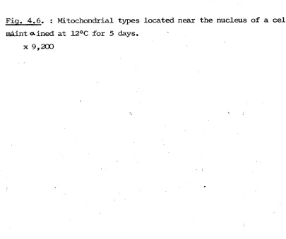
Normal cell culturing was controlled at 20 - 1°C. Where other growth temperatures were required amoebae were transferred to pre-set incubators or waterbaths for supra-room temperatures. The 10 and 12°C environments were ach^&ved by use of a small oven located within a 6°C cold-room. In all cases temperatures were monitored daily to ensure constancy.
2.2. Cell Treatments
The treatments of living cells with the various chemicals, incubation media and fixatives, used in the present study were ^f^®cted in solid watchglasses by the following standard procedure:
15
-incubation period might not be due so If.! ? to the treatment itself. Similarly wherever possible the test solution was withdrawn at the end of the exposure period and replaced with either normal Chalkley's or fixative with minimal disturbance to the cells.
Except in the case of the incubation media for cytochemical staining, the chemicals under consideration were prepared by the dilution of stock solutions with normal Chalkley's to the final concentrations required and adjusting the pH*
Where cell viability was to be studied after treatment, the amoebae were cultured singly. Two means of single culturing were enployed: either the watch glass technique generally used in Amoeba work, or the capillary tube method of Ord (1977).
In the watchglass technique cells are transferred by pipette to clean watchglasses three times a week and fed daily on tetrahymena. With the capillary method cells were transferred to clean sealed melting point capillary tubes (75 x 1mm) by a fine pipette. Dilute wheat food was introduced into the tubes to create a balanced system of Colpidia and Chilomonads. Such food was harvested from cultures two to three weeks old in order to maintain the correct proportions of the food organisms. The collected food was diluted with
Chalkley's medium prior to its introduction into the tubes with the pH at 5.9. The capillary tube method avoids excess handling of treated cells once set-up, reducing the possibility of mechanical damage on manipulation - a necessary consideration if the chemical or treatment has caused alteration of the fragility of the cell membrane in any way.
2.3. Cell Synchronisation
Where experimental design necessitated the uee of cells of a specific age, or when cells were to be starved, amoebae undergoing mitosis were selected from the mass cultures by use of a finely drawn-out pipette.
— 16 —
collected (- 20 minutes) and grouped together in watchglasses until use, so giving synchronous cell samples. It is considered that metabolic processes are less disturbed by this method of synchronisa-tion than in other techniques where tenperature, nutritive or
chemical shocks are involved (Mitchison, 1971).
2.4. Anaerobic Culturing
Anaerobic conditions were achieved in both hydrogen and nitrogen atmospheres by the use of two experimental set-ups.
1. Initially a microbial anaerobic culture jar (obtained from Baird and Tatlock) was used. This possessed a catalytic substance for use with an hydrogen atmosphere which functioned by reacting any traces of oxygen, remaining in the jar after the evacuation period, with the introduced hydrogen.
A redox indicator was included to check for any air leaks in the apparatus and to demonstrate that all detectable amounts of oxygen had been removed from the vessel prior to the introduction of the hydrogen or nitrogen. The indicator gel was made up as follows:
9% thioglycollic acid - 12 drops Phenol Red - 2 drops 2% Borax solution - 5 ml
Methylene Blue (9.5 mg/lOO ml) - 10 ml 2% melted agar - 10 ml
On boiling the above solution becomes colourless and was sealed in lengths of glass tubing until use in the apparatus. It is only in the presence of oxygen that the methylene blue is reoxidised resulting in the blue colouration, hence such a reaction in the anaerobic set-up indicated that evacuation had not been completed.
The commercial microbial culture jar was found to have two main disadvantages:
17
-(b) As the jar was constructed of stainless steel it was not possible to obsarvethe cells during the incubations. Thus changes in cell morphology and behaviour could not be followed.
2. In order to overcome these difficulties a small ejqDerimental arrangement was designed consisting of well-greased sections of Quickfit (Fig. 2.1). This apparatus allowed one to follow the cell's state by use of an inverted microscope. The smaller air space allowed a more satisfactory evacuation of the apparatus. It also permitted further replenishment with the substituted gas as the incubation proceeded so that a relatively greater amount of hydrogen or nitrogen could be passed through the apparatus. The redox indi-cator and the catalytic substance, for when hydrogen was used, were retained.
Cultures were set up as follows:
In the initial group of experiments cells were placed in the Quickfit culture dish and allowed to settle so that the Chalkley's medium could be withdrawn to a depth of not more than 5 mm. Ordin-ary aerated Chalkley's was included at a pH of 5.9. An attempt to reduce the quantities of dissolved oxygen from the medium by boiling for five minutes raised the pH to over 7.0, indicating that the ionic composition of the medium had been disturbed
This step was therefore not pursued. In later experiments Chalkley's, through which 95% nitrogen had been bubbled for one hour prior to use, was included. This precaution however was not considered crit-ical as the concentration of dissolved oxygen in the small quantity of medium should rapidly equilibrate with the gaseous phase above it and bfe removed on subsequent flushings with the oxygen-free atmosphere.
When a Quickfit culture dish was included in the apparatus the lid was firmly sealed to it by applying a layer of silicone greases. In later experiments the apparatus was further modified by permanently fixing the lid to a glass plate with Araldite, eliminating air leaks and improving cell visualisation. Amoebae were introduced into the chamber of this by pipette through inlet tube I,
1 8
-Fig 2.1. : The anaerobic-culturing set up.
— Inlet tap 1 to which the vacuum punp or replacement gas supply was fitted.
Tg — Tap 2.
I - Indicator gel. C - Catalyst
[image:26.513.11.501.15.671.2]— 19 —
commenced. The lead of a vacuum pump was attached to Tap 1 which then opened (all other taps being closed at this stage). After five minutes tap 1 was closed, the pump lead removed and the hydrogen or nitrogen source connected. Qn reopening Tap 1 the replacement gas was sucked into the apparatus. Tap 2 was then opened so that further gas could be passed through the system. This process of flushing hydrogen or nitrogen through the set-up was repeated five times in the first hour after the removal of the air; and then at periodic intervals throughout the rest of the incubation ensuring that the atmosphere remained oxygen-free.
2.5 Micrurqical Operations
These operations were kindly performed by Dr. M.J. Ord using a Fonbrune micromanipulator at mag. 200 x and the oil chamber tech-nique of Comandon and deFonbrune (1939). Glass needles, hooks and micro-pipettes were constructed on a microforge.
2.5.1. Nuclear Transfers
These involved the insertion of nuclei of DNP-treated cells into host control anucleated cytoplasm. The reciprocal transfer could not be effected due to the fragility of the treated cell membrane. Hybrid cells were cultured singly in watchglasses.
2.5.2. Microinjection
In addition to the Fonbrune manipulator, an Agla Microsyringe Outfit (Wellcome Reagents Ltd.), which can deliver volumes as small as 10 ul., was employed. The whole system was oil filled for increased sensitivity. An attempt to standardise the amount of a reagent in-jected into each amoeba was made in that, by eye, the amount of reagent delivered on each occasion occupied the same length of
- 20
-\
Figure 2.2 : The loading of the pipette tip for luicroinjection manipulations
< Standard length of the pipette ^ Plug of filled with injection fluid for
each operation i
2.6. Preparation of cells for the Electron Microscope
The principles involved in the fixation, dehydration and embedding stages necessary for specimen preparation for the
electron microscope have recently been clearly stated (Glauert, 1975). 2.6ol. Fixation
The primary fixative routinely used consisted of a glutaralde— hyde-formaldehyde mixture as proposed by Karnovsky (1965). This would seem to be a most suitable fixative as the formaldehyde is considered to penetrate the cells rapidly, initiating the cross-linkage of the cell's protein moieties; while the dialdehyde pene-trates less rapidly but subsequently stabilises and ramifies the protein cross-linking.
Williams et al. (1970) consider this fixative to be "eminently suitable for the preservation of mitochondrial configurational states present at the moment of fixation". The fixative was used as soon after preparation as possible and at -least within a week.
25 ml of fixative were prepared as follows:
1 gm of paraformaldehyde was heated to 70°C in 7 ml of distilled water. Single drops of IN NaOH were added until the white floccula-tions disappeared. This solution was allowed to cool before the addition of 5 ml of 25% ultrapure glutaraldehyde (EM scope). The
fixative was made up to 25 ml in a volumetric flask with 0.2M sodium cacody-late buffer. Extreme care should be followed in the preparation and
- 2 1
-The fixative possessed final concentrations of 4% formald-ehyde, 5% Glutaraldehyde and O.IM cacodylate. The pH was adjusted to 7.1 prior to use and normally stored at 4°C to retard the
degradation of the glutaraldehyde.
Cold unmodified fixative was added to the cells after the removal of as much Chalkley's or test solution from the amoebae as possible. The cells were left in the presence of the fixative for ^5—60 minutes. Cells were then washed briefly for 5 minutes with O, IM cacodylate buffer before post—fixation with 1% OsO^ buffered with O.IM cacodylate at pH 7.1 for one hour. Brevity of the washing stage is recommended to minimise any risk of lipid leeching (Busson—Mabillot, 1971) as the aldehyde fixatives do not stabilise these con^onents. Indeed some workers (Ockleford, 1975) dispense with all washing stages, applying the aldehyde and osmium simultaneously. The secondary fix-ative was also replaced by cacody^ate buffer prior to the cells being blocked in 2% agar for ease of further handling.
2.6.2. Agar blocking, dehydration and staining
The agar blocking of cells at this stage has many advantages in that the cells may be grouped together and oriented as desired to facilitate the sectioning and viewing of more than one cell on the E.M. at a time. It also reduces cell damage aind loss due to handling, for once fixed the cells become sticky and have a tendency to adhere to the pipette on transfer to subsequent fluids if not blocked.
Agar blocking has been used by several authors in fixation techniques for small cell populations (Kimball & Perdue, 1962; Stone & Cameron, 1964; Flickinger, 1965; Hirsch & Fedurko, 1968). In the procedure developed in this laboratory the disadvantages inherent in some of these methods have been eliminated. Many of these workers employed a centrifugation step to pellet the cells before blocking. This is considered undesirable as it introduces the possibility of
disrupting the internal organisation of the cell. The earlier techniques concentrated on trapping the cells in the middle of the agar block
whereas our method ensures the amoebae are on the surface of the block, thus facilitating penetration by the dehydrating and embedding solutions.
- 2 2
-amoebae to aid in sectioning, the cells were sometimes pipetted directly onto a glass surface rather than agar ( although this increases the risk of the cells drying out). Excess fluid was withdrawn by means of a finer pipette so that when a drop of molten agar was placed onto the cells from the end of a glass rod, they did not disperse from the group. Once the agar had set, the block was carefully cut out and excess agar removed by manipulation with the two needles.
Such blocks were dehydrated in a graded series of ethanol alcohols (20, 35, 50, 70, 90, 95 and two changes of absolute) for 10 minutes each except that they were left in either the 50% or 70% alcohol for staining purposes for one hour. Initially 3% uranyl acetate was prepared in 70% alcohol and millipore -filtered before use. Stain precipitate however was found to be reduced by using only 2% uranyl acetate in 50% alcohol and so this was adopted in the later stages of the work. Little differ-ence could be identified between the two stain concentrations. 2.6.3. Embedding
Wherever possible blocked cells were taken through the prep-aratory stages to Spurr resin (Spurr, 1969) on the same day that fixation had been performed. If cell treatments necessitated the cells being left overnight prior to this stage they were left in either 50 or 70% alcohol in preference to buffer or the lower
alcohols (Spector, 1975). This was also to minimise lipid extraction from the material (Korn & Weisman, 1966). Spurr resin is a low
viscosity epoxy resin and was prepared by thoroughly mixing the constituents together in a fume cupboard:
10 gm ERL 4206 (Vinyl cyclohexane dioxide)
6 gm DER 736 (diglycidyl ether of polypropylene glycol) 26 gm NSA (Nonenyl succinic anhydride)
0.4 gm SI (Dimethylaminoethanol)
Mixture was effected by means of a clean glass rod and the medium covered until use.
23
-Finally the blocks were lifted into, and suitably arranged in, plastic BEE'Ci capsules and filled with fresh Spurr resin after any of the previous resin had been removed by pipette. Polymerisat-ion was effected in a 70°C oven for two days.
2.6.3. Sectioning
Blocks were selected, trimmed and ultrathin sections with inter-ference colours of silver-gold (600-900 A) (Meek, 1970), were obtained on an LKB ultratome. Glass knives were constructed and used through-out the study.
Sections were stretched by xylene vapour while floating on the knife's watertrough and were mounted on grids. Normally 200^mesh copper grids were used, although nickel grids were also employed. Where serial sections were being studied, 2 x 1 mm copper grids covered with a film of Formavar were prepared and used. The grids were left to dry on a clean filter paper before storing in grid boxes until viewing.
Sections were examined on a Philips EM 3000 normally operated at 80kV.
2.7. Cytochemical method for the demonstration of cytochrome oxidase The DAB reaction has proved useful in the ultrastructural local-isation of peroxidative activities within many cells since its
introduction by GrahamfKarnovsky (1968). The following medium mod-ified from that of Spector (1975) was employed in the present study to demonstrate cytochrome oxidase activity in the mitochondria:
10 mg Na^PO^ (0.15M) buffer at pH 7.4 and containing 0.15M sucrose.
10 mg Diaminobenzidine hydro.chloride (DAB) (B.D.H.) 10 mg Cytochrome c Type IV. (Sigma)
lO Wg Catalase (Sigma)
This was shaken vigourously to dissolve the DAB. All incubation media were always made up freshly and kept away from direct light in order to minimise auto-oxidation of the DAB. The catalase was added to remove endogenous peroxide, while the cytochrome c
24
-(
oxidase activity, although the reaction has also been performed at lower pH»s as recommended by some (Opik, 1975). In an attempt
increase structural preservation of the material without the subsequent loss of enzyme activity, the concentration of sucrose was varied in many incubations(Litwin, 1975; Posakony et al., 1975).
Incubations were performed on both live and prefixed cells. The prefixation step, using formaldehyde solutions, was included to stabilise structural integrity of the amoebae and also to aid in the penetration of the DAB across the cell membrane.
In a number of experiments 5% dimethyl sulphoxide (DMSO) was added to the medium as a further means of increasing penetration. This was particularly necessary when unfixed cells were incubated in the medium as it was desiro-fele to obtain the shortest incubation time possible. DMSO, originally used as a cryogenic agent (Lovelock and Bishop, 1959), has more recently been developed in techniques requiring shortened incubation periods as it enhances permeability of membranes in animals, plants and micro-organisms (Reis, 1971; Ghajar & Harmon, 1968; Makita and Sandborn, 1971). DMSO is also beneficial in that it increases the solubility of the DAB in the incubation medium.
All incubations and subsequent rinses were performed at 20°C in a darkroom dimly illuminated by a yellow safety light. Even in these conditions a dark brown percipitate was occasionally noted in the incubations of more prolonged durations: presumably due to the auto-oxidation of the DAB. The incubation period extended from 20 minutes for some of the treatments of live cells to overnight for cells that had been given a relatively long period of prefixation.
Control incubations were included where either the cytochrome c and the catalase or the DAB were omitted from the complete medium. In other experiments KCN was added to the medium as an additional control.
After the required period of incubation the cells were
thoroughly washed for thirty minutes in two rinses of the phosphate/ sucrose buffer to remove any unreacted DAB. This is an iitportant consideration as the DAB itself will undergo osmication with the OsO^ and so must be removed to avoid erroneous results.
25
-been incubated, fixation with Karnovsky's fixative was carried out after the buffer rinses before the cells were handled further in the routine processes for E.M. Specimen preparation. Counter staining with uranyl acetate was not included.
2.8. E.M. Autoradiography 2.8.1. Labelling
Cells were sd.ected from mass cultures as division spheres and incubated at the required age by replacing the Chalkley*s medium with two drops of (CH*^)- Thymidine of specific activity 19 Ci/mM
(purchased from Radiochemicals Centre, Amersham) at a concentration of 0.5 mCi/ml.
After the desired exposure period the cells were washed in three rinses of Chalkley's medium prior to fixation and E.M. prep-aration as described in section 2.6, except that staining with uranyl acetate was not included.
2.8.2. Application of the photographic emulsion
A modification of the loop technique of Caro and Van Tubergen (1962) was followed (Kanobdee, 1975).
Prior to the application of the Ilford lA emulsion to the grids in the dark room, certain preparatory steps were performed. The sections from the ultratome were preferentially mounted on nickel grids, as in the initial experiments when copper grids had been used copper salts were deposited upon the sections in the final stages of the technique, making examination difficult. Two groups of five to ten grids were arranged on an alcohol-cleaned slide by means of securing the corner of each to a small strip of double sided
sello-tape. The grids were transferred through all subsequent stages attached to the slide in this manner.
The lA emulsion was transferred from the bottle to a clean beaker in the darkroom at a working distance of 4-5 feet from an amber safety light. The beaker was then placed in a water bath pre-heated to 45°C for 7-10 minutes, stirring occasionally. 10 ml
of the liquified emulsion was then pour c) into a measuring t cylinder and the volume made up to 25 ml with distilled water at the same
- 2 6
-introduction of excess air into the liquid. The dilute emulsion was transferred to a 32°C waterbath for five minutes to equili-brate and then left to cool at room temperature in a crystallizing dish.
A loop of silver wire was used which had been constructed of 0,6 mm diameter wire and had a loop diameter of approximately 4 cm
secured into the end of a glass pipette by means of dental wax. The loop was dipped into the emulsion and withdrawn to form a thin monolayer film of emulsion across it. The loop was held vertically so that the excess emulsion would drain to the bottom and could then be absorbed onto a piece of mediwipe. After approximately
30 seconds — 1 minute, intereference colours developed, beginning at the upper edge of the loop and moving downwards. When these had formed over the top half of the film, the loop was turned into the hor-izontal position and touched onto the surface of each slide containing grids.
The slides were dried vertically in a drying chamber before storage in a sealed black box containing crystals of silica gel as a drying agent. Exposure was at room temperature for 6 - 8 weeks. 2.8.3. Development
After the completion of the desired exposure period the slides were opened in the darkroom and transferred to Copland jars containing millipore-filtered D19 developer for 6 minutes at room temperature. This was followed by quickly rinsing the slides in three changes of distilled water prior to immersion in 20% sodium thiosulphate for lO minutes. This step dissolved away the non-exposed silver bromide crystals of the emulsion leaving only the reduced silver grains in position.
The grids were given three rinses in distilled water for 10 minutes before proceeding to the next stage.
2.8.4. Gelatin removal
- 27
followed by one where the slides were placed in 0.5M acetic acid for 15-30 minutes so that the softened gel was digested. The acetic acid was washed away with distilled water rinses aund the slides
dried so that the grids would be removed from the sellotape and stained individually.
2.8.5. Section staining for E.M. autoradioqraphv
In view of the radioactive decay of uranium salts, cells for autoradiographic studies were not stained until after the development of the photographic emulsion. The procedure for the staining of these thin sections involved the use of Millonig's lead stain followed
by a saturated solution of urainyl acetate.
Millonig (1961) suggested that lead hydroxide could be
stabilised in solution by the addition of sodium—potassium tartrate. This reduced the levels of stain precipitate. For use with amoebae the best results have been obtained in this laboratory by the
following method:
A stock solution was prepared with 20 gm NaOH and 1 gm K-Na-tartrate made up to 50 ml with distilled water. One ml of this was added to 5 ml of a 20% lead acetate solution in water. The resulting mixture was diluted five times with distilled water, shaken and filtered to remove the white precipitate which left a
clear colourless solution stable at room temperature for several weeks. Storage was normally effected at 4°C.
Drops of the lead stain were pipetted onto a sheet of dental wax and the grids placed section-side downwards in these for 10 minutes. They were given a brief rinse in distilled water before transferring them to 7.5% uranyl acetate in 50% alcohol for 1 minute. The stained grids were washed in three rinses of water and dried in a covered petri dish ready for examination.
2.9. Collection of data from the Electron Microscope
- 2 8
-were distinguished by differences in the matrical density, cristal organisation and gross shape. Means of quantifying these were considered.
Due to the non-randomness of mitochondrial distribution in amoebae, a consideration of a morphometric study was not pursued. The mitochondria tend to be more numerous at the cell periphery, in association with the contractile vacuole, and at regions close to the nucleus; other regions of the cytoplasm may be devoid of the organelle. As such stereological techniques were not considered a profitable approach for the present work.
Foir control and treated amoebae, estimates of the relative propor-tions of Type I and Type II were attempted to study whether the rela-tive numbers of the two forms altered at different times in the cell cycle or under different experimental conditions. Such counts were routinely sampled directly from the microscope. Random areas of cells were scored and the counts from at least four separate sections for each cell pooled. Thus sampling error due to any non-random associa-tion of a particular mitochondrial type with specific cellular local-isations was lessened. All individual experimental samples included some cells in which areas close to the nucleus and the contractile vacuole were studied.
Normally data was talcen from 12—25 cells for each experimental treatment group to give the mean values presented in the text. For the different sections of the work, this gave large total sanples. Thus in considering the various types throughout the cell cycle, over 220 cells were quantified aind a total of over 30,000 profiles scored. Other cells were also viewed quantato.l-»VS,ly in each case.
29
-Chapter Three
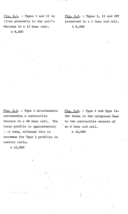
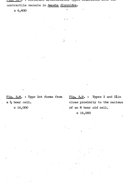
The structural characteristics of the mitochondrial types of A. proteus and their presence throughout the cell cycle.
3.1. Introduction
Early work indicated that A. prntpng contained a large number of small, discrete mitochondria (Mercer, 1959), which were concen-trated in the hyaline layer adjacent to the external membrane, and in association with the contractile vacuole (Mast ajid Doyle, 1935). As with similar work on Chaos (Torch, 1955; Andresen, 1956), this was considered to indicate that the organelles were located in areas where high levels of ATP were required. In other systems, in addition to distinct regions within the cell requiring high ATP levels, specific points in the cell cycle have been reported, such as mitosis and
during RNA and protein synthetic periods, where relatively greater energy demands existed (Amoore, 1963; ^ e l , 1963; Van»t Hof, 1966; Webster and Van't Pfof, 1969), The effect of such energetic differences on mitochondrial structure were not recorded.
The mitochondria of A. proteus are of interest because when ^^^'^o^sky's fixative, a glutaraldehyde—formaldehyde mixture, was
used, different morphological forms were ' (Flickinger, 1968a), These types were classified as light, dark and intermediate forms
depending on the density of the matrix and the orgsmisation of the cristae. In other systems when different mitochondrial forms were observed in separate cells, either in different tissues or activity phases, it was questioned whether such variations represented
genuine differences or not (due to the high osmplarity of aldehyde fixatives). This objection could not easily be applied to A. proteus where the coexistence of the two types in close proximity within the same cell indicates that the observation represents an actual chemical or physiological difference between the two types preserved by aldehyde fixation, rather than an artefact of fixation.
30
-vesicular structures in osmium—fixed cells, which had not been observed when an aldehyde prefixation had been carried out, needed clarifying. By fixing amoebae in sucrose-buffered osmium at the same osmotic strength as the Karnovsky's, Flickinger demonstrated that the osmolarity difference between Karnovsky's fixative and osmium tetroxide was not the crucial distinction between the fixation regimes. Since only one mitochondrial form was again preserved, as well as the vesicular structures, he concluded that the alde-hydes maintained a chemical difference which was not caused
primarily by osmotic factors. It was suggested that osmium tetroxide was a poor mitochondrial fixative and that the disrupted vesicf.. possibly represented the missing mitochondrial form. The control dark and light mitochondria have since been classed as Type I and Type II forms respectively in a report, where changes in mitochon-drial forms were recorded in A. proteus after treatment with certain carcinogens (Ord, 1976).
Tlie double fixation method of Karnovsky's fixative followed by postfixation with osmium tetroxide has been used throughout the major parts of this study. The nomenclature of Ord i.e. Type I and II, together with the introduction of Type Int to distinguish the trans itioi^3-l intermediate forms has been adopted. With the acceptance of the obvious dangers of attempting to draw inferences concerning the dynamic relationships within the living cell from the static sections viewed on the EM; the initial aim of this project was to clearly distinguish and categorise the various mitochondria preserved from healthy, untreated cells of A. proteus» Flickinger had studied the mitochondria in some detail, presenting measurements of 0.5-ip- diameter by a 2|1 long axis and mean cristal width of 660 A for the dark mitochondria; and estimates of 0.7-1.5(1 diameter and cristal
o