http://www.scirp.org/journal/fns ISSN Online: 2157-9458
ISSN Print: 2157-944X
DOI: 10.4236/fns.2018.94024 Apr. 26, 2018 314 Food and Nutrition Sciences
Physiological Functions of 70% Ethanol
Extracts of 6 Edible Flowers
in Vitro
: A
Comparative Study
Yukihiro Yamamoto
*, Masato Kono
Faculty of Life and Environmental Sciences, Prefectural University of Hiroshima, Hiroshima, Japan
Abstract
The physiological functions of 70% ethanol extracts of 6 edible flowers of dif-ferent colors—Chrysanthemum morifolium (Chry., yellow), Rosa miniature (Rosa, red), Dendrobium phalaenopsis (Dend., purple), Viola x wittrockiana cv. fluna (Fluna, white), Viola x wittrockiana cv. pansy (Pansy, orange), and Primula x juliana (Prim., blue)—were evaluated in vitro. Anti-oxidant activity was evaluated based on the DPPH radical scavenging activity. Anti-diabetic activity, anti-hypertensive activity, and anti-adiposity activity were evaluated based on α-amylase/α-glucosidase, ACE, and lipase inhibitory activity, respec-tively. The extracts showed 2 - 3-fold higher anti-oxidative activity than α-tocopherol, which was not in accordance with their polyphenol content and colors. The anti-diabetic activity of the extracts showed a unique pattern: the extracts from Rosa and Dend. showed only α-amylase inhibitory activity, where as those of Chry. and Prim. selectively showed α-glucosidase activity. The extracts from Fluna and Pansy showed both α-amylase and α-glucosidase inhibitory activity. Except for the extract from Dend., the ACE inhibitory ac-tivity of the extracts was relatively high, and that of Chry. showed the highest inhibition with an IC50 value of 69.1 ng/mL. None of the extracts showed
in-hibitory activity of lipase. These results suggested that daily consumption of the edible flowers used in this study would help prevent diabetes and hyper-tensive diseases.
Keywords
Edible Flower, Anti-Oxidant, α-Amylase, α-Glucosidase, ACE, Lipase
1. Introduction
Edible flowers are used to add special colors and flavors to dishes. The color of
How to cite this paper: Yamamoto, Y. and Kono, M. (2018) Physiological Functions of 70% Ethanol Extracts of 6 Edible Flowers in Vitro: A Comparative Study. Food and Nu-trition Sciences, 9, 314-324.
https://doi.org/10.4236/fns.2018.94024
Received: March 22, 2018 Accepted: April 23, 2018 Published: April 26, 2018
Copyright © 2018 by authors and Scientific Research Publishing Inc. This work is licensed under the Creative Commons Attribution International License (CC BY 4.0).
http://creativecommons.org/licenses/by/4.0/
DOI: 10.4236/fns.2018.94024 315 Food and Nutrition Sciences
edible flowers is an important factor that influences consumer purchasing [1]. Many countries have dietary cultures that utilize edible flowers, and we can see them in side dishes, cakes, drinks, and so on. The use of edible flowers is very popular in European countries, such as England, France, Spain, Italy, etc. In Ja-pan, the yellow-colored Chrysanthemum morifolium is a very popular edible flower that is used as an ornament or is sometimes cooked by boiling and sea-soned with soy source.
Studies on edible flowers have mainly analyzed the chemical compositions [2] [3], identified the chemical structures that result in their distinct colors [4] [5], and evaluated their physiological functions [6] [7] [8] [9]. For example, Rop et al. researched the chemical compositions of 12 edible flowers and found that Chrysanthemum, Dianthus, and Viola have the highest mineral content, of such minerals as potassium or sodium [2]. Pires et al. prepared hydromethanolic ex-tracts from four flowers of different colors: Dahlia mignon, Rosa damascene, Calendula officinalis L., and Centaurea cyanus L., and identified the main compounds as naringenin-3-O-glucoside, quercetin-3-O-glucoside, isorhamnetin-3-O-rutinoside, and apigenin-glucuronide, respectively [4]. In the same reports, they also found that their bioactive potentials, referring to the anti-oxidant, anti-proliferative, and anti-bacterial capacities, were different based on the polyphenolic composi-tions.
Concerning the physiological functions of edible flowers, anti-oxidative activ-ity has been well documented, while other activities have been less reported [10] [11] [12]. In the few reports regarding—physiological functions other than the anti-oxidative activity—anti-bacterial [4] [6], anti-adipogenic [7], anti-allergic [8], and anti-tumor activities [9] of edible flowers have been reported. Especially, as the interest in human health increases every year, it will be important to re-search the effects of edible flowers on lifestyle-related diseases, such as diabetes, hypertension, and adiposity.
As an abrupt increase in the blood glucose level is one of the risks of becom-ing diabetic, inhibition of α-amylase or α-glucosidase activities, which produce glucose from polysaccharides in the small intestine, is one of the effective strate-gies to protect against this disease. Hypertension is partially induced by the re-nin-angiotensin-aldosterone system. Angiotensin I converting enzyme (ACE) is a rate-controlling enzyme. It hydrolyzes angiotensin I to produce angiotensin II, which promotes the secretion of aldosterone in the adrenal gland. Aldosterone increases sodium reabsorption in the kidney, which results in hypertension. Li-pase hydrolyzes adipose (triacylglycerol) to release monoaclyglycerol and free fatty acids. As the absorption of adipose starts with the hydrolysis of triacylgly-cerolin the small intestine, the inhibition of lipase activity leads to suppression of the absorption of adipose.
DOI: 10.4236/fns.2018.94024 316 Food and Nutrition Sciences
(Fluna, white), Viola x wittrockiana cv. pansy (Pansy, orange), and Primula x ju-liana (Prim., blue). These flowers are available in commercial. In addition, the physiological functions of the extracts for their anti-diabetics, anti-hypertensive, and anti-adiposity, as well as the anti-oxidative activities were evaluated and compared in vitro.
2. Experimental Procedures
2.1. Materials
2.1.1. Edible Flowers
Chry. was purchased from a local supermarket in Shobara, Hiroshima, Japan. Rosa and Dend. were obtained by mail-order from Marche Co., Ltd. (Shizuoka, Japan). Fluna, Pansy, and Prim. were obtained by mail-order from Maro Co., Ltd (Osaka, Japan).
2.1.2. Enzymes
α-Amylase from Bacillus subtilis (200 U/mg) and α-glucosidase from yest (75 U/mg) were purchased from Wako Pure Chemical Industries, Ltd. (Osaka, Ja-pan). ACE from rabbit lung (2.0 U/mg) and lipase from porcine pancreas (100 - 500 U/mg) were purchased from Sigma Chemical Co. (St. Louis, MO, USA).
2.1.3. Chemicals
Phenol reagent (Folin-Ciocalteu reagent) was purchased from the Kishida Chemi-cal Co., Ltd. (Osaka, Japan). 1,1-diphenyl-2-picrylhydrazyl (DPPH), α-tocopherol, starch, hippuryl-histidyl-leucine, p-nitrophenyl laurate, chlorogenic acid, l-carnosineand captopril were purchased from Wako Pure Chemical Industries, Ltd. Ethyl gallate, 3,5-dinitrosalicylic acid, 4-nitrophenyl α-d-glucopyranoside, and o-phthalaldehyde were purchased from Tokyo Chemical Industry Co., Ltd. (Tokyo, Japan). Triton®X100 was purchased from nacalai tesque (Kyoto, Japan). Tannic acid was purchased from Sigma Chemical Co. Other reagents and sol-vents used in this study were of analysis grade.
2.2. Methods
2.2.1. Preparation of 70% Ethanol Extracts
Each flower petal (5 g) was cut smaller than 1 cm2 in area, and 100 mL of 70%
ethanol was added. The mixture was stirred with a magnetic stirrer for 3 h in the dark. The solution was filtered with filter paper (Advantec® No.1), and the fil-trate was evaporated to obtain the extract. The extract was dissolved in 70% ethanol and adjusted to an adequate concentration.
2.2.2. Polyphenol Content
The polyphenol content of the extracts was determined spectrophotometrically using Folin-Ciocalteu’s reagent, using a modified method of Folin and Ciocalteu [13].
DOI: 10.4236/fns.2018.94024 317 Food and Nutrition Sciences
4.3 mL of distilled water was added to each sample. After 30 min, the absorbance was measured at 765 nm with a spectrophotometer (U-2001, Hitachi High- Technologies Co., Tokyo, Japan). Ethyl gallate was used as a standard for the ca-libration curve (y = 0.073x + 0.0475, R2 = 0.9967), showing the relation of
poly-phenol content of sample solution and the absorbance of sample solution at 765 nm.
2.2.3. Polyphenol Composition
The polyphenol composition of the extracts was analyzed by high performance-liquid chromatography (HPLC) [14]. The HPLC system consisted of a Waters 2695 Separations module and a Waters 2487 Dual λ Absorbance Detector. An Inert Sustain C18 (4.6 × 250 mm, 5 μm, GL Science) column was used. The ex-tracts were eluted with a gradient elution of mobile phase A (5% acetonitrile in 0.035% trifluoroacetic acid) and B (80% acetonitrile in 0.025% trifluoroacetic acid), where B increased from 10% in 10 min to 50% by 30 min, maintained at 50% for 5 min, returned to the initial condition (10%) in 5 min, and remained there for 5 min before injection. The flow rate was 0.7 mL/min, and the column temperature was 30˚C. Elutes were detected at 280 nm.
2.2.4. Anti-Oxidative Activity
The anti-oxidative activity was evaluated as the ability to trap a DPPH radical. In this assay [15], 2 mL of extract was mixed with 2 mL of 0.1 M acetate buffer (pH 5.5) and 1 mL of 0.5 mM DPPH ethanol solution. Nest, the solutions were al-lowed to stand in a dark place at room temperature for 30 min. Absorbance was measured at 517 nm with a spectrophotometer (U-2001). The relative DPPH radical scavenging activity of the extract was evaluated and converted into the α-tocopherol equivalent (α-tocopherol = 1) with a calibration curve (y= −0.0067x + 0.8109, R2 = 0.9969).
2.2.5. Anti-Diabetic Activity
The anti-diabetic activity was evaluated as the ability to inhibit α-amylase [16] and α-glucosidase activities [17]. For the α-amylase inhibition system, 50 μL of extract was mixed with 50 μL of α-amylase solution (6 U/mL). After 10 min of incubation at 30˚C, 800 μL of 0.5% starch/1.0% NaCl in 0.1 M phosphate buffer (pH 6.8) was added. After 20 min of incubation at 30˚C, 125 μL of 2 M NaOH and 125 μL of 3,5-dinitrosalicylic acid was added. After 10 min of incubation at 99˚C, the absorbance of the solution at 540 nm was measured using a mi-cro-plate reader (Varioskan Flash, Thermo Fisher Scientific, Waltham, MA, USA). The inhibition was calculated from Equation (1), where S, B, Cs, and Cb means the absorbance of the sample solution, the solution without enzyme, the solution without sample, and the solution without enzyme and sample, respec-tively. The inhibition activity was expressed as the IC50 from the inhibition curve
(Figure 1).
( )
(
(
) (
)
)
DOI: 10.4236/fns.2018.94024 318 Food and Nutrition Sciences
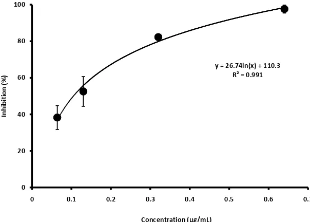
Figure 1. α-Glucosidase-inhibition curve obtained from the extract from Chry.
IC50 calculated from the equation inserted in this figure was 0.41 (μg/mL).
For the α-glucosidase inhibition system, 10 μL of extract was mixed with 40 μL of α-glucosidase solution (32 mU). After 5 min incubation at 37˚C, 950 μL of 0.7 mM 4-nitrophenyl α-d-glucopyranoside in 0.1 M phosphate buffer (pH 6.8) containing 100 mM NaCl was added. After 15 min of incubation at 37˚C, 500 μL of 1 M Tris was added, and the -absorbance of the solution at 400 nm was meas-ured using a micro-plate reader (Varioskan Flash). Calculation of the IC50 was
the same as the α-amylase inhibition system.
2.2.6. Anti-Hypertensive Activity
The anti-hypertensive activity was evaluated as ability to inhibit ACE [18]. Fifty microliters of the extract was mixed with 100 μL of ACE solution (10 mU/mL). After 10 min of incubation at 37˚C, 25 μL of 25 mM hippuryl-histidyl-leucine was added. After 40 min of incubation at 37˚C, 50 μL of 1 M NaOH was added, followed by 10 μL of 0.2% o-phthalaldehyde. After 15 min of incubation at room temperature, 15 μL of 3.6 M phosphate was added, and the fluorescence intensity (Ex: 360 nm, Em: 460 nm) was measured using a micro-plate reader (Varioskan Flash). Calculation of the IC50 was the same as the α-amylase inhibition system.
2.2.7. Anti-Adiposity Activity
The anti-adiposity activity was evaluated as the ability to inhibit lipase [19]. Fifty microliters of the extract was mixed with 50 μL of lipase solution (2 mg/mL) in 50 mL Tris-HCl buffer (pH 8.5). After 5 min of incubation at room temperature, 50 μL of 5 mM p-nitrophenyl laurate in dimethyl sulfoxide was added. After 5 min of incubation at 37˚C, 50 μL of 10% Triton®X100 was added, and the absor-bance of the solution at 410 nm was measured using a micro-plate reader (Vari-oskan Flash). Calculation of the IC50 was the same as the α-amylase inhibition
system.
y = 26.74ln(x) + 110.3 R² = 0.991
0 20 40 60 80 100
0 0.1 0.2 0.3 0.4 0.5 0.6 0.7
In
hi
bi
tio
n (
%)
DOI: 10.4236/fns.2018.94024 319 Food and Nutrition Sciences 2.2.8. Statistical Analysis
All experiments were conducted in triplicate. Data are shown as the means ± standard deviation. A significant difference was determined by Scheffe’s test (P < 0.01).
3. Results and Discussion
3.1. Yield, Colors, and Polyphenol Content of the Extracts
The yield, colors, and polyphenol content of the extracts are summarized in Table 1. The yields varied from 3.0% - 4.8%, based on their origin. As this study used fresh flowers, the yields were much lower those that have used dried flow-ers. For example, an approximate ~17% yield was obtained from a water or ethanol extraction of the dried daylily flower [10]. In addition, a 22% yield was obtained by methanol extraction of dried Rosa damascena [6]. However, Loizzo et al. reported 4.4% - 10.5% yield that was obtained by the ethanol extraction of several fresh edible flowers [20]. These values were slightly higher than those in this study.
The colors of the extracts were in accordance with their petal color before ex-traction, except for that of Fluna. As Fluna is primarily white color, but has a yellow color in the center of the petal, the yellow appeared in the extracts.
The polyphenol content of the extracts was highest for Fluna (18.6%) and lowest for Rosa (10.8%). Rocio et al. [3] reported that a polyphenol content of 15.2% was obtained for the Viola x wittrockiana cv. pansy, which is the same as the results of this study (14.4%). On the other hand, Özkan et al. reported a po-lyphenol content of 27.6% for Rosa damascena extracts [6]. It has been suggested that the polyphenol content differs even for the same genus.
3.2. Anti-Oxidative Activity of the Extracts
All of the extracts exerted more than 2-fold higher α-anti-oxidative activity, compared to α-tocopherol (Figure 2). Especially, the extracts from Fluna, which had highest polyphenol content (Table 1), exerted the highest activity. However, among the other extracts, there was no significant anti-oxidative activity. As the results showed that the polyphenol content of these extracts were varied in signi-ficance (Table 1), it was suggested that the composition of these polyphenols was varied. For example, Rosa, which had lowest polyphenol content, had the kind of polyphenols that have higher radical scavenging activity than the others.
3.3. Anti-Diabetics, Anti-Hypertensive, and Anti-Adiposity
Activities of the Extracts
DOI: 10.4236/fns.2018.94024 320 Food and Nutrition Sciences
Table 1. Yields, colors, and polyphenol content of the extracts from 6 edible flowers.
Samples (color) Yield (%) Color Polyphenol (%)
Chry. (yellow) 4.6 yellow 13.4 ± 0.7ad
Rosa (red) 4.6 red 10.8 ± 0.6bf
Dend. (purple) 3.2 purple 15.5 ± 0.5af
Fluna (white) 3.0 slightly yellow 18.6 ± 0.5c
Pansy (orange) 4.8 yellow 14.4 ± 0.4ad
Prim. (blue) 3.8 blue 12.7 ± 0.2bd
[image:7.595.210.538.267.461.2]adDifferent letters are significantly different in each other (Scheffe’s test, p < 0.01).
Figure 2. Anti-oxidative activity of the extracts from 6 edible flowers. a, b: different
letters are significantly different in each other (Scheffe’s test, p < 0.01).
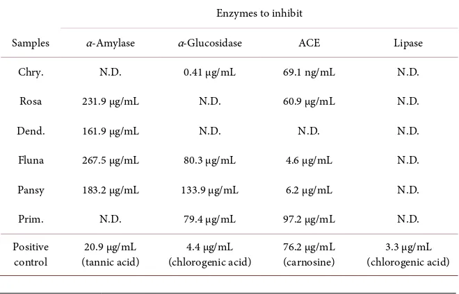
Table 2. Enzyme-inhibitory activities (IC50) of the extracts from 6 edible flowers.
Enzymes to inhibit
Samples α-Amylase α-Glucosidase ACE Lipase
Chry. N.D. 0.41 µg/mL 69.1 ng/mL N.D.
Rosa 231.9 µg/mL N.D. 60.9 µg/mL N.D.
Dend. 161.9 µg/mL N.D. N.D. N.D.
Fluna 267.5 µg/mL 80.3 µg/mL 4.6 µg/mL N.D.
Pansy 183.2 µg/mL 133.9 µg/mL 6.2 µg/mL N.D.
Prim. N.D. 79.4 µg/mL 97.2 µg/mL N.D.
Positive
[image:7.595.209.539.526.738.2]DOI: 10.4236/fns.2018.94024 321 Food and Nutrition Sciences
For anti-diabetes, the extracts from Rosa, Dend., Fluna, and Pansy showed dose-dependent inhibition α-amylase in which, Dend. showed the highest in-hibition with an IC50 value of 161.9 μg/mL, although it was only 1/8 of the
ac-tivity of tannic acid, a positive control. On the other hand, Chry., Fluna, Pan-sy, and Prim. showed a dose-dependent inhibition of α-glucosidase, in which Chry. showed the highest inhibition with an IC50 value of 0.41 μg/mL that was
10-fold higher than chlorogenic acid, a positive control. Loizzo et al. eva-luated the α-amylase/α-glucosidase inhibitory activities of 8 edible flower ex-tracts and reported that 7 of them excluding Sambucus nigra, showed inhibition of both of the two enzymes [20]. In this study, it was unique that Rosa and Dend. showed selective α-amylase inhibitory activity, and Chry. and Prim. showed selective α-glucosidase inhibitory activity. The inhibition mode of methanol/ethanol extracts of Rosa damascena Mill. and banana flower on α-glucosidase were non-competitive mode [21] [22]. Further studies are re-quired to determine the mechanism of inhibition of the extracts used in this study.
For anti-hypertension, the extracts, except for the extract from Dend., showed dose-dependent inhibition of ACE, in which Chry. showed the highest inhibi-tion with an IC50 value of 69.1 ng/mL that was 1000-fold higher than carnosine, a
positive control. As carnosine is a food derivative peptide, we evaluated the ac-tivity of captopril, an ACE-inhibitory drug and revealed that its IC50 was 1.9
ng/mL. The inhibitory activity of peptides for ACE is well known, and a lot of corresponding peptide sequences have also been identified [23]. However, the inhibition level of these peptides is in the order of μg/mL. To our knowledge, few papers describing the inhibitory activity of Chrysanthemum toward ACE have been published. Kim et al. [24] reported the ACE inhibitory activity of a water extract from Chrysanthemum boreale Makino with an IC50 value of 440 ng/mL.
They identified its structure as guanosine. Further studies are required to identi-fy the chemical structures and inhibition mechanism of the compounds eva-luated in this study.
For anti-adiposity, none of the extracts showed inhibitory activity for lipase in this assay.
3.4. Polyphenol Compositions of the Extracts
DOI: 10.4236/fns.2018.94024 322 Food and Nutrition Sciences
Figure 3. HPLC chlomatograms obtained from each sample. Concentration of the samples applied into HPLC were 4.6 mg/mL
(Chry.), 0.46 mg/mL (Rosa), 3.2 mg/mL (Dend.), 0.6 mg/mL (Fluna), 0.24 mg/mL (Pansy), and 0.38 mg/mL (Prim.), respectively.
4. Conclusion
As the color of edible flowers is an important factor that influences consumer purchasing, this study selected 6 edible flowers of different colors (yellow, red, orange, white, purple, and blue) and compared the physiological functions of their 70% ethanol extracts in vitro. Their anti-oxidative activities were 2 - 3-fold higher than α-tocopherol, which was not related to their color. All of the extracts from the edible flowers used in this study showed α-amylase/α-glucosidase bitory activity. Except for the extract from Dend., the extracts showed ACE inhi-bitory activity. Especially, Chry. showed strong inhiinhi-bitory activity of ACE. Fi-nally, none of them showed lipase inhibitory activity. These results suggested that daily consumption of the edible flowers used in this study will help prevent from diabetes and hypertensive diseases.
Acknowledgements
We would like to thank Editage (https://www.editage.jp/) for English language editing.
References
Prefe-DOI: 10.4236/fns.2018.94024 323 Food and Nutrition Sciences
rence for Edible-Flower Color, Container Size, and Price. HortScience, 36, 801-804. [2] Rop, O., Mlcek, J., Jurikova, T., Neugebauerova, J. and Vabkova, J. (2012) Edible
Flowers—A New Promising Source of Mineral Elements in Human Nutrition. Mo-lecules, 17, 6672-6683. https://doi.org/10.3390/molecules17066672
[3] Rocio, G.B., María, J.P, Cristina, L.R., Garcia-Alonso, F.J. and Inmaculada, N.G. (2018) Chemical Composition of the Edible Flowers, Pansy (Viola wittrockiana) and Snapdragon (Antirrhinum majus) as New Sources of Bioactive Compounds.
Food Chemistry, 252, 373-380. https://doi.org/10.1016/j.foodchem.2018.01.102
[4] Pires, T.G.S.P., Dias, M.I., Barros, L., Galhelha, R.G., Alves, M.J., Oliveira, M.B.P.P., Santos-Buelga, G. and Ferreira, I.G.F.R. (2018) Edible Flowers as Sources of Phe-nolic Compounds with Bioactive Potential. Food Research International, 105, 580-588. https://doi.org/10.1016/j.foodres.2017.11.014
[5] Maruyama, T., Yoda, H., Kobori, M., Shinmoto, H. and Tsushida, T. (2002) Evalua-tion of Three Antioxidants and Their IdentificaEvalua-tion and Radical Scavenging Activi-ties in Edible Chrysanthemums. Journal of the Japanese Society for Horitulcultural Science, 71, 236-242. https://doi.org/10.2503/jjshs.71.236
[6] Özkan, G., Sağdiç, O., Baydar, N.G. and Baydar, H. (2003) Antioxidant and Anti-bacterial Activities of Rosa Damascene Flower Extracts. Food Science and Technol-ogy International, 10, 277-281. https://doi.org/10.1177/1082013204045882
[7] Kim, G.C., Kim, J.S., Kim, G.M. and Choi, S.Y. (2017) Anti-Adipogenic Effects of
Tropaeolum majus (Nasturtium) Ethanol Extract on 3T3-L1 Cells. Food & Nutri-tion Research, 61, 1339555. https://doi.org/10.1080/16546628.2017.1339555
[8] Toyoda, M., Tanaka, K., Hoshino, K., Akiyama, H., Tanimura, A. and Saito, Y. (1997) Profiles of Potentially Antiallergic Flavonoids in 27 Kinds of Health Tea and Green Tea Infusions. Journal of Agricultural and Food Chemistry, 45, 2561-2564.
https://doi.org/10.1021/jf970024y
[9] Yasukawa, K., Akihisa, T., Oinuma, H., Kaminaga, T., Konno, H., Kasahara, Y., Tamura, T., Kumaki, K., Yamanouch, S. and Takido, M. (1996) Inhibitory Effect of Taraxastane-Type Triterpenes on Tumor Promotion by 12-O-tetradecanoylphorbol- 13-acetate in Two-Stage Carcinogenesis in Mouse Skin. Oncology, 53, 341-344.
https://doi.org/10.1159/000227584
[10] Mao, L.C., Pan, X., Que, F. and Fang, X.H. (2006) Antioxidant Properties of Water and Ethanol Extracts from Hot Air-Dried and Freeze-Dried Daylily Flowers. Euro-pean Food Research and Technology, 222, 236-241.
https://doi.org/10.1007/s00217-005-0007-0
[11] Tai, Z., Cai, L., Dai, L., Dong, L., Wang, M., Yang, Y., Cao, Q. and Ding, Z. (2011) Antioxidant Activity and Chemical Constituents of Edible Flower of Sophara Vicii-folia. Food Chemistry, 126, 1648-1654.
https://doi.org/10.1016/j.foodchem.2010.12.048
[12] Xiong, L., Yang, J., Jiang, Y., Lu, B., Hu, Y., Zhou, F., Mao, S. and Shen, C. (2014) Phenolic Compounds and Antioxidant Capacities of 10 Common Edible Flowers from China. Journal of Food Science, 79, 517-525.
https://doi.org/10.1111/1750-3841.12404
[13] Folin, O. and Ciocalteu, V. (1927) On Tyrosine and Tryptophane Determinations in Proteins. The Journal of Biological Chemistry, 73, 627-650.
[14] Yu, J., Ahmedna, M. and Goktepe, I. (2005) Effects of Processing Methods and Ex-traction Solvents on Concentration and Antioxidant Activity of Peanut Skin Phe-nolics. Food Chemistry, 90, 199-206.
DOI: 10.4236/fns.2018.94024 324 Food and Nutrition Sciences
[15] Yamamoto, Y., Hiyama, S., Takase, Y., Kadowaki, A. and Hara, S. (2014) Effects of Antioxidants and Additional Emulsifiers on the Stability of Emulsified Milk Fat in the Photo/Radical Oxidation System. Journal of Oleo Science, 63, 893-901.
https://doi.org/10.5650/jos.ess14111
[16] Hara, Y. and Honda, M. (1990) The Inhibition of Alpha-Amylase by Tea Polyphe-nols. Journal of Agricultural Biological Chemistry, 54, 1939-1945.
https://doi.org/10.1271/bbb1961.54.1939
[17] Matsui, T., Ueda, T., Oki, T., Sugita, K., Terahara, N. and Matsumoto, K. (2001)
α-Glucosidase Inhibitory Action of Natural Acylated Anthocyanins. 1. Survey of Natural Pigments with Potent Inhibitory Activity. Journal of Agricultural and Food Chemistry, 49, 1948-1951. https://doi.org/10.1021/jf001251u
[18] Ishiguro, K., Sameshima, Y., Kume, T., Ikeda, K.I., Matsumoto, J. and Yashimoto, M. (2012) Hypotensive Effect of a Seetpotato Protein Digest in Spontaneously Hypertensive Rats and Purification of Angiotensin I-Converting Enzyme Inhibitory Peptides. Food Chemistry, 131, 774-779.
https://doi.org/10.1016/j.foodchem.2011.09.038
[19] Gupta, N., Rathi, P. and Gupta, R. (2002) Simplified para-Nitropheny Palmitate Assay for Lipase and Esterases. Journal of Analytical Biochemistry, 311, 98-99.
https://doi.org/10.1016/S0003-2697(02)00379-2
[20] Loizzo, M.R., Pugliese, A., Bonesi, M., Tenuta, M.C., Manichini, F., Xiao, J. and Tundis, R. (2015) Edible Flowers: A Rich Source of Phytochemicals with Antioxi-dant and Hypoglycemic Properties. Journal of Agricultural and Food Chemistry, 64, 2467-2474. https://doi.org/10.1021/acs.jafc.5b03092
[21] Gholamhoseinian, A., Fallah, H. and Sharifi far, F. (2009) Inhibitory Effect of Me-thanol Extract of Rosa damascena Mill. Flowers on α-Glucosidase Activity and Postprandial Hyperglycemia in Normal and Diabetic Rats. Phytomedicine, 16, 935-941.https://doi.org/10.1016/j.phymed.2009.02.020
[22] Ramu, R., Shirahatti, P.S., Zameer, F., Ranganatha, L.V. and Prasad, M.N.N. (2014). Inhibitory Effect of Banana (Musa sp. var. Nanjangud rasa bale) Flower Extract and Its Constituents Umbelliferone and Lupeol on α-Glucosidase, Aldose Reductase and Glycation at Multiple Stages. South African Journal of Botany, 95, 54-63.
https://doi.org/10.1016/j.sajb.2014.08.001
[23] Lee, S.Y. and Hur, S.J. (2017) Antihypertensive Peptides from Animal Products, Marine Organisms, and Plants. Food Chemistry, 228, 506-517.
https://doi.org/10.1016/j.foodchem.2017.02.039
[24] Kim, J., Lee, S.H., Sun, N., Choung, D.H., Kim, W.K., Lee, S. and Song, K.B. (2006) Isolation of an Angiotensin Converting Enzyme Inhibitory Substance from Chry-santhemum boreale Makino. Food Chemistry and Toxicology, 68, 816-819.